

| Plane | Position | Flip |
| Show planes | Show edges |
0.0
M3#336
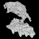
The specimen FSL848325 is separated in two fragments: the anterior part bears the incisors, the deciduous and permanent canines, while the posterior part bears the right P3, P4, M1 and M2. The P2 is isolated. When combined, the cranium length is approximatively 10.5 cm long. The anterior part is 6.9 cm long and 2.15 cm wide (taken at the level of the P1). The posterior part is 4.8 cm long. The anterior part of the cranium is very narrow.
Data citation:
Floréal Solé , Morgane Dubied
, KĂ©vin Le Verger
and Bastien Mennecart
, 2019. M3#336. doi: 10.18563/m3.sf.336
Main model solid/transparent
Show/Hide main model
Second model transparent/solid
Show/Hide second model

|
3D model related to the publication: Niche partitioning of the European carnivorous mammals during the paleogene.FlorĂ©al SolĂ©, Morgane Dubied, KĂ©vin Le Verger and Bastien MennecartPublished online: 21/01/2019Keywords: anatomy; France; juvenile; Oligocene; skull https://doi.org/10.18563/journal.m3.63 Abstract The present 3D Dataset contains the 3D model analyzed in the following publication: Solé et al. (2018), Niche partitioning of the European carnivorous mammals during the paleogene. Palaios. https://doi.org/10.2110/palo.2018.022 M3 article infos Published in Volume 05, issue 01 (2019) |
|