Holotype of Hamadasuchus rebouli
Inner ear morphology in wild vs laboratory mice
3D models of the endocranial anatomy of Voay robustus and comparative specimens
3D GM dataset of bird skeletal variation
Skeletal embryonic development in the catshark
Bony connexions of the petrosal bone of extant hippos
bony labyrinth (11) , inner ear (10) , Eocene (8) , South America (8) , skull (7) , brain (6) , Oligocene (6)
Maëva Judith Orliac (17) , Lionel Hautier (17) , Bastien Mennecart (12) , Laurent Marivaux (11) , Pierre-Olivier Antoine (11) , Leonardo Kerber (10) , Renaud Lebrun (9)
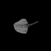
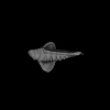
computationally reconstructed stomach of the human embryo (M3#59_KC-CS19STM17998) at Carnegie Stage 19 (Crown Rump Length was unmeasured ).
Data citation:
Ami Nako, Norihito Kaigai, Naoto Shiraki, Shigehito Yamada ![]() , Chigako Uwabe, Katsumi Kose
, Chigako Uwabe, Katsumi Kose ![]() and Tetsuya Takakuwa
and Tetsuya Takakuwa ![]() , 2016. M3#59. doi: 10.18563/m3.sf59
, 2016. M3#59. doi: 10.18563/m3.sf59
Model solid/transparent
Flags:
body of stomach, cardiac incisure, fundus, greater curvature, lesser curvature, pyloric part