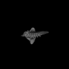
Holotype of Hamadasuchus rebouli
3D models related to the publication: Shape diversity in conodont elements, a quantitative study using 3D topography
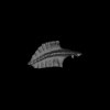
Inner ear morphology in wild vs laboratory mice
3D GM dataset of bird skeletal variation
Skeletal embryonic development in the catshark
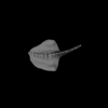
Bony connexions of the petrosal bone of extant hippos
bony labyrinth (11) , inner ear (10) , South America (8) , Eocene (8) , skull (7) , brain (6) , Oligocene (6)
Maëva Judith Orliac (17) , Lionel Hautier (17) , Bastien Mennecart (12) , Laurent Marivaux (11) , Pierre-Olivier Antoine (11) , Leonardo Kerber (10) , Renaud Lebrun (9)
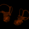
Human liver at Carnegie Stage (CS) 21
Data citation:
Ayumi Hirose ![]() , Takashi Nakashima, Naoto Shiraki, Shigehito Yamada
, Takashi Nakashima, Naoto Shiraki, Shigehito Yamada ![]() , Chigako Uwabe, Katsumi Kose
, Chigako Uwabe, Katsumi Kose ![]() and Tetsuya Takakuwa
and Tetsuya Takakuwa ![]() , 2016. M3#71. doi: 10.18563/m3.sf.71
, 2016. M3#71. doi: 10.18563/m3.sf.71
Model solid/transparent
Flags:
horizontal plane by pyloric antrum, imprint of stomach, indentation by right adrenal gland, IVC, IVC, PV, UV