

| Plane | Position | Flip |
| Show planes | Show edges |
0.0
M3#34
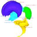
Computationally reconstructed cerebral parenchyma and ventricle of the human embryo at Carnegie Stage 23.
Data citation:
Naoki Shiraishi , Airi Katayama, Takashi Nakashima, Naoto Shiraki, Shigehito Yamada
, Chigako Uwabe, Katsumi Kose
and Tetsuya Takakuwa
, 2015. M3#34. doi: 10.18563/m3.sf34
Tag legend:
aqueduct of midbrain, diencephalon, fourth ventricle, lateral ventricle, mesencephalon, metencephalon, myelencephalon, rhombencephalon, telencephalon, third ventricle
Model solid/transparent
Flags:
aqueduct of midbrain, cerebral hemisphere, chiasmatic plate, diencephalon, epiphysis, fourth ventricle, inferior horn of lateral ventricle, infundibular recess, isthmic groove, isthmic groove, lateral ventricle, mesencephalon, metencephalon(cerebellum), nerve 10, nerve 3, nerve 5, nerve 8, neurohypophysis, olfactory bulb, olfactory recess, postoptic recess, supramamillary recess, synencephalon(medulla oblongata), third ventricle

|
3D models related to the publication: Morphology of the human embryonic brain and ventriclesNaoki Shiraishi, Airi Katayama, Takashi Nakashima, Naoto Shiraki, Shigehito Yamada, Chigako Uwabe, Katsumi Kose and Tetsuya TakakuwaPublished online: 27/07/2015Keywords: human brain; human embryo; magnetic resonance imaging; three-dimensional reconstruction https://doi.org/10.18563/m3.1.3.e3 Abstract This contribution contains the 3D models described and figured in the following publication: Shiraishi N et al. Morphology and morphometry of the human embryonic brain: A three-dimensional analysis NeuroImage 115, 2015, 96-103, DOI: 10.1016/j.neuroimage.2015.04.044. See original publication M3 article infos Published in Volume 01, Issue 03 (2015) |
|