

| Plane | Position | Flip |
| Show planes | Show edges |
Measured length
0.0
0.0
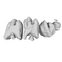
M3#392
Left cheek teeth and incisors at 18 dpf
Data citation:
Ludivine Bertonnier-Brouty , Laurent Viriot
, Thierry Joly
and Cyril Charles
, 2019. M3#392. doi: 10.18563/m3.sf.392
Model solid/transparent

|
3D reconstructions of dental epithelium during Oryctolagus cuniculus embryonic development related to the publication ”Morphological features of tooth development and replacement in the rabbit Oryctolagus cuniculus”Ludivine Bertonnier-Brouty, Laurent Viriot, Thierry Joly and Cyril CharlesPublished online: 30/09/2019Keywords: dental development; Oryctolagus cuniculus; rabbit teeth; tooth replacement https://doi.org/10.18563/journal.m3.90 Abstract The present 3D Dataset contains the 3D models analyzed in ”Morphological features of tooth development and replacement in the rabbit Oryctolagus cuniculus”, Archives of Oral Biology, https://doi.org/10.1016/j.archoralbio.2019.104576 M3 article infos Published in Volume 05, issue 04 (2019) |
|