

| Plane | Position | Flip |
| Show planes | Show edges |
0.0
M3#24
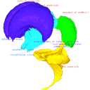
Computationally reconstructed cerebral parenchyma and ventricle of the human embryo at Carnegie Stage 13.
Data citation:
Naoki Shiraishi , Airi Katayama, Takashi Nakashima, Naoto Shiraki, Shigehito Yamada
, Chigako Uwabe, Katsumi Kose
and Tetsuya Takakuwa
, 2015. M3#24. doi: 10.18563/m3.sf24
Tag legend:
aqueduct of midbrain, diencephalon, fourth ventricle, lateral ventricle, mesencephalon, metencephalon, myelencephalon, rhombencephalon, telencephalon, third ventricle
Model solid/transparent
Flags:
aqueduct of midbrain, chiasmatic plate, fourth ventricle, isthmus, mamillary region, mesencephalon, nerve 5, optic ventricle, optic vesicle, prosencephalon, rhombencephalon, third ventricle

|
3D models related to the publication: Morphology of the human embryonic brain and ventriclesNaoki Shiraishi, Airi Katayama, Takashi Nakashima, Naoto Shiraki, Shigehito Yamada, Chigako Uwabe, Katsumi Kose and Tetsuya TakakuwaPublished online: 27/07/2015Keywords: human brain; human embryo; magnetic resonance imaging; three-dimensional reconstruction https://doi.org/10.18563/m3.1.3.e3 Abstract This contribution contains the 3D models described and figured in the following publication: Shiraishi N et al. Morphology and morphometry of the human embryonic brain: A three-dimensional analysis NeuroImage 115, 2015, 96-103, DOI: 10.1016/j.neuroimage.2015.04.044. See original publication M3 article infos Published in Volume 01, Issue 03 (2015) |
|