

| Plane | Position | Flip |
| Show planes | Show edges |
0.0
M3#347
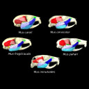
.ply surfaces of the skull and masticatory muscles of Mus minutoides. Created with MorphoDig, .pos and .ntw files also included. Scans were obtained thanks to the Institut des Sciences de l'Evolution de Montpellier MRI platform.
Data citation:
Samuel Ginot , Julien Claude
and Lionel Hautier
, 2018. M3#347. doi: 10.18563/m3.sf.347
Model solid/transparent
Flags:
Anterior deep masseter, Anterior zygomaticomandibularis, External pterygoid, Infra-orbital zygomaticomandibularis, Internal pterygoid, Lateral temporal, Medial temporal, Posterior deep masseter, Posterior zygomaticomandibularis, Skull, Superficial masseter

|
3D models related to the publication: One skull to rule them all? Descriptive and comparative anatomy of the masticatory apparatus in five mice species based on traditional and digital dissections.Samuel Ginot, Julien Claude and Lionel HautierPublished online: 04/09/2018Keywords: Dissection; iodine-enhanced CT-scan; Masticatory musculature; Murinae; skull myology https://doi.org/10.18563/journal.m3.65 Abstract The present 3D Dataset contains the 3D models analyzed in the article entitled "One skull to rule them all? Descriptive and comparative anatomy of the masticatory apparatus in five mice species based on traditional and digital dissections" (Ginot et al. 2018, Journal of Morphology, https://doi.org/10.1002/jmor.20845). M3 article infos Published in Volume 04, issue 02 (2018) |
|