

| Plane | Position | Flip |
| Show planes | Show edges |
0.0
M3#38
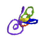
Computationally reconstructed membranous labyrinth of a human embryo (KC-CS19IER16127) at Carnegie Stage 19 (Crown Rump Length= 13mm).
Data citation:
Saki Toyoda, Naoto Shiraki, Shigehito Yamada , Chigako Uwabe, Hirohiko Imai
, Tetsuya Matsuda
, Akio Yoneyama
, Tohoru Takeda and Tetsuya Takakuwa
, 2015. M3#38. doi: 10.18563/m3.sf38
Tag legend:
anterior SD, cochlear duct, lateral SD, posterior SD, utriculus
Model solid/transparent
Flags:
Anterior semicircular duct, Cochlear duct, Common crus, Endolymphatic duct, Endolymphatic sac, Lateral semicircular duct, Posterior semicircular duct, Saccule, Utricle

|
3D models related to the publication: Morphogenesis of the inner ear at different stages of normal human developmentSaki Toyoda, Naoto Shiraki, Shigehito Yamada, Chigako Uwabe, Hirohiko Imai, Tetsuya Matsuda, Akio Yoneyama, Tohoru Takeda and Tetsuya TakakuwaPublished online: 22/10/2015Keywords: human embryo; human inner ear; magnetic resonance imaging; phase-contrast X-ray CT; three-dimensional reconstruction https://doi.org/10.18563/m3.1.3.e6 Abstract The present 3D Dataset contains the 3D models analyzed in: Toyoda S et al., 2015, Morphogenesis of the inner ear at different stages of normal human development. The Anatomical Record. doi : 10.1002/ar.23268 See original publication M3 article infos Published in Volume 01, Issue 03 (2015) |
|