

| Plane | Position | Flip |
| Show planes | Show edges |
Measured length
0.0
0.0
M3#63
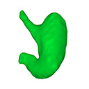
computationally reconstructed stomach of the human embryo (M3#63_KC-CS23STM20018) at Carnegie Stage 23 (Crown Rump Length= 23.1 mm).
Data citation:
Ami Nako, Norihito Kaigai, Naoto Shiraki, Shigehito Yamada , Chigako Uwabe, Katsumi Kose
and Tetsuya Takakuwa
, 2016. M3#63. doi: 10.18563/m3.sf63
Model solid/transparent
Flags:
body of stomach, cardiac incisure, fundus, greater curvature, lesser curvature, pyloric part

|
3D models related to the publication: Morphogenesis of the stomach during the human embryonic periodAmi Nako, Norihito Kaigai, Naoto Shiraki, Shigehito Yamada, Chigako Uwabe, Katsumi Kose and Tetsuya TakakuwaPublished online: 16/11/2015Keywords: human embryo; human stomach; magnetic resonance imaging; three-dimensional reconstruction https://doi.org/10.18563/m3.1.4.e3 Abstract The present 3D Dataset contains the 3D models analyzed in: Kaigai N et al. Morphogenesis and three-dimensional movement of the stomach during the human embryonic period, Anat Rec (Hoboken). 2014 May;297(5):791-797. doi: 10.1002/ar.22833. See original publication M3 article infos Published in Volume 01, Issue 04 (2016) |
|