

| Plane | Position | Flip |
| Show planes | Show edges |
Measured length
0.0
0.0
M3#727
UFAC 1840, left astragalus.
Data citation:
Leonardo Kerber , Adriana M. Candela
, José D. Ferreira
, Flávio A. Pretto
, Jamile Bubadué
and Francisco R. Negri
, 2021. M3#727. doi: 10.18563/m3.sf.727
Model solid/transparent

|
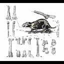
3D models related to the publication: Postcranial morphology of the extinct rodent Neoepiblema (Rodentia: Chinchilloidea): insights into the paleobiology of neoepiblemidsLeonardo Kerber, Adriana M. Candela, JosĂ© D. Ferreira, Flávio A. Pretto, Jamile BubaduĂ© and Francisco R. NegriPublished online: 20/10/2021Keywords: Chinchilloidea; functional morphology; Giant rodents; Neogene; Solimões Formation. https://doi.org/10.18563/journal.m3.140 Abstract This contribution contains the 3D models of postcranial bones (humerus, ulna, innominate, femur, tibia, astragalus, navicular, and metatarsal III) described and figured in the following publication: “Postcranial morphology of the extinct rodent Neoepiblema (Rodentia: Chinchilloidea): insights into the paleobiology of neoepiblemids”. See original publication M3 article infos Published in Volume 07, issue 04 (2021) |
|