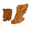
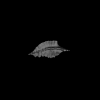
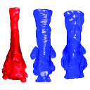
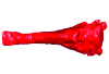
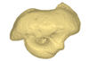
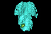
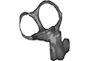
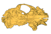
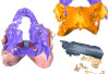
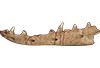
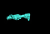
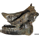
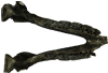
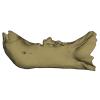
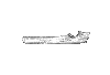
Holotype of Hamadasuchus rebouli
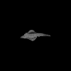
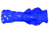
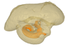
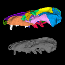
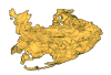
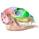
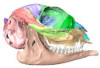
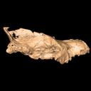
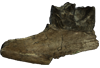
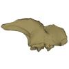
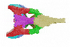
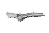
3D model of the holotype specimen of Pebanista yacuruna
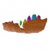
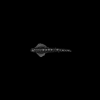
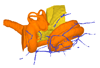
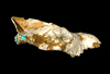
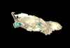
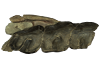
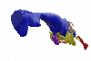
3D models of Eocene–Miocene anuran fossils from Peruvian Amazonia
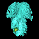
3D GM dataset of bird skeletal variation
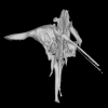
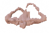
Skeletal embryonic development in the catshark
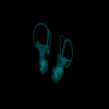
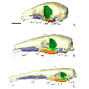
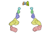
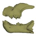
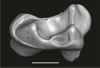
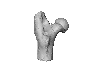
Bony connexions of the petrosal bone of extant hippos
bony labyrinth (11) , inner ear (10) , South America (8) , Eocene (8) , skull (7) , Paleobiogeography (6) , brain (6)
Maëva Judith Orliac (17) , Lionel Hautier (17) , Bastien Mennecart (12) , Pierre-Olivier Antoine (11) , Laurent Marivaux (11) , Leonardo Kerber (10) , Renaud Lebrun (9)
|
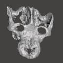
3D models related to the publication: Virtual reconstruction of cranial endocasts of traversodontid cynodonts (Eucynodontia: Gomphodontia) from the upper Triassic of Southern Brazil.Ane E. B. Pavanatto
Published online: 10/09/2019 |
|
M3#4253D model of the brain endocast Type: "3D_surfaces"doi: 10.18563/m3.sf.425 state:published |
Download 3D surface file |
Exaeretodon riograndensis CAPPA/UFSM 0030 View specimen

|
M3#4263D model of the brain endocast Type: "3D_surfaces"doi: 10.18563/m3.sf.426 state:published |
Download 3D surface file |
Exaeretodon riograndensis CAPPA/UFSM 0227 View specimen
|
M3#4273D model of the brain endocast Type: "3D_surfaces"doi: 10.18563/m3.sf.427 state:published |
Download 3D surface file |
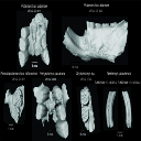
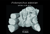
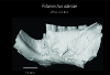
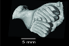
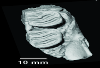
This contribution contains 3D models of extinct rodents Dinomyidae from Miocene and Quaternary of Brazil. The Miocene specimens that were digitalized include the holotypes of Potamarchus adamiae, Pseudopotamarchus villanuevai, and Ferigolomys pacarana collected in the Solimões Formation (Upper Miocene), northern Brazil. The Quaternary specimens are the holotype and paratype of Niedemys piauiensis, found in Upper Pleistocene deposits from northeast Brazil.
Potamarchus adamiae UFAC-CS 011 View specimen
|
M3#410UFAC-CS 011 – holotype, palatal region of the skull with cheek teeth Type: "3D_surfaces"doi: 10.18563/m3.sf.410 state:published |
Download 3D surface file |
Potamarchus adamiae UFAC-CS 043 View specimen
|
M3#411UFAC-CS 043, left dentary with cheek teeth Type: "3D_surfaces"doi: 10.18563/m3.sf.411 state:published |
Download 3D surface file |
Pseudopotamarchus villanuevai UFAC 4762 View specimen
|
M3#412UFAC 4762 – holotype, incomplete right maxilla with cheek teeth Type: "3D_surfaces"doi: 10.18563/m3.sf.412 state:published |
Download 3D surface file |
Ferigolomys pacarana UFAC 6460 View specimen
|
M3#413UFAC 6460 – holotype, palatal region of the skull with cheek teeth Type: "3D_surfaces"doi: 10.18563/m3.sf.413 state:published |
Download 3D surface file |
Drytomomys sp. UFAC 2742 View specimen

|
M3#414UFAC 2742, right dentary with cheek teeth Type: "3D_surfaces"doi: 10.18563/m3.sf.414 state:published |
Download 3D surface file |
Niedemys piauiensis FUMDHAM 113-146365-2 View specimen
|
M3#418FUMDHAM 113-146365-2 - holotype, upper right tooth Type: "3D_surfaces"doi: 10.18563/m3.sf.418 state:published |
Download 3D surface file |
Niedemys piauiensis FUMDHAM 113-145304-2 View specimen
|
M3#419FUMDHAM 113-145304-2 - paratype, left lower molar Type: "3D_surfaces"doi: 10.18563/m3.sf.419 state:published |
Download 3D surface file |
In this contribution, we describe the external and internal morphology of a delphinid petrosal bone collected from Ahu Tahai, a burial site located on the Southwestern coast of Easter Island, at Hangaroa. We discuss the taxonomic attribution of this archaeological item and describe its internal structures based on µCT data, including the bony labyrinth and the nerve and vein patterns. Identification of the nerves exists lead us to relocate the identification of the foramen singulare in delphinid petrosals.
indet. indet. AT1 View specimen
|
M3#420Stapes Type: "3D_surfaces"doi: 10.18563/m3.sf.420 state:published |
Download 3D surface file |
|
M3#421petrosal bone Type: "3D_surfaces"doi: 10.18563/m3.sf.421 state:published |
Download 3D surface file |
|
M3#422in situ bony labyrinth Type: "3D_surfaces"doi: 10.18563/m3.sf.422 state:published |
Download 3D surface file |
|
M3#423bony labyrinth and associated nerves and blood vessels Type: "3D_surfaces"doi: 10.18563/m3.sf.423 state:published |
Download 3D surface file |
The present 3D Dataset contains the 3D models of the holotype (NMB Sth. 833) of the new species Micromeryx? eiselei analysed in the article Aiglstorfer, M., Costeur, L., Mennecart, B., Heizmann, E.P.J.. 2017. Micromeryx? eiselei - a new moschid species from Steinheim am Albuch, Germany, and the first comprehensive description of moschid cranial material from the Miocene of Central Europe. PlosOne https://doi.org/10.1371/journal.pone.0185679
Micromeryx? eiselei NMB Sth. 833 View specimen
|
M3#284The 3 D surfaces comprises the skull, petrosal, and bony labyrinth of NMB Sth.833, the holotype of Micromeryx? eiselei Type: "3D_surfaces"doi: 10.18563/m3.sf.284 state:published |
Download 3D surface file |
Here, the semicircular canals of the most aquatic seal, the rare Antarctic Ross Seal (Ommatophoca rossii), are presented for the first time, along with representatives of every species in the Lobodontini: the leopard seal (Hydrurga leptonyx), Weddell seal (Leptonychotes weddellii), and crabeater seal (Lobodon carcinophagus). Because encounters with wild Ross seal are rare, and few specimens are available in collections worldwide, this dataset increases accessibility to a rare species. For further comparison, we present the bony labyrinths of other carnivorans, the elephant seal (Mirounga leonina), harbor seal (Phoca vitulina), walrus (Odobenus rosmarus), South American sea lion (Otaria byronia).
Odobenus rosmarus MVZ 125566 View specimen
|
M3#173Surface of the semicircular canals and cochlea of the walrus, Odobenus rosmarus Type: "3D_surfaces"doi: 10.18563/m3.sf.173 state:published |
Download 3D surface file |
Phoca vitulina UZNH 17973 View specimen
|
M3#174Endocast surface of the semicircular canals and cochlea of the harbor seal, Phoca vitulina. Type: "3D_surfaces"doi: 10.18563/m3.sf.174 state:published |
Download 3D surface file |
Hydrurga leptonyx MLP 14.IV.48.11 View specimen
|
M3#285Endocast surface of the semicircular canals and cochlea of the leopard seal, Hydrurga leptonyx. Type: "3D_surfaces"doi: 10.18563/m3.sf.285 state:published |
Download 3D surface file |
Leptonychotes weddellii IAA 02-13 View specimen
|
M3#288Endocast surface of the semicircular canals and cochlea of the Weddell seal Leptonychotes weddellii. Type: "3D_surfaces"doi: 10.18563/m3.sf.288 state:published |
Download 3D surface file |
Lobodon carcinophagus IAA 530 View specimen
|
M3#286Endocast surface of the semicircular canals and cochlea of the crabeater seal, Lobodon carcinophagus. Type: "3D_surfaces"doi: 10.18563/m3.sf.286 state:published |
Download 3D surface file |
Ommatophoca rossii MACN 48259 View specimen
|
M3#176Endocast surface of the semicircular canals and cochlea of the Ross seal Ommatophoca rossii. Type: "3D_surfaces"doi: 10.18563/m3.sf.176 state:published |
Download 3D surface file |
Mirounga leonina IAA 03-5 View specimen
|
M3#287Right endocast surface of the semicircular canals and cochlea of the elephant seal, Mirounga leonina. Type: "3D_surfaces"doi: 10.18563/m3.sf.287 state:published |
Download 3D surface file |
The present 3D Dataset contains the 3D model analyzed in the following publication: Carolina A. Hoffmann, A. G. Martinelli & M. B. Andrade. 2023. Anatomy of the holotype of “Probelesodon” kitchingi revisited, a chiniquodontid cynodont (Synapsida, Probainognathia) from the early Late Triassic of southern Brazil, Journal of Paleontology
Probelesodon kitchingi MCP 1600 PV View specimen

|
M3#11513D models of the skull with segmented bones and without the segmentation. colormap and orientation files also added. Type: "3D_surfaces"doi: 10.18563/m3.sf.1151 state:published |
Download 3D surface file |
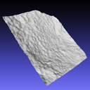
The present 3D Dataset contains the 3D model of the skin of Allosaurus described in Hendrickx, C. et al. in press. Morphology and distribution of scales, dermal ossifications, and other non-feather integumentary structures in non-avialan theropod dinosaurs. Biological Reviews.
Allosaurus jimmadseni UMNH VP C481 View specimen

|
M3#902The material consists of a 3D reconstruction of the counterpart of a 30 cm2 patch of skin impression associated with the anterior dorsal ribs/pectoral region of the specimen of Allosaurus jimmadseni UMNH VP C481. The skin shows a semi-uniform basement of 1-2 mm diameter pebbles with a smaller number of slightly larger (up to 3 mm) ovoid scales. The irregular shape, distribution, and overall small size of these larger scales suggest that they are not classifiable as feature scales but rather as variations in the basement scales. Type: "3D_surfaces"doi: 10.18563/m3.sf.902 state:published |
Download 3D surface file |
The present 3D Dataset contains the 3D models described in “Comparative masticatory myology in anteaters and its implications for interpreting morphological convergence in myrmecophagous placentals”.
Cyclopes didactylus M1571_JAG View specimen
|
M3#522Skull, mandible, and muscles of Cyclopes didactylus Type: "3D_surfaces"doi: 10.18563/m3.sf.522 state:published |
Download 3D surface file |
Tamandua tetradactyla M3075_JAG View specimen
|
M3#524Skull, left mandibles, and muscles of Tamandua tetradactyla. Type: "3D_surfaces"doi: 10.18563/m3.sf.524 state:published |
Download 3D surface file |
Myrmecophaga tridactyla M3023_JAG View specimen
|
M3#523Skull, left mandible and muscles of Myrmecophaga tridactyla. Type: "3D_surfaces"doi: 10.18563/m3.sf.523 state:published |
Download 3D surface file |
The present 3D Dataset contains the 3D models described and figured in the following publication: Grohé C., Bonis L. de, Chaimanee Y., Chavasseau O., Rugbumrung M., Yamee C., Suraprasit K., Gibert C., Surault J., Blondel C., Jaeger J.-J. 2020. The late middle Miocene Mae Moh Basin of northern Thailand: the richest Neogene assemblage of Carnivora from Southeast Asia and a paleobiogeographic analysis of Miocene Asian carnivorans. American Museum Novitates. http://digitallibrary.amnh.org/handle/2246/7223
Siamogale bounosa MM-54 View specimen
|
M3#5053D model of the skull of Siamogale bounosa The zip file contains: - the 3D surface in PLY - the orientation files in .pos and .ori - the project in .ntw Type: "3D_surfaces"doi: 10.18563/m3.sf.505 state:published |
Download 3D surface file |
Vishnuonyx maemohensis MM-78 View specimen
|
M3#5063D model of the skull of Vishnuonyx maemohensis The zip file contains: - the 3D surface in PLY - the orientation files in .pos and .ori - the project in .ntw Type: "3D_surfaces"doi: 10.18563/m3.sf.506 state:published |
Download 3D surface file |
|
M3#5073D model of the reconstructed upper teeth of Vishnuonyx maemohensis The zip file contains: - the 3D surface in PLY - the orientation files in .pos and .ori - the project in .ntw Type: "3D_surfaces"doi: 10.18563/m3.sf.507 state:published |
Download 3D surface file |
The present 3D Dataset contains the 3D models of a skull and lower jaw of the holotype of Santagnathus mariensis, described in “Old fossil findings in the Upper Triassic rocks of southern Brazil improve diversity of traversodontid cynodonts (Therapsida, Cynodontia)”
Santagnathus mariensis UFRGS-PV-1419-T View specimen

|
M3#1157Skull Type: "3D_surfaces"doi: 10.18563/m3.sf.1157 state:published |
Download 3D surface file |

|
M3#1158Lower jaw Type: "3D_surfaces"doi: 10.18563/m3.sf.1158 state:published |
Download 3D surface file |
The present 3D Dataset contains the 3D models analyzed in Keppeler, H., Schultz, J. A., Ruf, I., & Martin, T., 2023. Cranial anatomy of Hypisodus minimus (Artiodactyla: Ruminantia) from the Oligocene Brule Formation of North America. Palaeontographica Abteilung A.
Hypisodus minimus SMNK-PAL 27212 View specimen
|
M3#1031CT image stack of a skull of Hypisodus minimus. Also includes a lumbar vertebra and a probable proximal phalanx of digit III or IV. Type: "3D_CT"doi: 10.18563/m3.sf.1031 state:published |
Download CT data |
|
M3#10363D surface models of a skull of Hypisodus minimus (SMNK-PAL27212). The data includes a surface model for: basisphenoid, tympanic bullae, ethmoid (lamina perpendicularis), frontals, jugal (left), jugal (right), lacrimals, lower dentition, mandibles, mastoid processes, maxillaries, maxilloturbinals, nasals, occipital, palatine, parietals, petrosals, presphenoid, squamosals, turbinates, upper dentition, and the vomer. Type: "3D_surfaces"doi: 10.18563/m3.sf.1036 state:published |
Download 3D surface file |
Hypisodus minimus SMNK-PAL 27213 View specimen
|
M3#1033CT image stack of a skull of Hypisodus minimus. Also shows numerous postcranial material including an atlas articulated with the occipital bone, the distal part of a left humerus articulated to radius and ulna, a part of a femur, a part of a tibia and fibula, unidentifiable tarsal bones, parts of the metatarsals II, III, IV and V and their phalanges, a proximal phalanx of digit III or IV, a middle phalanx of digit III or IV, a possible patella and calcaneus, as well as numerous unidentifiable broken bony fragments. Type: "3D_CT"doi: 10.18563/m3.sf.1033 state:published |
Download CT data |
|
M3#10353D surface models of a skull of Hypisodus minimus (SMNK-PAL27213). The data includes a surface model for: atlas, basisphenoid, tympanic bullae, nasals, occipital, the petrosals, and the inner ear. Type: "3D_surfaces"doi: 10.18563/m3.sf.1035 state:published |
Download 3D surface file |
The present 3D Dataset contains the 3D models of the holotype mandible and referred fragmented skull of the new species Amphimoschus xishuiensis analyzed in the article Li, Y.-K., Mennecart, B., Aiglstorfer, M., Ni, X.-J., Li, Q., Deng, T. 2021. The early evolution of cranial appendages in Bovoidea revealed by new species of Amphimoschus (Mammalia: Ruminantia) from China. Zoological Journal of the Linnean Society https://doi.org/10.1093/zoolinnean/zlab053
Amphimoschus xishuiensis IVPP V 25521.1 View specimen
|
M3#803the holotype, a right hemimandible with tooth row p2 to m3 Type: "3D_surfaces"doi: 10.18563/m3.sf.803 state:published |
Download 3D surface file |
Amphimoschus xishuiensis IVPP V 25521.2 View specimen
|
M3#804referred material, anterior part of a skull with right P4-M3 and left P3-M2 Type: "3D_surfaces"doi: 10.18563/m3.sf.804 state:published |
Download 3D surface file |
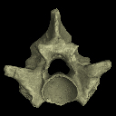
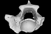
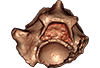
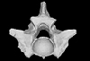
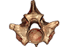
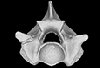
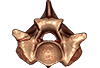
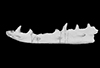
The present 3D Dataset contains the 3D models analyzed in the following publication: Georgalis, G. L., and T. M. Scheyer. A new species of Palaeopython (Serpentes) and other extinct squamates from the Eocene of Dielsdorf (Zurich, Switzerland). Swiss Journal of Geosciences (in press). https://doi.org/10.1007/s00015-019-00341-6
Palaeopython helveticus PIMUZ A/III 631 View specimen
|
M3#399ZIP file containing .ply of vertebra PIMUZ A/III 631 from Palaeopython helveticus n. sp. Type: "3D_surfaces"doi: 10.18563/m3.sf.399 state:published |
Download 3D surface file |
|
M3#403dataset of snake vertebra PIMUZ A/III 631 Type: "3D_CT"doi: 10.18563/m3.sf.403 state:published |
Download CT data |
Palaeopython helveticus PIMUZ A/III 634 View specimen
|
M3#400ZIP file containing .ply of vertebra PIMUZ A/III 634 from Palaeopython helveticus n. sp. (holotype) Type: "3D_surfaces"doi: 10.18563/m3.sf.400 state:published |
Download 3D surface file |
|
M3#404dataset of snake vertbra PIMUZ A/III 634 (holotype) Type: "3D_CT"doi: 10.18563/m3.sf.404 state:published |
Download CT data |
Palaeopython helveticus PIMUZ A/III 636 View specimen
|
M3#401ZIP file containing .ply of vertebra PIMUZ A/III 636 from Palaeopython helveticus n. sp. Type: "3D_surfaces"doi: 10.18563/m3.sf.401 state:published |
Download 3D surface file |
|
M3#406dataset of snake vertebra PIMUZ A/III 636 Type: "3D_CT"doi: 10.18563/m3.sf.406 state:published |
Download CT data |
Palaeovaranus sp. PIMUZ A/III 234 View specimen
|
M3#402ZIP file containing .ply of dentary PIMUZ A/III 234 of Palaeovaranus sp. Type: "3D_surfaces"doi: 10.18563/m3.sf.402 state:published |
Download 3D surface file |
|
M3#405dataset of dentary of Palaeovaranus sp. (PIMUZ A/III 234) Type: "3D_CT"doi: 10.18563/m3.sf.405 state:published |
Download CT data |
The present 3D Dataset contains the 3D model analyzed in the article : Dubied et al. (2021), Endocranium and ecology of Eurotherium theriodis, a European hyaenodont mammal from the Lutetian. Acta Palaeontologica Polonica 2021, https://doi.org/10.4202/app.00771.2020
Eurotherium theriodis NMB.Em12 View specimen
|
M3#381NMB.Em12 unprepared specimen Type: "3D_surfaces"doi: 10.18563/m3.sf.381 state:published |
Download 3D surface file |
|
M3#382NMB.Em12 cranium Type: "3D_surfaces"doi: 10.18563/m3.sf.382 state:published |
Download 3D surface file |
|
M3#383NMB.Em12 endocast Type: "3D_surfaces"doi: 10.18563/m3.sf.383 state:published |
Download 3D surface file |
This contribution contains the 3D models described and figured in: The Neogene record of northern South American native ungulates. Smithsonian Contributions to Paleobiology. Doi: 10.5479/si.1943-6688.101
Hilarcotherium miyou IGMp 881327 View specimen
|
M3#318Right upper M2 Type: "3D_surfaces"doi: 10.18563/m3.sf.318 state:published |
Download 3D surface file |
Hilarcotherium miyou MUN-STRI 34216 View specimen
|
M3#319Right upper P4 Type: "3D_surfaces"doi: 10.18563/m3.sf.319 state:published |
Download 3D surface file |
|
M3#320Right upper M2 Type: "3D_surfaces"doi: 10.18563/m3.sf.320 state:published |
Download 3D surface file |
Falcontoxodon aguilerai AMU-CURS 585 View specimen
|
M3#321Maxilla with left M3-P2 and right I2 Type: "3D_surfaces"doi: 10.18563/m3.sf.321 state:published |
Download 3D surface file |
This contribution contains the 3D models described and figured in the following publication: Tissier et al. (in prep.).
Sellamynodon zimborensis UBB MPS 15795 View specimen
|
M3#297Incomplete skull with left M3. Type: "3D_surfaces"doi: 10.18563/m3.sf.297 state:published |
Download 3D surface file |
Sellamynodon zimborensis UBB MPS 15795 View specimen
|
M3#298Mandible with complete molar and premolar rows, lacking symphysis. Type: "3D_surfaces"doi: 10.18563/m3.sf.298 state:published |
Download 3D surface file |
Amynodontopsis aff. bodei UBB MPS V545 View specimen
|
M3#299Maxillary fragment with M1-3. Type: "3D_surfaces"doi: 10.18563/m3.sf.299 state:published |
Download 3D surface file |
Amynodontopsis aff. bodei UBB MPS V546 View specimen
|
M3#300Unworn m1/2 on mandible fragment. Type: "3D_surfaces"doi: 10.18563/m3.sf.300 state:published |
Download 3D surface file |
This contribution contains the 3D models described and figured in the following publication: Bonis et al. 2023. A new large pantherine and a sabre-toothed cat (Mammalia, Carnivora, Felidae) from the late Miocene hominoid-bearing Khorat sand pits, Nakhon Ratchasima Province, northeastern Thailand. The Science of Nature 110(5):42. https://doi.org/10.1007/s00114-023-01867-4
Pachypanthera piriyai CUF-KR-1 View specimen
|
M3#1209Holotype of Pachypanthera piriyai, a left hemi-mandible with alveoli for i1-i3 and canine, roots of p3, p4 and partially broken off m1 crown. Type: "3D_surfaces"doi: 10.18563/m3.sf.1209 state:published |
Download 3D surface file |
Pachypanthera piriyai CUF-KR-2 View specimen
|
M3#1210Paratype of Pachypanthera piriyai, a right hemi-maxilla with P3-P4, alveoli of C and M1, root of P2 Type: "3D_surfaces"doi: 10.18563/m3.sf.1210 state:published |
Download 3D surface file |
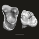
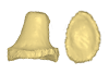
This contribution contains the three-dimensional digital model of one isolated fossil tooth of an anthropoid primate (Ashaninkacebus simpsoni), discovered in sedimentary deposits located on the upper Rio Juruá in State of Acre, Brazil (Western Amazonia). This fossil was described, figured and discussed in the following publication: Marivaux et al. (2023), An eosimiid primate of South Asian affinities in the Paleogene of Western Amazonia and the origin of New World monkeys. Proceedings of the National Academy of Sciences USA. https://doi.org/10.1073/pnas.2301338120
Ashaninkacebus simpsoni UFAC-CS 066 View specimen
|
M3#1114Right first upper molar (rM1), pristine. Type: "3D_surfaces"doi: 10.18563/m3.sf.1114 state:published |
Download 3D surface file |
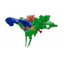
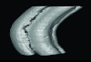
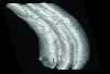
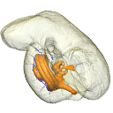
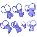
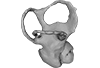
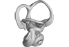
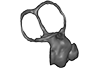
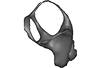
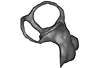
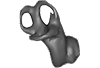
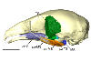
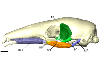
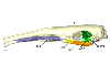
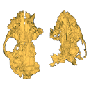
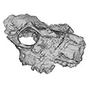
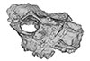
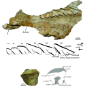
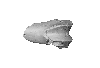
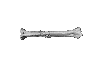
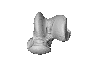
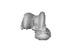
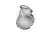
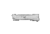
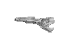
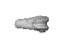
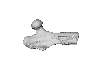
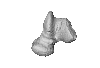
The present 3D Dataset contains the 3D models analyzed in Pochat-Cottilloux Y., Rinder N., Perrichon G., Adrien J., Amiot R., Hua S. & Martin J. E. (2023). The neuroanatomy and pneumaticity of Hamadasuchus from the Cretaceous of Morocco and its significance for the paleoecology of Peirosauridae and other altirostral crocodylomorphs. Journal of Anatomy, https://doi.org/10.1111/joa.13887
Hamadasuchus sp. UCBL-FSL 532408 View specimen
|
M3#10943D volume reconstruction of the braincase osteology Type: "3D_surfaces"doi: 10.18563/m3.sf.1094 state:published |
Download 3D surface file |
|
M3#10963D volume reconstruction of the endocast Type: "3D_surfaces"doi: 10.18563/m3.sf.1096 state:published |
Download 3D surface file |
|
M3#10973D volume reconstruction of the labyrinths Type: "3D_surfaces"doi: 10.18563/m3.sf.1097 state:published |
Download 3D surface file |
|
M3#10983D volume reconstruction of the pneumatic cavities Type: "3D_surfaces"doi: 10.18563/m3.sf.1098 state:published |
Download 3D surface file |
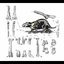
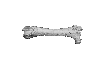
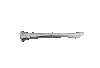
This contribution contains the 3D models of postcranial bones (humerus, ulna, innominate, femur, tibia, astragalus, navicular, and metatarsal III) described and figured in the following publication: “Postcranial morphology of the extinct rodent Neoepiblema (Rodentia: Chinchilloidea): insights into the paleobiology of neoepiblemids”.
Neoepiblema acreensis UFAC 3549 View specimen
|
M3#719UFAC 3549, left humerus missing the proximal region. Type: "3D_surfaces"doi: 10.18563/m3.sf.719 state:published |
Download 3D surface file |
Neoepiblema acreensis UFAC 5076 View specimen
|
M3#720UFAC 5076, right humerus missing the proximal region. Type: "3D_surfaces"doi: 10.18563/m3.sf.720 state:published |
Download 3D surface file |
Neoepiblema acreensis UFAC 1939 View specimen
|
M3#721UFAC 1939, right ulna missing the olecranon epiphysis and the distal region. Type: "3D_surfaces"doi: 10.18563/m3.sf.721 state:published |
Download 3D surface file |
Neoepiblema acreensis UFAC 3697 View specimen
|
M3#722UFAC 3697, right innominate bone. Type: "3D_surfaces"doi: 10.18563/m3.sf.722 state:published |
Download 3D surface file |
Neoepiblema acreensis UFAC 2574 View specimen
|
M3#723UFAC 2574, proximal region of a left femur. Type: "3D_surfaces"doi: 10.18563/m3.sf.723 state:published |
Download 3D surface file |
Neoepiblema acreensis UFAC 2937 View specimen
|
M3#724UFAC 2937, right femur with damaged proximal region. Type: "3D_surfaces"doi: 10.18563/m3.sf.724 state:published |
Download 3D surface file |
Neoepiblema acreensis UFAC 2210 View specimen
|
M3#725UFAC 2210, distal region of a right femur. Type: "3D_surfaces"doi: 10.18563/m3.sf.725 state:published |
Download 3D surface file |
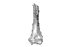
Neoepiblema acreensis UFAC 1887 View specimen
|
M3#726UFAC 1887, right tibia Type: "3D_surfaces"doi: 10.18563/m3.sf.726 state:published |
Download 3D surface file |
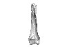
Neoepiblema acreensis UFAC 1840 View specimen
|
M3#727UFAC 1840, left astragalus. Type: "3D_surfaces"doi: 10.18563/m3.sf.727 state:published |
Download 3D surface file |
Neoepiblema acreensis UFAC 2549 View specimen
|
M3#728UFAC 2549, right astragalus. Type: "3D_surfaces"doi: 10.18563/m3.sf.728 state:published |
Download 3D surface file |
Neoepiblema acreensis UFAC 3672 View specimen
|
M3#729UFAC 3672, right navicular. Type: "3D_surfaces"doi: 10.18563/m3.sf.729 state:published |
Download 3D surface file |
Neoepiblema acreensis UFAC 2116 View specimen
|
M3#730UFAC 2116, left metatarsal III. Type: "3D_surfaces"doi: 10.18563/m3.sf.730 state:published |
Download 3D surface file |
Neoepiblema horridula UFAC 3260 View specimen
|
M3#731UFAC 3260, fragmented left innominate. Type: "3D_surfaces"doi: 10.18563/m3.sf.731 state:published |
Download 3D surface file |
Neoepiblema horridula UFAC 2620 View specimen
|
M3#732UFAC 2620, distal region of a right femur. Type: "3D_surfaces"doi: 10.18563/m3.sf.732 state:published |
Download 3D surface file |
Neoepiblema horridula UFAC 2737 View specimen
|
M3#733UFAC 2737, proximal region of right femur. Type: "3D_surfaces"doi: 10.18563/m3.sf.733 state:published |
Download 3D surface file |
Neoepiblema horridula UFAC 3202 View specimen
|
M3#734UFAC 3202, right tibia, missing the proximalmost and distal portions. Type: "3D_surfaces"doi: 10.18563/m3.sf.734 state:published |
Download 3D surface file |
Neoepiblema horridula UFAC 3212 View specimen
|
M3#735UFAC 3212, left astragalus. Type: "3D_surfaces"doi: 10.18563/m3.sf.735 state:published |
Download 3D surface file |