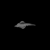
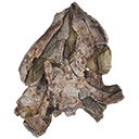
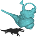
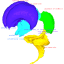
Holotype of Hamadasuchus rebouli
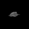
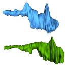
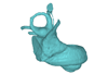
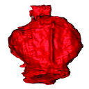
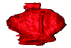
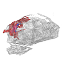
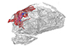
3D model of the holotype specimen of Pebanista yacuruna
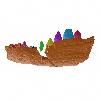
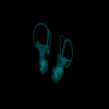
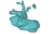
3D models of Eocene–Miocene anuran fossils from Peruvian Amazonia
3D GM dataset of bird skeletal variation
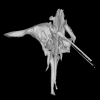
Skeletal embryonic development in the catshark
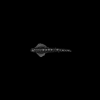
Bony connexions of the petrosal bone of extant hippos
bony labyrinth (11) , inner ear (10) , South America (8) , Eocene (8) , skull (7) , Paleobiogeography (6) , brain (6)
Maëva Judith Orliac (17) , Lionel Hautier (17) , Bastien Mennecart (12) , Laurent Marivaux (11) , Pierre-Olivier Antoine (11) , Leonardo Kerber (10) , Renaud Lebrun (9)
|
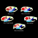
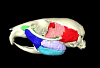
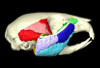
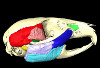
3D models related to the publication: One skull to rule them all? Descriptive and comparative anatomy of the masticatory apparatus in five mice species based on traditional and digital dissections.Samuel Ginot
Published online: 04/09/2018 |
|
M3#343.ply surfaces of the skull and masticatory muscles of Mus cervicolor. Created with MorphoDig, .pos and .ntw files also included. Scans were obtained thanks to the Institut des Sciences de l'Evolution de Montpellier MRI platform. Type: "3D_surfaces"doi: 10.18563/m3.sf.343 state:published |
Download 3D surface file |
Mus caroli R7264 View specimen
|
M3#344.ply surfaces of the skull and masticatory muscles of Mus caroli. Created with MorphoDig, .pos and .ntw files also included. Scans were obtained thanks to the Institut des Sciences de l'Evolution de Montpellier MRI platform. Type: "3D_surfaces"doi: 10.18563/m3.sf.344 state:published |
Download 3D surface file |
Mus fragilicauda R7260 View specimen
|
M3#345.ply surfaces of the skull and masticatory muscles of Mus fragilicauda. Created with MorphoDig, .pos and .ntw files also included. Scans were obtained thanks to the Institut des Sciences de l'Evolution de Montpellier MRI platform. Type: "3D_surfaces"doi: 10.18563/m3.sf.345 state:published |
Download 3D surface file |
Mus pahari R7226 View specimen
|
M3#346.ply surfaces of the skull and masticatory muscles of Mus pahari. Created with MorphoDig, .pos and .ntw files also included. Scans were obtained thanks to the Institut des Sciences de l'Evolution de Montpellier MRI platform. Type: "3D_surfaces"doi: 10.18563/m3.sf.346 state:published |
Download 3D surface file |
Mus minutoides minutoides-1 View specimen
|
M3#347.ply surfaces of the skull and masticatory muscles of Mus minutoides. Created with MorphoDig, .pos and .ntw files also included. Scans were obtained thanks to the Institut des Sciences de l'Evolution de Montpellier MRI platform. Type: "3D_surfaces"doi: 10.18563/m3.sf.347 state:published |
Download 3D surface file |
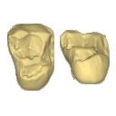
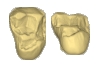
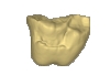
This contribution contains the 3D models of the isolated teeth of Canaanimico amazonensis, a new stem platyrrhine primate, described and figured in the following publication: Marivaux et al. (2016), Neotropics provide insights into the emergence of New World monkeys: new dental evidence from the late Oligocene of Peruvian Amazonia. Journal of Human Evolution. http://dx.doi.org/10.1016/j.jhevol.2016.05.011
Canaanimico amazonensis MUSM-2499 View specimen
|
M3#2893D model of left upper M2 Type: "3D_surfaces"doi: 10.18563/m3.sf.289 state:published |
Download 3D surface file |
Canaanimico amazonensis MUSM-2500 View specimen
|
M3#2903D model of left upper M1 (lingual part) Type: "3D_surfaces"doi: 10.18563/m3.sf.290 state:published |
Download 3D surface file |
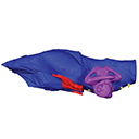
The present publication contains the µCT dataset and the 3D models analyzed in the following publication: Mautner, A.-K., A. E. Latimer, U. Fritz, and T. M. Scheyer. An updated description of the osteology of the pancake tortoise Malacochersus tornieri (Testudines: Testudinidae) with special focus on intraspecific variation. Journal of Morphology. https://doi.org/10.1002/jmor.20640
Malacochersus tornieri ZM 100.102 View specimen

|
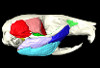
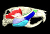
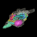
M3#129Virtual brain and inner ear endocast of Malacochersus tornieri (ZM 100.102; Zoological Museum of The University of Zurich). This virtual model is accompanied by the 3D dataset. Blue, endocranium; red, blood vessels; purple, semicircular canals; yellow, cranial nerves. Type: "3D_surfaces"doi: 10.18563/m3.sf.129 state:published |
Download 3D surface file |

|
M3#1303D dataset of skull of Malacochersus tornieri (ZM 100.102) Type: "3D_CT"doi: 10.18563/m3.sf.130 state:published |
Download CT data |
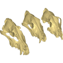
Archaeozoological studies are increasingly using new methods and approaches to explore questions about domestication. Here, we provide 3D models of three archaeological Canis lupus skulls from Belgium originating from the sites of Goyet (31,680±250BP; 31,890+240/-220BP), Trou des Nutons (21,810±90BP) and Trou Balleux (postglacial). Since their identification as either wolves or early dogs is still debated, we present these models as additional tools for further investigating their evolutionary history and the history of dog domestication.
Canis lupus Goyet 2860 View specimen

|
M3#213D surface model of the cranium of the Late Pleistocene Canis lupus "Goyet 2860" from the Royal Belgian Institute of Natural Sciences. Type: "3D_surfaces"doi: 10.18563/m3.sf21 state:published |
Download 3D surface file |
Canis lupus Trou Balleux no-nr View specimen

|
M3#223D surface model of the cranium of the Late Pleistocene Canis lupus "Trou Balleux no-nr" from the University of Liège, Belgium Type: "3D_surfaces"doi: 10.18563/m3.sf22 state:published |
Download 3D surface file |
Canis lupus Trou des Nutons 2559-1 View specimen

|
M3#233D surface model of the cranium of the Late Pleistocene Canis lupus "Trou des Nutons 2559-1" from the Royal Belgian Institute of Natural Sciences. Type: "3D_surfaces"doi: 10.18563/m3.sf23 state:published |
Download 3D surface file |
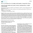
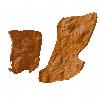
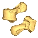
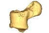
This contribution contains the 3D model described and figured in the following publication: Hautier L, Sarr R, Lihoreau F, Tabuce R, Marwan Hameh P. 2014. First record of the family Protocetidae in the Lutetian of Senegal (West Africa). Palaeovertebrata 38(2)-e2
indet. indet. SN103 View specimen

|
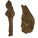
M3#5SN103, partial left innominate. Age and occurrence – Taïba Formation, Lutetian of the near Taïba Ndiaye, quarry of the Industries Chimiques du Sénégal (ICS) Type: "3D_surfaces"doi: 10.18563/m3.sf5 state:published |
Download 3D surface file |
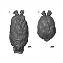
This contribution contains the 3D model(s) described and figured in the following publication: Carolina A. Hoffmann, P. G. Rodrigues, M. B. Soares & M. B. Andrade. 2021. Brain endocast of two non-mammaliaform cynodonts from southern Brazil: an ontogenetic and evolutionary approach, Historical Biology, 33:8, 1196-1207, https://doi.org/10.1080/08912963.2019.1685512
Probelesodon kitchingi MCP 1600 PV View specimen

|
M3#9783D model of the brain endocast of Probelesodon kitchingi. Type: "3D_surfaces"doi: 10.18563/m3.sf.978 state:published |
Download 3D surface file |
Massetognathus ochagaviae MCP 3871 PV View specimen

|
M3#9793D model of the brain endocast of Massetognathus ochagaviae. Type: "3D_surfaces"doi: 10.18563/m3.sf.979 state:published |
Download 3D surface file |
The present 3D Dataset contains the 3D models of the brain endocast analyzed in “Virtual brain endocast of Antifer (Mammalia: Cervidae), an extinct large cervid from South America”.
Antifer ensenadensis U-4922 View specimen
|
M3#550Brain endocast Type: "3D_surfaces"doi: 10.18563/m3.sf.550 state:published |
Download 3D surface file |
Antifer ensenadensis MCN-PV 943 View specimen
|
M3#551Brain endocast Type: "3D_surfaces"doi: 10.18563/m3.sf.551 state:published |
Download 3D surface file |
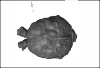
This contribution contains the 3D surface model of the holotype cranium of the Late Jurassic thalassochelydian turtle Solnhofia brachyrhyncha described and figured in the publication of Anquetin and Püntener (2020).
Solnhofia brachyrhyncha MJSN BAN001-2.1 View specimen

|
M3#536Textured 3D surface model of the holotype cranium of the Late Jurassic turtle Solnhofia brachyrhyncha Type: "3D_surfaces"doi: 10.18563/m3.sf.536 state:published |
Download 3D surface file |
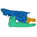
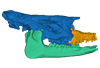
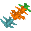
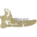
The present 3D Dataset contains two 3D models described in Tissier et al. (https://doi.org/10.1098/rsos.200633): the only known complete mandible of the early-branching rhinocerotoid Epiaceratherium magnum Uhlig, 1999, and a hypothetical reconstruction of the complete archetypic skull of Epiaceratherium Heissig, 1969, created by merging three cranial parts from three distinct Epiaceratherium species.
Epiaceratherium magnum NMB.O.B.928 View specimen
|
M3#5343D surface model of the mandible NMB.O.B.928 of Epiaceratherium magnum, with texture file. Type: "3D_surfaces"doi: 10.18563/m3.sf.534 state:published |
Download 3D surface file |
Epiaceratherium magnum NMB.O.B.928 + MJSN POI007–245 + NMB.I.O.43 View specimen
|
M3#535Archetypal reconstruction of the skull of Epiaceratherium, generated by 3D virtual association of the cranium of E. delemontense (MJSN POI007–245, in blue), mandible of E. magnum (NMB.O.B.928, green) and snout of E. bolcense (NMB.I.O.43, in orange). Type: "3D_surfaces"doi: 10.18563/m3.sf.535 state:published |
Download 3D surface file |
This contribution contains the 3D models of the bony labyrinths of two protocetid archaeocetes from the locality of Kpogamé, Togo, described and figured in the publication of Mourlam and Orliac (2017). https://doi.org/10.1016/j.cub.2017.04.061
?Carolinacetus indet. UM KPG-M 164 View specimen
|
M3#149bony labyrinth of ? Carolinacetus sp. from Kpogamé, Togo Type: "3D_surfaces"doi: 10.18563/m3.sf.149 state:published |
Download 3D surface file |
indet. indet. UM KPG-M 73 View specimen
|
M3#150bony labyrinth of Protocetidae indet. from Kpogamé, Togo Type: "3D_surfaces"doi: 10.18563/m3.sf.150 state:published |
Download 3D surface file |
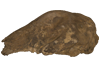
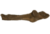
The present 3D Dataset contains the 3D models analyzed in Neogene sloth assemblages (Mammalia, Pilosa) of the Cocinetas Basin (La Guajira, Colombia): implications for the Great American Biotic Interchange. Palaeontology. doi: 10.1111/pala.12244
cf. Nothrotherium indet. MUN STRI 12924 View specimen
|
M3#106Fragmentary basicranium with posterior portion of the skull roof. Type: "3D_surfaces"doi: 10.18563/m3.sf.106 state:published |
Download 3D surface file |
indet. indet. MUN STRI 16535 View specimen
|
M3#107Complete left ulna of a Scelidotheriinae gen. et sp. indet. Type: "3D_surfaces"doi: 10.18563/m3.sf.107 state:published |
Download 3D surface file |
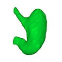
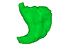
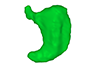
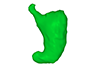
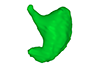
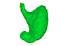
The present 3D Dataset contains the 3D models analyzed in: Kaigai N et al. Morphogenesis and three-dimensional movement of the stomach during the human embryonic period, Anat Rec (Hoboken). 2014 May;297(5):791-797. doi: 10.1002/ar.22833.
Homo sapiens KC-CS16STM27159 View specimen
|
M3#56computationally reconstructed stomach of the human embryo (M3#56_KC-CS16STM27159) at Carnegie Stage 16 (Crown Rump Length= 9.9mm). Type: "3D_surfaces"doi: 10.18563/m3.sf56 state:published |
Download 3D surface file |
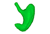
Homo sapiens KC-CS17STM20383 View specimen
|
M3#57computationally reconstructed stomach of the human embryo (M3#57_KC-CS17STM20383) at Carnegie Stage 17 (Crown Rump Length= 12.3mm). Type: "3D_surfaces"doi: 10.18563/m3.sf57 state:published |
Download 3D surface file |
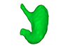
Homo sapiens KC-CS18STM21807 View specimen
|
M3#58computationally reconstructed stomach of the human embryo (M3#58_KC-CS18STM21807) at Carnegie Stage 18 (Crown Rump Length= 14.7mm). Type: "3D_surfaces"doi: 10.18563/m3.sf58 state:published |
Download 3D surface file |
Homo sapiens KC-CS19STM17998 View specimen
|
M3#59computationally reconstructed stomach of the human embryo (M3#59_KC-CS19STM17998) at Carnegie Stage 19 (Crown Rump Length was unmeasured ). Type: "3D_surfaces"doi: 10.18563/m3.sf59 state:published |
Download 3D surface file |
Homo sapiens KC-CS20STM20785 View specimen
|
M3#60computationally reconstructed stomach of the human embryo (M3#60_KC-CS20STM20785) at Carnegie Stage 20 (Crown Rump Length= 18.7 mm). Type: "3D_surfaces"doi: 10.18563/m3.sf60 state:published |
Download 3D surface file |
Homo sapiens KC-CS21STM24728 View specimen
|
M3#61computationally reconstructed stomach of the human embryo (M3#61_KC-CS21STM24728) at Carnegie Stage 21 (Crown Rump Length= 20.9 mm). Type: "3D_surfaces"doi: 10.18563/m3.sf61 state:published |
Download 3D surface file |
Homo sapiens KC-CS22STM26438 View specimen
|
M3#62computationally reconstructed stomach of the human embryo (M3#62_KC-CS22STM26438) at Carnegie Stage 22 (Crown Rump Length= 21.5 mm). Type: "3D_surfaces"doi: 10.18563/m3.sf62 state:published |
Download 3D surface file |
Homo sapiens KC-CS23STM20018 View specimen
|
M3#63computationally reconstructed stomach of the human embryo (M3#63_KC-CS23STM20018) at Carnegie Stage 23 (Crown Rump Length= 23.1 mm). Type: "3D_surfaces"doi: 10.18563/m3.sf63 state:published |
Download 3D surface file |
This contribution contains the 3D models described and figured in the following publication: Molnar, JL, Pierce, SE, Bhullar, B-A, Turner, AH, Hutchinson, JR (accepted). Morphological and functional changes in the crocodylomorph vertebral column with increasing aquatic adaptation. Royal Society Open Science.
Protosuchus richardsoni AMNH-VP 3024 View specimen

|
M3#448th and 9th dorsal vertebrae, 1st and 2nd lumbar vertebrae, and 5th lumbar and sacral vertebrae. Type: "3D_surfaces"doi: 10.18563/m3.sf44 state:published |
Download 3D surface file |
Terrestrisuchus gracilis NHM-PV R 7562 View specimen

|
M3#451st and 2nd lumbar vertebrae, and 5th lumbar and sacral vertebrae Type: "3D_surfaces"doi: 10.18563/m3.sf45 state:published |
Download 3D surface file |
Pelagosaurus typus NHM-PV OR 32598 View specimen

|
M3#467th and 8th dorsal vertebrae, 11th and 12th dorsal vertebrae, 15th dorsal vertebra and sacral vertebra. Type: "3D_surfaces"doi: 10.18563/m3.sf46 state:published |
Download 3D surface file |
Metriorhynchus superciliosus NHM-PV R 2054 View specimen

|
M3#476th and 7th dorsal vertebrae, 10th and 11th dorsal vertebrae, 17th dorsal vertebra and sacral vertebra Type: "3D_surfaces"doi: 10.18563/m3.sf47 state:published |
Download 3D surface file |
Crocodylus niloticus FNC0 View specimen

|
M3#487th and 8th dorsal vertebrae, 1st and 2nd lumbar vertebrae, 5th lumbar vertebra and sacral vertebra. Type: "3D_surfaces"doi: 10.18563/m3.sf48 state:published |
Download 3D surface file |
This contribution contains the 3D models described and figured in the following publication: Shiraishi N et al. Morphology and morphometry of the human embryonic brain: A three-dimensional analysis NeuroImage 115, 2015, 96-103, DOI: 10.1016/j.neuroimage.2015.04.044.
Homo sapiens KC-CS13BRN50455 View specimen

|
M3#24Computationally reconstructed cerebral parenchyma and ventricle of the human embryo at Carnegie Stage 13. Type: "3D_surfaces"doi: 10.18563/m3.sf24 state:published |
Download 3D surface file |
Homo sapiens KC-CS14BRN18834 View specimen

|
M3#25Computationally reconstructed cerebral parenchyma and ventricle of the human embryo at Carnegie Stage 14. Type: "3D_surfaces"doi: 10.18563/m3.sf25 state:published |
Download 3D surface file |
Homo sapiens KC-CS15BRN19975 View specimen

|
M3#26Computationally reconstructed cerebral parenchyma and ventricle of the human embryo at Carnegie Stage 15. Type: "3D_surfaces"doi: 10.18563/m3.sf26 state:published |
Download 3D surface file |
Homo sapiens KC-CS16BRN7870 View specimen

|
M3#27Computationally reconstructed cerebral parenchyma and ventricle of the human embryo at Carnegie Stage 16. Type: "3D_surfaces"doi: 10.18563/m3.sf27 state:published |
Download 3D surface file |
Homo sapiens KC-CS17BRN26702 View specimen

|
M3#28Computationally reconstructed cerebral parenchyma and ventricle of the human embryo at Carnegie Stage 17. Type: "3D_surfaces"doi: 10.18563/m3.sf28 state:published |
Download 3D surface file |
Homo sapiens KC-CS18BRN25914 View specimen

|
M3#29Computationally reconstructed cerebral parenchyma and ventricle of the human embryo at Carnegie Stage 18. Type: "3D_surfaces"doi: 10.18563/m3.sf29 state:published |
Download 3D surface file |
Homo sapiens KC-CS19BRN16508 View specimen

|
M3#30Computationally reconstructed cerebral parenchyma and ventricle of the human embryo at Carnegie Stage 19. Type: "3D_surfaces"doi: 10.18563/m3.sf30 state:published |
Download 3D surface file |
Homo sapiens KC-CS20BRN26581 View specimen

|
M3#31Computationally reconstructed cerebral parenchyma and ventricle of the human embryo at Carnegie Stage 20. Type: "3D_surfaces"doi: 10.18563/m3.sf31 state:published |
Download 3D surface file |
Homo sapiens KC-CS21BRN33434 View specimen

|
M3#32Computationally reconstructed cerebral parenchyma and ventricle of the human embryo at Carnegie Stage 21. Type: "3D_surfaces"doi: 10.18563/m3.sf32 state:published |
Download 3D surface file |
Homo sapiens KC-CS22BRN27960 View specimen

|
M3#33Computationally reconstructed cerebral parenchyma and ventricle of the human embryo at Carnegie Stage 22. Type: "3D_surfaces"doi: 10.18563/m3.sf33 state:published |
Download 3D surface file |
Homo sapiens KC-CS23BRN28189 View specimen

|
M3#34Computationally reconstructed cerebral parenchyma and ventricle of the human embryo at Carnegie Stage 23. Type: "3D_surfaces"doi: 10.18563/m3.sf34 state:published |
Download 3D surface file |
This project presents a µCT dataset and an associated 3D surface model of the holotype of Donrussellia magna (UM PAT 17; Primates, Adapiformes). UM PAT17 is the only known specimen for the species and consists of a well-preserved left lower jaw with p4-m3. It documents one of the oldest European primates, eventually dated near the Paleocene Eocene Thermal Maximum.
Donrussellia magna UM PAT 17 View specimen

|
M3#173D surface file model of UM PAT 17 (type specimen of Donrussellia magna), which is a well preserved left lower jaw with p4-m3. The teeth (and roots) were manually segmented. Type: "3D_surfaces"doi: 10.18563/m3.sf17 state:published |
Download 3D surface file |
|
M3#18CT Scan Data of Donrussellia magna UM PAT 17. Voxel size (in µm): 36µm (isotropic voxels). Dimensions in x,y,z : 594 pixels, 294 pixels, 1038 pixels. Image type : 8-bit voxels. Image format : raw data format (no header). Type: "3D_CT"doi: 10.18563/m3.sf18 state:published |
Download CT data |
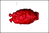
The present Dataset contains the 3D model of the male genital organs of greater horseshoe bat, Rhinolophus ferrumequinum. This is the first detailed 3D structure of the soft-tissue genital organs of bats. The 3D model was generated using microCT and techniques of virtual reconstruction.
Rhinolophus ferrumequinum JP18-006 View specimen

|
M3#521The genital organs of male greater horseshoe bat. Type: "3D_surfaces"doi: 10.18563/m3.sf.521 state:published |
Download 3D surface file |
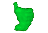
The present 3D Dataset contains the 3D model of the brain endocast of Neoepiblema acreensis analyzed in “Small within the largest: Brain size and anatomy of the extinct Neoepiblema acreensis, a giant rodent from the Neotropics”. The 3D model was generated using CT-Scanning and techniques of virtual reconstruction.
Neoepiblema acreensis UFAC 4515 View specimen
|
M3#502Brain endocast of Neoepiblema acreensis Type: "3D_surfaces"doi: 10.18563/m3.sf.502 state:published |
Download 3D surface file |
The present 3D Dataset contains the 3D models analyzed in the following publication: Paulina-Carabajal, A., Ezcurra, M., Novas, F., 2019. New information on the braincase and endocranial morphology of the Late Triassic neotheropod Zupaysaurus rougieri using Computed Tomography data. Journal of Vertebrate Paleontology. https://doi.org/10.1080/02724634.2019.1630421
Zupaysaurus rougieri PULR 076 View specimen
|
M3#424The Zip contains 3 files, which correspond to: PULR_076-M1: Zupaysaurus rougieri skull, braincase and cranial endocast PULR_076-M2: Zupaysaurus rougieri braincase PULR_076-M1: Zupaysaurus rougieri brain and inner ear Type: "3D_surfaces"doi: 10.18563/m3.sf.424 state:published |
Download 3D surface file |
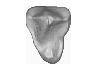
This contribution contains the 3D model of the fossil talus of a small-bodied anthropoid primate (Platyrrhini, Cebidae, Cebinae) discovered from lower Miocene deposits of Peruvian Amazonia (MD-61 locality, Upper Madre de Dios Basin). This fossil was described and figured in the following publication: Marivaux et al. (2012), A platyrrhine talus from the early Miocene of Peru (Amazonian Madre de Dios Sub-Andean Zone). Journal of Human Evolution. http://dx.doi.org/10.1016/j.jhevol.2012.07.005
Cebinae indet. sp. MUSM-2024 View specimen
|
M3#380Right talus 3D surface of a Miocene Cebinae indet. primate Type: "3D_surfaces"doi: 10.18563/m3.sf.380 state:published |
Download 3D surface file |
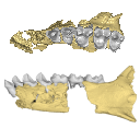
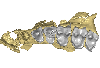
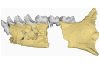
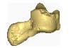
This contribution contains the 3D models of the fossil remains (maxilla, dentary, and talus) attributed to Djebelemur martinezi, a ca. 50 Ma primate from Tunisia (Djebel Chambi), described and figured in the following publication: Marivaux et al. (2013), Djebelemur, a tiny pre-tooth-combed primate from the Eocene of Tunisia: a glimpse into the origin of crown strepsirhines. PLoS ONE. https://doi.org/10.1371/journal.pone.0080778
Djebelemur martinezi CBI-1-544 View specimen
|
M3#365CBI-1-544, left maxilla preserving P3-M3 and alveoli for P2 and C1 Type: "3D_surfaces"doi: 10.18563/m3.sf.365 state:published |
Download 3D surface file |
Djebelemur martinezi CBI-1-567 View specimen
|
M3#363Isolated left upper P4 Type: "3D_surfaces"doi: 10.18563/m3.sf.363 state:published |
Download 3D surface file |
Djebelemur martinezi CBI-1-565-577-587-580 View specimen
|
M3#366- CBI-1-565, a damaged right mandible, which consists of three isolated pieces found together and reassembled here: the anterior part of the dentary bears the p3 and m1, and alveoli for p4, p2 and c, while the posterior part preserves m3 and a portion of the ascending ramus; the m2 was found isolated but in the same small calcareous block treated by acid processing. - CBI-1-577, isolated right lower p4. - CBI-1-587, isolated left lower p2 (reversed). - CBI-1-580, isolated left lower canine (reversed). Type: "3D_surfaces"doi: 10.18563/m3.sf.366 state:published |
Download 3D surface file |
Djebelemur martinezi CBI-1-545 View specimen
|
M3#364Right Talus Type: "3D_surfaces"doi: 10.18563/m3.sf.364 state:published |
Download 3D surface file |