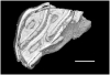
Holotype of Hamadasuchus rebouli
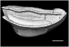
3D model of the holotype specimen of Pebanista yacuruna
3D models of Eocene–Miocene anuran fossils from Peruvian Amazonia
3D GM dataset of bird skeletal variation
Skeletal embryonic development in the catshark
Bony connexions of the petrosal bone of extant hippos
bony labyrinth (11) , inner ear (10) , South America (8) , Eocene (8) , skull (7) , brain (6) , Oligocene (6)
Maëva Judith Orliac (17) , Lionel Hautier (17) , Bastien Mennecart (12) , Laurent Marivaux (11) , Pierre-Olivier Antoine (11) , Leonardo Kerber (10) , Renaud Lebrun (9)
|
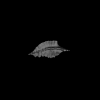
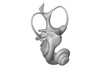
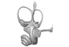
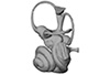
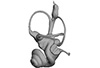
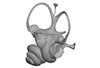
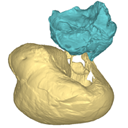
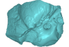
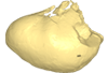
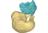
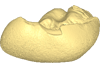
3D model related to the publication: The cranium of Proviverra typica (Mammalia, Hyaenodonta) and its impact on hyaenodont phylogeny and endocranial evolutionMorgane Dubied
Published online: 26/08/2019 |
|
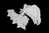
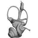
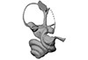
M3#355The file contain the cranium (yellow) and the endocast (blue) of the facial part and the brain case part of the type specimen of Proviverra typica (NMB Em18). Type: "3D_surfaces"doi: 10.18563/m3.sf.355 state:published |
Download 3D surface file |
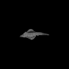
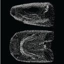
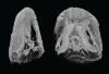
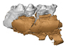
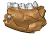
This contribution contains the 3D model of the holotype of Chambius kasserinensis, the basalmost ‘elephant-shrew’ figured in the following publication: New remains of Chambius kasserinensis from the Eocene of Tunisia and evaluation of proposed affinities for Macroscelidea (Mammalia, Afrotheria). https://doi.org/10.1080/08912963.2017.1297433
Chambius kasserinensis CBI-1-06 View specimen
|
M3#1463D model of the holotype maxilla of Chambius kasserinensis. The 3D surface was extracted manually from the limestone matrix within AVIZO 9.2 Type: "3D_surfaces"doi: 10.18563/m3.sf.146 state:published |
Download 3D surface file |
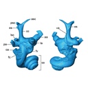
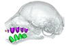
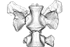
The present 3D Dataset contains the 3D models analyzed in Costeur L., Mennecart B., Müller B., Schulz G., 2016. Prenatal growth stages show the development of the ruminant bony labyrinth and petrosal bone. Journal of Anatomy. https://doi.org/10.1111/joa.12549
Bos taurus NMB3038 View specimen
|
M3#124Right bony labyrinth of a Bos taurus foetus (gestational age 115 days) Type: "3D_surfaces"doi: 10.18563/m3.sf.124 state:published |
Download 3D surface file |
Bos taurus NMB3367 View specimen
|
M3#125Right bony labyrinth of a Bos taurus foetus (gestational age 165 days) Type: "3D_surfaces"doi: 10.18563/m3.sf.125 state:published |
Download 3D surface file |
Bos taurus NMB3365 View specimen
|
M3#126Right bony labyrinth of a Bos taurus foetus (gestational age 210 days) Type: "3D_surfaces"doi: 10.18563/m3.sf.126 state:published |
Download 3D surface file |
Bos taurus NMB2855 View specimen
|
M3#127Right bony labyrinth of a Bos taurus foetus (gestational age 260 days) Type: "3D_surfaces"doi: 10.18563/m3.sf.127 state:published |
Download 3D surface file |
Bos taurus NMB1037 View specimen
|
M3#128Left bony labyrinth of an adult Bos taurus Type: "3D_surfaces"doi: 10.18563/m3.sf.128 state:published |
Download 3D surface file |
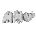
This contribution contains the 3D models described and figured in the following publication: Hautier L., Gomes Rodrigues H., Billet G., Asher R.J., 2016. The hidden teeth of sloths: evolutionary vestiges and the development of a simplified dentition. Scientific Reports. doi: 10.1038/srep27763
Bradypus variegatus ZMB 33812 View specimen
|
M3#110Three-dimensional reconstruction of the teeth, mandibles, maxillary and premaxillary bones Type: "3D_surfaces"doi: 10.18563/m3.sf.110 state:published |
Download 3D surface file |
Bradypus variegatus ZMB 41122 View specimen
|
M3#109Three-dimensional reconstruction of the teeth, mandibles, maxillary and premaxillary bones Type: "3D_surfaces"doi: 10.18563/m3.sf.109 state:published |
Download 3D surface file |
Bradypus variegatus MNHN-ZM-MO-1995-326A View specimen
|
M3#111Three-dimensional reconstruction of the teeth, mandibles, maxillary and premaxillary bones Type: "3D_surfaces"doi: 10.18563/m3.sf.111 state:published |
Download 3D surface file |
Bradypus variegatus MNHN-ZM-MO-1995-326B View specimen
|
M3#112Three-dimensional reconstruction of the teeth, mandibles, maxillary and premaxillary bones Type: "3D_surfaces"doi: 10.18563/m3.sf.112 state:published |
Download 3D surface file |
Bradypus sp. MNHN-ZM-MO-1902-325 View specimen
|
M3#113Three-dimensional reconstruction of the teeth, mandibles, maxillary, and premaxillary bones Type: "3D_surfaces"doi: 10.18563/m3.sf.113 state:published |
Download 3D surface file |
Bradypus sp. MNHN-ZM-MO-1995-327 View specimen
|
M3#114Three-dimensional reconstruction of the teeth, mandibles, maxillary and premaxillary bones Type: "3D_surfaces"doi: 10.18563/m3.sf.114 state:published |
Download 3D surface file |
Choloepus didactylus MNHN-ZM-MO-1882-625 View specimen
|
M3#115Three-dimensional reconstruction of the teeth, mandibles, maxillary and premaxillary bones Type: "3D_surfaces"doi: 10.18563/m3.sf.115 state:published |
Download 3D surface file |
The present 3D Dataset contains the 3D models analyzed in: Toyoda S et al., 2015, Morphogenesis of the inner ear at different stages of normal human development. The Anatomical Record. doi : 10.1002/ar.23268
Homo sapiens KC-CS17IER29248 View specimen

|
M3#36Computationally reconstructed membranous labyrinth of a human embryo (KC-CS17IER29248) at Carnegie Stage 17 (Crown Rump Length= 7mm). Type: "3D_surfaces"doi: 10.18563/m3.sf36 state:published |
Download 3D surface file |
Homo sapiens KC-CS18IER17746 View specimen

|
M3#37Computationally reconstructed membranous labyrinth of a human embryo (KC-CS18IER17746) at Carnegie Stage 18 (Crown Rump Length= 12mm). Type: "3D_surfaces"doi: 10.18563/m3.sf37 state:published |
Download 3D surface file |
Homo sapiens KC-CS19IER16127 View specimen

|
M3#38Computationally reconstructed membranous labyrinth of a human embryo (KC-CS19IER16127) at Carnegie Stage 19 (Crown Rump Length= 13mm). Type: "3D_surfaces"doi: 10.18563/m3.sf38 state:published |
Download 3D surface file |
Homo sapiens KC-CS20IER20268 View specimen

|
M3#39Computationally reconstructed membranous labyrinth of a human embryo (KC-CS20IER20268) at Carnegie Stage 20 (Crown Rump Length= 13.7mm). Type: "3D_surfaces"doi: 10.18563/m3.sf39 state:published |
Download 3D surface file |
Homo sapiens KC-CS21IER28066 View specimen

|
M3#40Computationally reconstructed membranous labyrinth of a human embryo (KC-CS21IER28066) at Carnegie Stage 21 (Crown Rump Length= 16.7mm). Type: "3D_surfaces"doi: 10.18563/m3.sf40 state:published |
Download 3D surface file |
Homo sapiens KC-CS22IER35233 View specimen

|
M3#41Computationally reconstructed membranous labyrinth of a human embryo (KC-CS22IER35233) at Carnegie Stage 22 (Crown Rump Length= 22mm). Type: "3D_surfaces"doi: 10.18563/m3.sf41 state:published |
Download 3D surface file |
Homo sapiens KC-CS23IER15919 View specimen

|
M3#42Computationally reconstructed membranous labyrinth of a human embryo (KC-CS23IER15919) at Carnegie Stage 23 (Crown Rump Length= 32.3mm). Type: "3D_surfaces"doi: 10.18563/m3.sf42 state:published |
Download 3D surface file |
Homo sapiens KC-FIER52730 View specimen

|
M3#43Computationally reconstructed human membranous labyrinth in post embryonic phase (KC-FIER52730). Crown Rump Length: 43.5mm. Type: "3D_surfaces"doi: 10.18563/m3.sf43 state:published |
Download 3D surface file |
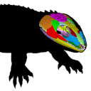
The present 3D Dataset contains the 3D models analyzed in: Abel P., Pommery Y., Ford D. P., Koyabu D., Werneburg I. 2022. Skull sutures and cranial mechanics in the Permian reptile Captorhinus aguti and the evolution of the temporal region in early amniotes. Frontiers in Ecology and Evolution. https://doi.org/10.3389/fevo.2022.841784
Captorhinus aguti OMNH 44816 View specimen

|
M3#965Segmented cranial bone surfaces of OMNH 44816 Type: "3D_surfaces"doi: 10.18563/m3.sf.965 state:published |
Download 3D surface file |
This contribution contains the 3D models described and figured in the following publication: Hautier L, Tabuce R, Kassegne KE, Amoudji YZ, Mourlam M, Orliac M, Quillévéré F, Charruault A-L, Johnson AKC, Guinot G. 2021. New middle Eocene proboscidean from Togo illuminates the early evolution of the elephantiform-like dental pattern.
Dagbatitherium tassyi ULDG-DAG1 View specimen
|
M3#7693D model of a molar of Dagbatitherium tassyi. Type: "3D_surfaces"doi: 10.18563/m3.sf.769 state:published |
Download 3D surface file |
|
M3#771µCT scan of a molar of Dagbatitherium tassyi. Type: "3D_CT"doi: 10.18563/m3.sf.771 state:published |
Download CT data |
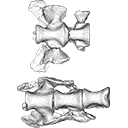
The present 3D Dataset contains the 3D models of the sacral vertebrae analyzed in “Sacral co-ossification in dinosaurs: The oldest record of fused sacral vertebrae in Dinosauria and the diversity of sacral co-ossification patterns in the group”.
Buriolestes schultzi CAPPA/UFSM 0035 View specimen
|
M3#705Sacral vertebrae of Buriolestes schultzi Type: "3D_surfaces"doi: 10.18563/m3.sf.705 state:published |
Download 3D surface file |
indet indet CAPPA/UFSM 0228 View specimen
|
M3#706Sacral vertebrae of a saurischian dinosaur indet. Type: "3D_surfaces"doi: 10.18563/m3.sf.706 state:published |
Download 3D surface file |
The present 3D Dataset contains the 3D models analyzed in ”Morphological features of tooth development and replacement in the rabbit Oryctolagus cuniculus”, Archives of Oral Biology, https://doi.org/10.1016/j.archoralbio.2019.104576
Oryctogalus cuniculus E14 View specimen

|
M3#390Right cheek teeth, Left and right incisors at 14 dpf Type: "3D_surfaces"doi: 10.18563/m3.sf.390 state:published |
Download 3D surface file |
Oryctogalus cuniculus E16 View specimen

|
M3#391Left cheek teeth, Left and right incisors at 16 dpf Type: "3D_surfaces"doi: 10.18563/m3.sf.391 state:published |
Download 3D surface file |
Oryctogalus cuniculus E18 View specimen

|
M3#392Left cheek teeth and incisors at 18 dpf Type: "3D_surfaces"doi: 10.18563/m3.sf.392 state:published |
Download 3D surface file |
Oryctogalus cuniculus E20 View specimen

|
M3#393Left cheek teeth and incisors at 20 dpf Type: "3D_surfaces"doi: 10.18563/m3.sf.393 state:published |
Download 3D surface file |
Oryctogalus cuniculus E22 View specimen

|
M3#394Left lower cheek teeth and incisors, right upper cheek teeth and incisors at 22 dpf Type: "3D_surfaces"doi: 10.18563/m3.sf.394 state:published |
Download 3D surface file |
Oryctogalus cuniculus E24 View specimen

|
M3#395Left cheek teeth and incisors at 24 dpf Type: "3D_surfaces"doi: 10.18563/m3.sf.395 state:published |
Download 3D surface file |
Oryctogalus cuniculus E28 View specimen

|
M3#396Right cheek teeth and incisors at 28 dpf Type: "3D_surfaces"doi: 10.18563/m3.sf.396 state:published |
Download 3D surface file |
Oryctogalus cuniculus E26 View specimen

|
M3#397Right cheek teeth and incisors at 26 dpf Type: "3D_surfaces"doi: 10.18563/m3.sf.397 state:published |
Download 3D surface file |
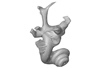
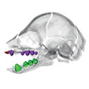
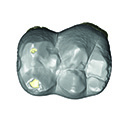
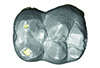
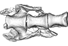
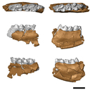
The present 3D Dataset contains the 3D model analyzed in the following publication: Solé et al. (2018), Niche partitioning of the European carnivorous mammals during the paleogene. Palaios. https://doi.org/10.2110/palo.2018.022
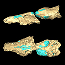
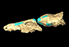
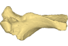
Hyaenodon leptorhynchus FSL848325 View specimen
|
M3#336The specimen FSL848325 is separated in two fragments: the anterior part bears the incisors, the deciduous and permanent canines, while the posterior part bears the right P3, P4, M1 and M2. The P2 is isolated. When combined, the cranium length is approximatively 10.5 cm long. The anterior part is 6.9 cm long and 2.15 cm wide (taken at the level of the P1). The posterior part is 4.8 cm long. The anterior part of the cranium is very narrow. Type: "3D_surfaces"doi: 10.18563/m3.sf.336 state:published |
Download 3D surface file |
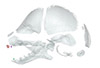
The present contribution contains the 3D model and dataset analyzed in the following publication: Scheyer, T. M., J. M. Neenan, T. Bodogan, H. Furrer, C. Obrist, and M. Plamondon. 2017. A new, exceptionally preserved juvenile specimen of Eusaurosphargis dalsassoi (Diapsida) and implications for Mesozoic marine diapsid phylogeny. Scientific Reports, https://doi.org/10.1038/s41598-017-04514-x .
Eusaurosphargis dalsassoi PIMUZ A/III 4380 View specimen

|
M3#17994 extracted surfaces of skeletal elements of PIMUZ A/III 4380 Type: "3D_surfaces"doi: 10.18563/m3.sf.179 state:published |
Download 3D surface file |

|
M3#180Accompanying CT scan dataset Type: "3D_CT"doi: 10.18563/m3.sf.180 state:published |
Download CT data |
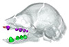
The present 3D Dataset contains the 3D models analyzed in the article Mennecart, B., and L. Costeur. 2016. A Dorcatherium (Mammalia, Ruminantia, Middle Miocene) petrosal bone and the tragulid ear region. Journal of Vertebrate Paleontology 36(6), 1211665(1)-1211665(7). DOI: 10.1080/02724634.2016.1211665.
Tragulus javanicus 10028 View specimen
|
M3#1193D surface of the left bony labyrinth of Tragulus javanicus NMB 10028 Type: "3D_surfaces"doi: 10.18563/m3.sf.119 state:published |
Download 3D surface file |
Moschiola meminna C.2453 View specimen
|
M3#1203D surface of the left bony labyrinth of Moschiola meminna NMB C.2453 Type: "3D_surfaces"doi: 10.18563/m3.sf.120 state:published |
Download 3D surface file |
Hyemoschus aquaticus C.1930 View specimen
|
M3#1223D surface of the right bony labyrinth of Hyemoschus aquaticus NMB C.1930 Type: "3D_surfaces"doi: 10.18563/m3.sf.122 state:published |
Download 3D surface file |
Dorcatherium crassum San.15053 View specimen
|
M3#1233D surface of the right bony labyrinth of Dorcatherium crassum NMB San.15053 Type: "3D_surfaces"doi: 10.18563/m3.sf.123 state:published |
Download 3D surface file |
This contribution contains the 3D models described and figured in the following publication: Mourlam, M., Orliac, M. J. (2017), Protocetid (Cetacea, Artiodactyla) bullae and petrosals from the Middle Eocene locality of Kpogamé, Togo: new insights into the early history of cetacean hearing. Journal of Systematic Palaeontology https://doi.org/10.1080/14772019.2017.1328378
?Carolinacetus indet. UM KPG-M 164 View specimen
|
M3#132left petrosal of ?Carolinacetus sp. from the locality of Kpogamé, Togo Type: "3D_surfaces"doi: 10.18563/m3.sf.132 state:published |
Download 3D surface file |
indet. indet. UM KPG-M 73 View specimen

|
M3#133labelled surface of the left petrosal Type: "3D_surfaces"doi: 10.18563/m3.sf.133 state:published |
Download 3D surface file |
|
M3#134left bullaof Protocetidae indeterminate from Kpogamé, Togo Type: "3D_surfaces"doi: 10.18563/m3.sf.134 state:published |
Download 3D surface file |
|
M3#135petrotympanic complex of Protocetidae indeterminate from Kpogamé, Togo Type: "3D_surfaces"doi: 10.18563/m3.sf.135 state:published |
Download 3D surface file |
?Carolinacetus indet. UM KPG-M 33 View specimen
|
M3#136left auditory bulla of a juvenile specimen of ?Carolinacetus sp. from Kpogamé, Togo Type: "3D_surfaces"doi: 10.18563/m3.sf.136 state:published |
Download 3D surface file |
Togocetus traversei UM KPG-M 80 View specimen

|
M3#137fragmentary right auditory bulla of Togocetus traversei from Kpogamé, Togo Type: "3D_surfaces"doi: 10.18563/m3.sf.137 state:published |
Download 3D surface file |
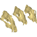
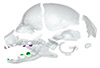
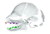
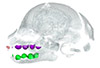
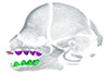
Archaeozoological studies are increasingly using new methods and approaches to explore questions about domestication. Here, we provide 3D models of three archaeological Canis lupus skulls from Belgium originating from the sites of Goyet (31,680±250BP; 31,890+240/-220BP), Trou des Nutons (21,810±90BP) and Trou Balleux (postglacial). Since their identification as either wolves or early dogs is still debated, we present these models as additional tools for further investigating their evolutionary history and the history of dog domestication.
Canis lupus Goyet 2860 View specimen

|
M3#213D surface model of the cranium of the Late Pleistocene Canis lupus "Goyet 2860" from the Royal Belgian Institute of Natural Sciences. Type: "3D_surfaces"doi: 10.18563/m3.sf21 state:published |
Download 3D surface file |
Canis lupus Trou Balleux no-nr View specimen

|
M3#223D surface model of the cranium of the Late Pleistocene Canis lupus "Trou Balleux no-nr" from the University of Liège, Belgium Type: "3D_surfaces"doi: 10.18563/m3.sf22 state:published |
Download 3D surface file |
Canis lupus Trou des Nutons 2559-1 View specimen

|
M3#233D surface model of the cranium of the Late Pleistocene Canis lupus "Trou des Nutons 2559-1" from the Royal Belgian Institute of Natural Sciences. Type: "3D_surfaces"doi: 10.18563/m3.sf23 state:published |
Download 3D surface file |
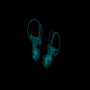
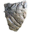
This contribution contains the 3D model described and figured in the following publication: Crochet, J.-Y., Hautier, L., Lehmann, T., 2015. A pangolin (Manidae, Pholidota, Mammalia) from the French Quercy phosphorites (Pech du Fraysse, Saint-Projet, Tarn-et-Garonne, late Oligocene, MP 28). Palaeovertebrata 39(2)-e4. doi: 10.18563/pv.39.2.e4
Necromanis franconica UM PFY 4051 View specimen
|
M3#12A partial left humerus from Pech du Fraysse (Saint-Projet, Tarn-et-Garonne, France), MP 28 (late Oligocene) Type: "3D_surfaces"doi: 10.18563/m3.sf12 state:published |
Download 3D surface file |
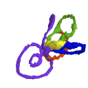
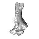
This contribution contains the 3D model(s) described and figured in the following publication: Carolina A. Hoffmann, P. G. Rodrigues, M. B. Soares & M. B. Andrade. 2021. Brain endocast of two non-mammaliaform cynodonts from southern Brazil: an ontogenetic and evolutionary approach, Historical Biology, 33:8, 1196-1207, https://doi.org/10.1080/08912963.2019.1685512
Probelesodon kitchingi MCP 1600 PV View specimen

|
M3#9783D model of the brain endocast of Probelesodon kitchingi. Type: "3D_surfaces"doi: 10.18563/m3.sf.978 state:published |
Download 3D surface file |
Massetognathus ochagaviae MCP 3871 PV View specimen

|
M3#9793D model of the brain endocast of Massetognathus ochagaviae. Type: "3D_surfaces"doi: 10.18563/m3.sf.979 state:published |
Download 3D surface file |
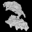
The present dataset contains the 3D models analyzed in Berio, F., Bayle, Y., Baum, D., Goudemand, N., and Debiais-Thibaud, M. 2022. Hide and seek shark teeth in Random Forests: Machine learning applied to Scyliorhinus canicula. It contains the head surfaces of 56 North Atlantic and Mediterranean small-spotted catsharks Scyliorhinus canicula, from which tooth surfaces were further extracted to perform geometric morphometrics and machine learning.
Scyliorhinus canicula 081118A View specimen

|
M3#941Head of a 10.6 cm long Scyliorhinus canicula female from a North Atlantic population. Type: "3D_surfaces"doi: 10.18563/m3.sf.941 state:published |
Download 3D surface file |
Scyliorhinus canicula 081118B View specimen

|
M3#942Head of a 11.0 cm long Scyliorhinus canicula female from a North Atlantic population. Type: "3D_surfaces"doi: 10.18563/m3.sf.942 state:published |
Download 3D surface file |
Scyliorhinus canicula 200118I View specimen

|
M3#959Head of a 45.0 cm long Scyliorhinus canicula female from a Mediterranean population. Type: "3D_surfaces"doi: 10.18563/m3.sf.959 state:published |
Download 3D surface file |
Scyliorhinus canicula 200118H View specimen

|
M3#958Head of a 47.0 cm long Scyliorhinus canicula female from a Mediterranean population. Type: "3D_surfaces"doi: 10.18563/m3.sf.958 state:published |
Download 3D surface file |
Scyliorhinus canicula 200118G View specimen

|
M3#957Head of a 40.0 cm long Scyliorhinus canicula female from a Mediterranean population. Type: "3D_surfaces"doi: 10.18563/m3.sf.957 state:published |
Download 3D surface file |
Scyliorhinus canicula 081118C View specimen

|
M3#940Head of a 11.2 cm long Scyliorhinus canicula female from a North Atlantic population. Type: "3D_surfaces"doi: 10.18563/m3.sf.940 state:published |
Download 3D surface file |
Scyliorhinus canicula 081118D View specimen

|
M3#939Head of a 10.2 cm long Scyliorhinus canicula female from a North Atlantic population. Type: "3D_surfaces"doi: 10.18563/m3.sf.939 state:published |
Download 3D surface file |
Scyliorhinus canicula 081118E View specimen

|
M3#938Head of a 12.0 cm long Scyliorhinus canicula male from a North Atlantic population. Type: "3D_surfaces"doi: 10.18563/m3.sf.938 state:published |
Download 3D surface file |
Scyliorhinus canicula 081118F View specimen

|
M3#937Head of a 10.7 cm long Scyliorhinus canicula male from a North Atlantic population. Type: "3D_surfaces"doi: 10.18563/m3.sf.937 state:published |
Download 3D surface file |
Scyliorhinus canicula 081118G View specimen

|
M3#936Head of a 10.8 cm long Scyliorhinus canicula male from a North Atlantic population. Type: "3D_surfaces"doi: 10.18563/m3.sf.936 state:published |
Download 3D surface file |
Scyliorhinus canicula 200118F View specimen

|
M3#935Head of a 41.5 cm long Scyliorhinus canicula female from a Mediterranean population. Type: "3D_surfaces"doi: 10.18563/m3.sf.935 state:published |
Download 3D surface file |
Scyliorhinus canicula 200118E View specimen

|
M3#934Head of a 40.0 cm long Scyliorhinus canicula female from a Mediterranean population. Type: "3D_surfaces"doi: 10.18563/m3.sf.934 state:published |
Download 3D surface file |
Scyliorhinus canicula 200118D View specimen

|
M3#933Head of a 42.0 cm long Scyliorhinus canicula male from a Mediterranean population. Type: "3D_surfaces"doi: 10.18563/m3.sf.933 state:published |
Download 3D surface file |
Scyliorhinus canicula 200118C View specimen

|
M3#943Head of a 41.0 cm long Scyliorhinus canicula male from a Mediterranean population. Type: "3D_surfaces"doi: 10.18563/m3.sf.943 state:published |
Download 3D surface file |
Scyliorhinus canicula 200118B View specimen

|
M3#945Head of a 44.0 cm long Scyliorhinus canicula male from a Mediterranean population. Type: "3D_surfaces"doi: 10.18563/m3.sf.945 state:published |
Download 3D surface file |
Scyliorhinus canicula 200118A View specimen

|
M3#944Head of a 46.0 cm long Scyliorhinus canicula male from a Mediterranean population. Type: "3D_surfaces"doi: 10.18563/m3.sf.944 state:published |
Download 3D surface file |
Scyliorhinus canicula 030418A View specimen

|
M3#956Head of a 13.9 cm long Scyliorhinus canicula female from a North Atlantic population. Type: "3D_surfaces"doi: 10.18563/m3.sf.956 state:published |
Download 3D surface file |
Scyliorhinus canicula 030418B View specimen

|
M3#955Head of a 13.6 cm long Scyliorhinus canicula female from a North Atlantic population. Type: "3D_surfaces"doi: 10.18563/m3.sf.955 state:published |
Download 3D surface file |
Scyliorhinus canicula 030418C View specimen

|
M3#954Head of a 13.4 cm long Scyliorhinus canicula male from a North Atlantic population. Type: "3D_surfaces"doi: 10.18563/m3.sf.954 state:published |
Download 3D surface file |
Scyliorhinus canicula 030418D View specimen

|
M3#953Head of a 13.2 cm long Scyliorhinus canicula male from a North Atlantic population. Type: "3D_surfaces"doi: 10.18563/m3.sf.953 state:published |
Download 3D surface file |
Scyliorhinus canicula 071118A View specimen

|
M3#952Head of a 36.0 cm long Scyliorhinus canicula female from a North Atlantic population. Type: "3D_surfaces"doi: 10.18563/m3.sf.952 state:published |
Download 3D surface file |
Scyliorhinus canicula 071118B View specimen

|
M3#951Head of a 33.0 cm long Scyliorhinus canicula female from a North Atlantic population. Type: "3D_surfaces"doi: 10.18563/m3.sf.951 state:published |
Download 3D surface file |
Scyliorhinus canicula 071118C View specimen

|
M3#950Head of a 32.0 cm long Scyliorhinus canicula female from a North Atlantic population. Type: "3D_surfaces"doi: 10.18563/m3.sf.950 state:published |
Download 3D surface file |
Scyliorhinus canicula 071118D View specimen

|
M3#949Head of a 35.0 cm long Scyliorhinus canicula male from a North Atlantic population. Type: "3D_surfaces"doi: 10.18563/m3.sf.949 state:published |
Download 3D surface file |
Scyliorhinus canicula 071118E View specimen
|
M3#948Head of a 35.0 cm long Scyliorhinus canicula male from a North Atlantic population. Type: "3D_surfaces"doi: 10.18563/m3.sf.948 state:published |
Download 3D surface file |
Scyliorhinus canicula 071118F View specimen

|
M3#947Head of a 33.0 cm long Scyliorhinus canicula male from a North Atlantic population. Type: "3D_surfaces"doi: 10.18563/m3.sf.947 state:published |
Download 3D surface file |
Scyliorhinus canicula 121118G View specimen

|
M3#946Head of a 36.0 cm long Scyliorhinus canicula female from a North Atlantic population. Type: "3D_surfaces"doi: 10.18563/m3.sf.946 state:published |
Download 3D surface file |
Scyliorhinus canicula 121118H View specimen

|
M3#932Head of a 35.0 cm long Scyliorhinus canicula female from a North Atlantic population. Type: "3D_surfaces"doi: 10.18563/m3.sf.932 state:published |
Download 3D surface file |
Scyliorhinus canicula 121118I View specimen

|
M3#931Head of a 33.0 cm long Scyliorhinus canicula male from a North Atlantic population. Type: "3D_surfaces"doi: 10.18563/m3.sf.931 state:published |
Download 3D surface file |
Scyliorhinus canicula 121118J View specimen

|
M3#917Head of a 36.0 cm long Scyliorhinus canicula male from a North Atlantic population. Type: "3D_surfaces"doi: 10.18563/m3.sf.917 state:published |
Download 3D surface file |
Scyliorhinus canicula 180118A View specimen

|
M3#916Head of a 57.0 cm long Scyliorhinus canicula female from a North Atlantic population. Type: "3D_surfaces"doi: 10.18563/m3.sf.916 state:published |
Download 3D surface file |
Scyliorhinus canicula 180118B View specimen

|
M3#915Head of a 58.0 cm long Scyliorhinus canicula female from a North Atlantic population. Type: "3D_surfaces"doi: 10.18563/m3.sf.915 state:published |
Download 3D surface file |
Scyliorhinus canicula 180118C View specimen

|
M3#911Head of a 58.5 cm long Scyliorhinus canicula female from a North Atlantic population. Type: "3D_surfaces"doi: 10.18563/m3.sf.911 state:published |
Download 3D surface file |
Scyliorhinus canicula 180118D View specimen

|
M3#914Head of a 56.0 cm long Scyliorhinus canicula male from a North Atlantic population. Type: "3D_surfaces"doi: 10.18563/m3.sf.914 state:published |
Download 3D surface file |
Scyliorhinus canicula 180118E View specimen

|
M3#913Head of a 58.0 cm long Scyliorhinus canicula male from a North Atlantic population. Type: "3D_surfaces"doi: 10.18563/m3.sf.913 state:published |
Download 3D surface file |
Scyliorhinus canicula 180118F View specimen

|
M3#912Head of a 59.0 cm long Scyliorhinus canicula male from a North Atlantic population. Type: "3D_surfaces"doi: 10.18563/m3.sf.912 state:published |
Download 3D surface file |
Scyliorhinus canicula 270918A View specimen

|
M3#910Head of a 56.0 cm long Scyliorhinus canicula male from a North Atlantic population. Type: "3D_surfaces"doi: 10.18563/m3.sf.910 state:published |
Download 3D surface file |
Scyliorhinus canicula 270918B View specimen

|
M3#908Head of a 59.5 cm long Scyliorhinus canicula male from a North Atlantic population. Type: "3D_surfaces"doi: 10.18563/m3.sf.908 state:published |
Download 3D surface file |
Scyliorhinus canicula 270918C View specimen

|
M3#909Head of a 63.0 cm long Scyliorhinus canicula female from a North Atlantic population. Type: "3D_surfaces"doi: 10.18563/m3.sf.909 state:published |
Download 3D surface file |
Scyliorhinus canicula 270918D View specimen

|
M3#907Head of a 64.0 cm long Scyliorhinus canicula female from a North Atlantic population. Type: "3D_surfaces"doi: 10.18563/m3.sf.907 state:published |
Download 3D surface file |
Scyliorhinus canicula 12111931 View specimen

|
M3#905Head of a 9.5 cm long Scyliorhinus canicula male from a Mediterranean population. Type: "3D_surfaces"doi: 10.18563/m3.sf.905 state:published |
Download 3D surface file |
Scyliorhinus canicula 12111933 View specimen

|
M3#906Head of a 9.5 cm long Scyliorhinus canicula female from a Mediterranean population. Type: "3D_surfaces"doi: 10.18563/m3.sf.906 state:published |
Download 3D surface file |
Scyliorhinus canicula 190118A View specimen

|
M3#918Head of a 8.8 cm long Scyliorhinus canicula female from a Mediterranean population. Type: "3D_surfaces"doi: 10.18563/m3.sf.918 state:published |
Download 3D surface file |
Scyliorhinus canicula 190118C View specimen

|
M3#930Head of a 9.0 cm long Scyliorhinus canicula female from a Mediterranean population. Type: "3D_surfaces"doi: 10.18563/m3.sf.930 state:published |
Download 3D surface file |
Scyliorhinus canicula 190118D View specimen

|
M3#929Head of a 8.9 cm long Scyliorhinus canicula male from a Mediterranean population. Type: "3D_surfaces"doi: 10.18563/m3.sf.929 state:published |
Download 3D surface file |
Scyliorhinus canicula 190118F View specimen

|
M3#928Head of a 9.1 cm long Scyliorhinus canicula male from a Mediterranean population. Type: "3D_surfaces"doi: 10.18563/m3.sf.928 state:published |
Download 3D surface file |
Scyliorhinus canicula 060718A View specimen

|
M3#927Head of a 25.5 cm long Scyliorhinus canicula male from a Mediterranean population. Type: "3D_surfaces"doi: 10.18563/m3.sf.927 state:published |
Download 3D surface file |
Scyliorhinus canicula 060718B View specimen

|
M3#926Head of a 23.0 cm long Scyliorhinus canicula female from a Mediterranean population. Type: "3D_surfaces"doi: 10.18563/m3.sf.926 state:published |
Download 3D surface file |
Scyliorhinus canicula 060718C View specimen

|
M3#925Head of a 28.0 cm long Scyliorhinus canicula male from a Mediterranean population. Type: "3D_surfaces"doi: 10.18563/m3.sf.925 state:published |
Download 3D surface file |
Scyliorhinus canicula 060718D View specimen

|
M3#924Head of a 21.0 cm long Scyliorhinus canicula male from a Mediterranean population. Type: "3D_surfaces"doi: 10.18563/m3.sf.924 state:published |
Download 3D surface file |
Scyliorhinus canicula 060718E View specimen

|
M3#923Head of a 23.5 cm long Scyliorhinus canicula male from a Mediterranean population. Type: "3D_surfaces"doi: 10.18563/m3.sf.923 state:published |
Download 3D surface file |
Scyliorhinus canicula 060718F View specimen

|
M3#922Head of a 22.5 cm long Scyliorhinus canicula female from a Mediterranean population. Type: "3D_surfaces"doi: 10.18563/m3.sf.922 state:published |
Download 3D surface file |
Scyliorhinus canicula 121218A View specimen

|
M3#921Head of a 31.0 cm long Scyliorhinus canicula female from a Mediterranean population. Type: "3D_surfaces"doi: 10.18563/m3.sf.921 state:published |
Download 3D surface file |
Scyliorhinus canicula 121218B View specimen

|
M3#920Head of a 31.0 cm long Scyliorhinus canicula female from a Mediterranean population. Type: "3D_surfaces"doi: 10.18563/m3.sf.920 state:published |
Download 3D surface file |
Scyliorhinus canicula 121218C View specimen

|
M3#919Head of a 31.0 cm long Scyliorhinus canicula female from a Mediterranean population. Type: "3D_surfaces"doi: 10.18563/m3.sf.919 state:published |
Download 3D surface file |
Scyliorhinus canicula 121218D View specimen

|
M3#904Head of a 31.0 cm long Scyliorhinus canicula male from a Mediterranean population. Type: "3D_surfaces"doi: 10.18563/m3.sf.904 state:published |
Download 3D surface file |
The present 3D Dataset contains the 3D model analyzed in Hendrickx, C. and Bell, P. R. 2021. The scaly skin of the abelisaurid Carnotaurus sastrei (Theropoda: Ceratosauria) from the Upper Cretaceous of Patagonia. Cretaceous Research. https://doi.org/10.1016/j.cretres.2021.104994
Carnotaurus sastrei MACN 894 View specimen

|
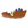
M3#8023D reconstruction of the biggest patch of skin (~1200 cm2) from the anterior tail region of the holotype of Carnotaurus, which is the largest single patch of squamous integument available for any saurischian. The skin consists of medium to large (up to 65 mm in diameter) conical feature scales surrounded by a network of low and small (< 14 mm) irregular basement scales separated by narrow interstitial tissue. Type: "3D_surfaces"doi: 10.18563/m3.sf.802 state:published |
Download 3D surface file |
The present 3D Dataset contains the 3D models analyzed in Mennecart B., Wazir W.A., Sehgal R.K., Patnaik R., Singh N.P., Kumar N, and Nanda A.C. 2021. New remains of Nalamaeryx (Tragulidae, Mammalia) from the Ladakh Himalaya and their phylogenetical and palaeoenvironmental implications. Historical Biology. https://doi.org/10.1080/08912963.2021.2014479
Nalameryx savagei WIMF/A4801 View specimen
|
M3#766Nalameryx savagei, Partial lower right jaw preserving m2 and m3. Type: "3D_surfaces"doi: 10.18563/m3.sf.766 state:published |
Download 3D surface file |
Nalameryx savagei WIMF/A4802 View specimen
|
M3#767Nalameryx savagei, partial lower right jaw preserving m2 and m3 Type: "3D_surfaces"doi: 10.18563/m3.sf.767 state:published |
Download 3D surface file |
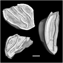
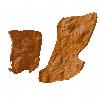
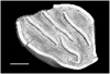
This contribution contains the 3D models of the fossil teeth of two chinchilloid caviomorph rodents (Borikenomys praecursor and Chinchilloidea gen. et sp. indet.) discovered from lower Oligocene deposits of Puerto Rico, San Sebastian Formation (locality LACM Loc. 8060). These fossils were described and figured in the following publication: Marivaux et al. (2020), Early Oligocene chinchilloid caviomorphs from Puerto Rico and the initial rodent colonization of the West Indies. Proceedings of the Royal Society B. http://dx.doi.org/10.1098/rspb.2019.2806
Borikenomys praecursor LACM 162447 View specimen
|
M3#638Right lower m3. This isolated tooth was scanned with a resolution of 6 µm using a μ-CT-scanning station EasyTom 150 / Rx Solutions (Montpellier RIO Imaging, ISE-M, Montpellier, France). AVIZO 7.1 (Visualization Sciences Group) software was used for visualization, segmentation, and 3D rendering. The specimen was prepared within a “labelfield” module of AVIZO, using the segmentation threshold selection tool. Type: "3D_surfaces"doi: 10.18563/m3.sf.638 state:published |
Download 3D surface file |
Borikenomys praecursor LACM 162446 View specimen
|
M3#639Fragment of lower molar (most of the mesial part). This isolated broken tooth was scanned with a resolution of 6 µm using a μ-CT-scanning station EasyTom 150 / Rx Solutions (Montpellier RIO Imaging, ISE-M, Montpellier, France). AVIZO 7.1 (Visualization Sciences Group) software was used for visualization, segmentation, and 3D rendering. The specimen was prepared within a “labelfield” module of AVIZO, using the segmentation threshold selection tool. Type: "3D_surfaces"doi: 10.18563/m3.sf.639 state:published |
Download 3D surface file |
indet indet LACM 162448 View specimen
|
M3#640Fragment of either an upper tooth (mesial laminae) or a lower tooth (distal laminae). The specimen was scanned with a resolution of 6 µm using a μ-CT-scanning station EasyTom 150 / Rx Solutions (Montpellier RIO Imaging, ISE-M, Montpellier, France). AVIZO 7.1 (Visualization Sciences Group) software was used for visualization, segmentation, and 3D rendering. This fragment of tooth was prepared within a “labelfield” module of AVIZO, using the segmentation threshold selection tool. Type: "3D_surfaces"doi: 10.18563/m3.sf.640 state:published |
Download 3D surface file |