

| Plane | Position | Flip |
| Show planes | Show edges |
Measured length
0.0
0.0
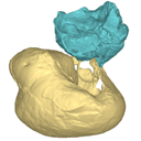
M3#137
fragmentary right auditory bulla of Togocetus traversei from Kpogamé, Togo
Data citation:
Mickaël Mourlam and Maëva J. Orliac
, 2017. M3#137. doi: 10.18563/m3.sf.137
Model solid/transparent
Flags:
contact with the basioccipital, Eustachian tube, internal pedicle of the posterior process of tympanic (broken), t1, transinvolucrum sulcus

|
3D models related to the publication: Protocetid (Cetacea, Artiodactyla) bullae and petrosals from the Middle Eocene locality of Kpogamé, Togo: new insights into the early history of cetacean hearingMickaĂ«l Mourlam and MaĂ«va J. OrliacPublished online: 31/05/2017Keywords: archaeocete; auditory region; Lutetian; petrotympanic complex https://doi.org/10.18563/m3.3.1.e2 Abstract This contribution contains the 3D models described and figured in the following publication: Mourlam, M., Orliac, M. J. (2017), Protocetid (Cetacea, Artiodactyla) bullae and petrosals from the Middle Eocene locality of Kpogamé, Togo: new insights into the early history of cetacean hearing. Journal of Systematic Palaeontology https://doi.org/10.1080/14772019.2017.1328378 See original publication M3 article infos Published in Volume 03, Issue 01 (2017) |
|