

| Plane | Position | Flip |
| Show planes | Show edges |
0.0
M3#78
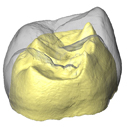
Outer enamel surface (OES) and enamel-dentine junction (EDJ) of Neolithic upper permanent right second molar
Data citation:
Mona Le Luyer , Michael Coquerelle
, Stéphane Rottier
and Priscilla Bayle
, 2016. M3#78. doi: 10.18563/m3.sf.78
Main model solid/transparent
Show/Hide main model
Second model transparent/solid
Show/Hide second model

|
3D models related to the publication: Internal tooth structure and burial practices: insights into the Neolithic necropolis of Gurgy (France, 5100-4000 cal. BC).Mona Le Luyer, Michael Coquerelle, Stéphane Rottier and Priscilla BaylePublished online: 25/07/2016Keywords: modern humans; Neolithic; upper permanent second molars https://doi.org/10.18563/m3.2.1.e1 Abstract The present 3D Dataset contains the 3D models of external and internal aspects of human upper permanent second molars from the Neolithic necropolis analyzed in the following publication: Le Luyer M., Coquerelle M., Rottier S., Bayle P. (2016): Internal tooth structure and burial practices: insights into the Neolithic necropolis of Gurgy (France, 5100-4000 cal. BC). Plos One 11(7): e0159688. doi: 10.1371/journal.pone.0159688. M3 article infos Published in Volume 02, Issue 01 (2016) |
|