

| Plane | Position | Flip |
| Show planes | Show edges |
Measured length
0.0
0.0
M3#355
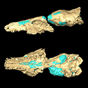
The file contain the cranium (yellow) and the endocast (blue) of the facial part and the brain case part of the type specimen of Proviverra typica (NMB Em18).
Data citation:
Morgane Dubied , Bastien Mennecart
and Floréal Solé
, 2019. M3#355. doi: 10.18563/m3.sf.355
Tag legend:
Endocranium, Skull
Main model solid/transparent
Show/Hide main model
Second model transparent/solid
Show/Hide second model

|
3D model related to the publication: The cranium of Proviverra typica (Mammalia, Hyaenodonta) and its impact on hyaenodont phylogeny and endocranial evolutionMorgane Dubied, Bastien Mennecart and FlorĂ©al SolĂ©Published online: 26/08/2019Keywords: brain; microtomography; Middle Eocene; Proviverrinae; skull https://doi.org/10.18563/journal.m3.74 Abstract This contribution contains the 3D model described and figured in the following publication: Dubied, M., Mennecart, B. and Solé, F. 2019. The cranium of Proviverra typica (Mammalia, Hyaenodonta) and its impact on hyaenodont phylogeny and endocranial evolution. Palaeontology. https://doi.org/10.1111/pala.12437 M3 article infos Published in Volume 05, issue 03 (2019) |
|