

| Plane | Position | Flip |
| Show planes | Show edges |
Measured length
0.0
0.0
M3#65
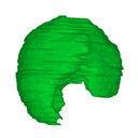
Human liver at Carnegie Stage (CS) 15
Data citation:
Ayumi Hirose , Takashi Nakashima, Naoto Shiraki, Shigehito Yamada
, Chigako Uwabe, Katsumi Kose
and Tetsuya Takakuwa
, 2016. M3#65. doi: 10.18563/m3.sf.65
Model solid/transparent
Flags:
imprint of stomach

|
3D models related to the publication: Morphogenesis of the liver during the human embryonic periodAyumi Hirose, Takashi Nakashima, Naoto Shiraki, Shigehito Yamada, Chigako Uwabe, Katsumi Kose and Tetsuya TakakuwaPublished online: 17/03/2016Keywords: human embryo; human liver; magnetic resonance imaging; three-dimensional reconstruction https://doi.org/10.18563/m3.1.4.e1 Abstract The present 3D Dataset contains the 3D models analyzed in: Hirose, A., Nakashima, T., Yamada, S., Uwabe, C., Kose, K., Takakuwa, T. 2012. Embryonic liver morphology and morphometry by magnetic resonance microscopic imaging. Anat Rec (Hoboken) 295, 51-59. doi: 10.1002/ar.21496 See original publication M3 article infos Published in Volume 01, Issue 04 (2016) |
|