

| Plane | Position | Flip |
| Show planes | Show edges |
0.0
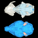
M3#963
Left bony labyrinth
Data citation:
Camille Grohé , Jérôme Surault
, Axelle Gardin
and Louis de Bonis
, 2022. M3#963. doi: 10.18563/m3.sf.963
Model solid/transparent
Flags:
Anterior ampulla, Anterior semicircular canal, Cochlear aqueduct, Cochlear spiral apex, Common crus, Fenestra cochleae, Fenestra vestibuli, Lateral ampulla, Lateral semicircular canal, Posterior ampulla, Posterior semicircular canal, Secondary bony lamina, Secondary common crus

|
3D models related to the publication: Description of the first cranium and endocranial structures of Stenoplesictis minor (Mammalia, Carnivora), an early aeluroid from the Oligocene of the Quercy Phosphorites (southwestern France)Camille Grohé, Jérôme Surault, Axelle Gardin and Louis de BonisPublished online: 08/05/2022Keywords: Aeluroidea; bony labyrinth; brain endocast; stapes; Stenoplesictoid https://doi.org/10.18563/m3.166 Abstract This contribution contains the 3D models described and figured in the following publication: Bonis, L. de, Grohé, C., Surault, J., Gardin, A. 2022. Description of the first cranium and endocranial structures of Stenoplesictis minor (Mammalia, Carnivora), an early aeluroid from the Oligocene of the Quercy Phosphorites (southwestern France). Historical Biology. https://doi.org/10.1080/08912963.2022.2045980 See original publication M3 article infos Published in Volume 08, issue 02 (2022) |
|