3D models of Cainotheriids Ossicular chain
3D models of Kalakocetus, the earliest Cetacea
The specimens of Speothos pacivorus
3D GM dataset of bird skeletal variation
Skeletal embryonic development in the catshark
Bony connexions of the petrosal bone of extant hippos
bony labyrinth (14) , inner ear (11) , Eocene (11) , geometric morphometrics (10) , CT-scan (10) , Oligocene (9) , Micro-CT (9)
Lionel Hautier (25) , Maëva Judith Orliac (24) , Laurent Marivaux (18) , Renaud Lebrun (15) , Rodolphe Tabuce (15) , Pierre-Olivier Antoine (13) , Bastien Mennecart (13)
Page 1 of 1, showing 4 record(s) out of 4 total
|
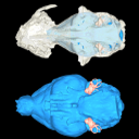
3D models related to the publication: Description of the first cranium and endocranial structures of Stenoplesictis minor (Mammalia, Carnivora), an early aeluroid from the Oligocene of the Quercy Phosphorites (southwestern France)Camille Grohé
Published online: 08/05/2022 |
|
M3#961Endocranium Type: "3D_surfaces"doi: 10.18563/m3.sf.961 state:published |
Download 3D surface file |
|
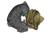
M3#962Right bony labyrinth Type: "3D_surfaces"doi: 10.18563/m3.sf.962 state:published |
Download 3D surface file |

|
M3#963Left bony labyrinth Type: "3D_surfaces"doi: 10.18563/m3.sf.963 state:published |
Download 3D surface file |
|
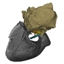
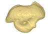
M3#964Cranium in transparency with endocranial structures Type: "3D_surfaces"doi: 10.18563/m3.sf.964 state:published |
Download 3D surface file |
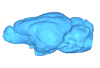
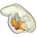
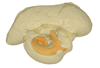
This contribution includes the 3D models of the reconstructed ossicular chain of the cainotheriid Caenomeryx filholi from the late Oligocene locality of Pech Desse (MP28, Quercy, France) described and figured in the publication of Assemat et al. (2020). It represents the oldest ossicular chain reconstruction for a Paleogene terrestrial artiodactyl species.
Caenomeryx filholi UM PDS 3353 View specimen
|
M3#508reconstruction of the middle ear with petrosal, bulla, stapes, incus, malleus Type: "3D_surfaces"doi: 10.18563/m3.sf.508 state:published |
Download 3D surface file |
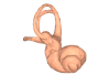
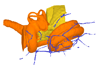
In this contribution, we describe the external and internal morphology of a delphinid petrosal bone collected from Ahu Tahai, a burial site located on the Southwestern coast of Easter Island, at Hangaroa. We discuss the taxonomic attribution of this archaeological item and describe its internal structures based on µCT data, including the bony labyrinth and the nerve and vein patterns. Identification of the nerves exists lead us to relocate the identification of the foramen singulare in delphinid petrosals.
indet. indet. AT1 View specimen
|
M3#420Stapes Type: "3D_surfaces"doi: 10.18563/m3.sf.420 state:published |
Download 3D surface file |
|
M3#421petrosal bone Type: "3D_surfaces"doi: 10.18563/m3.sf.421 state:published |
Download 3D surface file |
|
M3#422in situ bony labyrinth Type: "3D_surfaces"doi: 10.18563/m3.sf.422 state:published |
Download 3D surface file |
|
M3#423bony labyrinth and associated nerves and blood vessels Type: "3D_surfaces"doi: 10.18563/m3.sf.423 state:published |
Download 3D surface file |
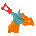
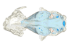
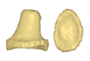
This contribution contains the 3D models of the ossicles of a protocetid archaeocete from the locality of Kpogamé, Togo, described and figured in the publication of Mourlam and Orliac (2019).
indet. indet. UM KPG-M 73 View specimen

|
M3#407stapes Type: "3D_surfaces"doi: 10.18563/m3.sf.407 state:published |
Download 3D surface file |

|
M3#408Incus Type: "3D_surfaces"doi: 10.18563/m3.sf.408 state:published |
Download 3D surface file |

|
M3#409Malleus Type: "3D_surfaces"doi: 10.18563/m3.sf.409 state:published |
Download 3D surface file |
Page 1 of 1, showing 4 record(s) out of 4 total


