3D models of Cainotheriids Ossicular chain
Explodable 3D Dog Skull for Veterinary Education
3D models of Kalakocetus, the earliest Cetacea
3D GM dataset of bird skeletal variation
Skeletal embryonic development in the catshark
Bony connexions of the petrosal bone of extant hippos
bony labyrinth (14) , inner ear (11) , Eocene (11) , geometric morphometrics (10) , CT-scan (10) , Oligocene (9) , Micro-CT (9)
Lionel Hautier (25) , Maëva Judith Orliac (24) , Laurent Marivaux (18) , Renaud Lebrun (15) , Rodolphe Tabuce (15) , Pierre-Olivier Antoine (13) , Bastien Mennecart (13)
Page 1 of 1, showing 13 record(s) out of 13 total
|
3D models related to the publication: Hidden diversity of Palaeogene metatherians: a new family of polydolopimorphian marsupials from Peruvian AmazoniaNarla Stutz
Published online: 17/04/2026 |

|
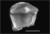
M3#1874Pozodolops manuelorum, fragmentary left dentary with p3–m1 Type: "3D_surfaces"doi: 10.18563/m3.sf.1874 state:published |
Download 3D surface file |
Wamradolops telloi MUSM 4032 View specimen
|
M3#1875Wamradolops telloi, fragmentary right dentary with m1–m2 Type: "3D_surfaces"doi: 10.18563/m3.sf.1875 state:published |
Download 3D surface file |
Pozodolops manuelorum MUSM 4036 View specimen
|
M3#1876Pozodolops manuelorum, holotype, fragmentary right maxilla with M1 Type: "3D_surfaces"doi: 10.18563/m3.sf.1876 state:published |
Download 3D surface file |
Pozodolops manuelorum MUSM 4041 View specimen

|
M3#1877Pozodolops manuelorum, fragmentary right dentary with m1 Type: "3D_surfaces"doi: 10.18563/m3.sf.1877 state:published |
Download 3D surface file |
Pozodolops manuelorum MUSM 4058 View specimen

|
M3#1878Pozodolops manuelorum, fragmentary left P3 Type: "3D_surfaces"doi: 10.18563/m3.sf.1878 state:published |
Download 3D surface file |
Wamradolops telloi MUSM 4144 View specimen

|
M3#1879Wamradolops telloi, fragmentary left dentary with p2–m1 Type: "3D_surfaces"doi: 10.18563/m3.sf.1879 state:published |
Download 3D surface file |
Wamradolops telloi MUSM 4179 View specimen

|
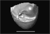
M3#1880Wamradolops telloi, holotype, partial skull, with right P2–M4 and left I4–M3, plus boneless lower teeth below upper teeth, belonging to the same individual Type: "3D_surfaces"doi: 10.18563/m3.sf.1880 state:published |
Download 3D surface file |
Wamradolops telloi MUSM 4221 View specimen

|
M3#1881Wamradolops telloi, fragmentary of right dentary with p3–m2 Type: "3D_surfaces"doi: 10.18563/m3.sf.1881 state:published |
Download 3D surface file |
This contribution contains the 3D models described and figured in the following publication: Pujos F., Hautier L., Antoine P-O., Boivin M., Moison B, Salas-Gismondi R, Tejada J.V. , Varas-Malca R.M., Yans J., Marivaux L. (2025). Unexpected pampatheriid from the early Oligocene of Peruvian Amazonia: insights into the tropical differentiation of cingulate xenarthrans. Historical Biology.
Bradypus tridactylus UM-ZOOL-V69 View specimen
|
M3#1600Molariform and associated dentinal microstructure Type: "3D_surfaces"doi: 10.18563/m3.sf.1600 state:published |
Download 3D surface file |
Choloepus didactylus UM-ZOOL-V12 View specimen
|
M3#1601Molariform and associated dentinal microstructure Type: "3D_surfaces"doi: 10.18563/m3.sf.1601 state:published |
Download 3D surface file |
Dasypus mexicanus UM-ZOOL-2787 View specimen

|
M3#1602Molariform and associated dentinal microstructure Type: "3D_surfaces"doi: 10.18563/m3.sf.1602 state:published |
Download 3D surface file |
Tolypeutes matacus UM-ZOOL-2789 View specimen
|
M3#1603Molariform and associated dentinal microstructure Type: "3D_surfaces"doi: 10.18563/m3.sf.1603 state:published |
Download 3D surface file |
Euphractus sexcinctus UM-ZOOL-2790 View specimen
|
M3#1604Molariform and associated dentinal microstructure Type: "3D_surfaces"doi: 10.18563/m3.sf.1604 state:published |
Download 3D surface file |
Holmesina septrionalis UM-FLD-1 View specimen
|
M3#1605Molariform and associated dentinal microstructure Type: "3D_surfaces"doi: 10.18563/m3.sf.1605 state:published |
Download 3D surface file |
Megatherium sp. UM-TAR-1 View specimen
|
M3#1607Molariform and associated dentinal microstructure Type: "3D_surfaces"doi: 10.18563/m3.sf.1607 state:published |
Download 3D surface file |
Indet indet MUSM-3965 View specimen
|
M3#1606Molariform and associated dentinal microstructure Type: "3D_surfaces"doi: 10.18563/m3.sf.1606 state:published |
Download 3D surface file |
The present contribution contains the 3D models of fossil humeri and ilia of anurans from various Eocene-Miocene deposits of Peruvian Amazonia. These fossils were described and figured in the following publication: Jansen et al. (2023), First Eocene–Miocene anuran fossils from Peruvian Amazonia: insights into Neotropical frog evolution and diversity. Papers in Palaeontology, The Palaeontological Association.
Indet. indet. MUSM 4746 View specimen
|
M3#1231Humeral fragment (distal end) Type: "3D_surfaces"doi: 10.18563/m3.sf.1231 state:published |
Download 3D surface file |
Indet. indet. MUSM 4747 View specimen
|
M3#1232Humeral fragment (distal end) Type: "3D_surfaces"doi: 10.18563/m3.sf.1232 state:published |
Download 3D surface file |
Indet. indet. MUSM 4748 View specimen
|
M3#1233Humeral fragment (distal end) Type: "3D_surfaces"doi: 10.18563/m3.sf.1233 state:published |
Download 3D surface file |
Indet. indet. MUSM 4755 View specimen
|
M3#1234Humeral fragment (distal end) Type: "3D_surfaces"doi: 10.18563/m3.sf.1234 state:published |
Download 3D surface file |
Indet. indet. MUSM 4756 View specimen
|
M3#1235Humeral fragment (distal end) Type: "3D_surfaces"doi: 10.18563/m3.sf.1235 state:published |
Download 3D surface file |
Indet. indet. MUSM 4757 View specimen

|
M3#1236Humeral fragment (distal end) Type: "3D_surfaces"doi: 10.18563/m3.sf.1236 state:published |
Download 3D surface file |
Indet. indet. MUSM 4761 View specimen
|
M3#1237Humeral fragment (distal end) Type: "3D_surfaces"doi: 10.18563/m3.sf.1237 state:published |
Download 3D surface file |
Indet. indet. MUSM 4763 View specimen
|
M3#1238Humeral fragment (distal end) Type: "3D_surfaces"doi: 10.18563/m3.sf.1238 state:published |
Download 3D surface file |
Indet. indet. MUSM 4765 View specimen
|
M3#1239Humeral fragment (distal end) Type: "3D_surfaces"doi: 10.18563/m3.sf.1239 state:published |
Download 3D surface file |
Indet. indet. MUSM 4766 View specimen
|
M3#1240Humeral fragment (distal end) Type: "3D_surfaces"doi: 10.18563/m3.sf.1240 state:published |
Download 3D surface file |
Indet. indet. MUSM 4775 View specimen
|
M3#1241Humeral fragment (distal end) Type: "3D_surfaces"doi: 10.18563/m3.sf.1241 state:published |
Download 3D surface file |
cf. Pipa sp. MUSM 4776 View specimen
|
M3#1242Humeral fragment (distal end) Type: "3D_surfaces"doi: 10.18563/m3.sf.1242 state:published |
Download 3D surface file |
Indet. indet. MUSM 4788 View specimen
|
M3#1243Ilial fragment Type: "3D_surfaces"doi: 10.18563/m3.sf.1243 state:published |
Download 3D surface file |
Indet. indet. MUSM 4789 View specimen
|
M3#1244Ilial fragment Type: "3D_surfaces"doi: 10.18563/m3.sf.1244 state:published |
Download 3D surface file |
Indet. indet. MUSM 4790 View specimen
|
M3#1245Ilial fragment Type: "3D_surfaces"doi: 10.18563/m3.sf.1245 state:published |
Download 3D surface file |
Indet. indet. MUSM 4792 View specimen
|
M3#1246Ilial fragment Type: "3D_surfaces"doi: 10.18563/m3.sf.1246 state:published |
Download 3D surface file |
Indet. indet. MUSM 4793 View specimen
|
M3#1247Ilial fragment Type: "3D_surfaces"doi: 10.18563/m3.sf.1247 state:published |
Download 3D surface file |
Indet. indet. MUSM 4794 View specimen
|
M3#1249Ilial fragment Type: "3D_surfaces"doi: 10.18563/m3.sf.1249 state:published |
Download 3D surface file |
Indet. indet. MUSM 4795 View specimen
|
M3#1250Ilial fragment Type: "3D_surfaces"doi: 10.18563/m3.sf.1250 state:published |
Download 3D surface file |
cf. Pipa sp. MUSM 4796 View specimen
|
M3#1251Ilial fragment Type: "3D_surfaces"doi: 10.18563/m3.sf.1251 state:published |
Download 3D surface file |
cf. Pipa sp. MUSM 4797 View specimen
|
M3#1252Ilial fragment Type: "3D_surfaces"doi: 10.18563/m3.sf.1252 state:published |
Download 3D surface file |
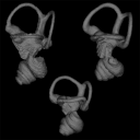
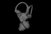
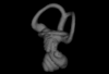
This contribution contains the three-dimensional models of the inner ear of the hetaxodontid rodents Amblyrhiza, Clidomys and Elasmodontomys from the West Indies. These specimens were analyzed and discussed in : The inner ear of caviomorph rodents: phylogenetic implications and application to extinct West Indian taxa.
Amblyrhiza inundata 11842 View specimen
|
M3#11543D surface of the left-oriented inner ear of Amblyrhiza. Type: "3D_surfaces"doi: 10.18563/m3.sf.1154 state:published |
Download 3D surface file |
Clidomys sp NA View specimen
|
M3#11553D surface of the left-oriented inner ear of Clidomys sp. Type: "3D_surfaces"doi: 10.18563/m3.sf.1155 state:published |
Download 3D surface file |
Elasmodontomys obliquus 17127 View specimen
|
M3#11563D surface of the left-oriented inner ear of Elasmodontomys obliquus. Type: "3D_surfaces"doi: 10.18563/m3.sf.1156 state:published |
Download 3D surface file |
This contribution contains the three-dimensional digital models of a part of the dental fossil material (the large specimens) of caviomorph rodents, discovered in late middle Miocene detrital deposits of the TAR-31 locality in Peruvian Amazonia (San Martín, Peru). These fossils were described, figured and discussed in the following publication: Boivin, Marivaux et al. (2021), Late middle Miocene caviomorph rodents from Tarapoto, Peruvian Amazonia. PLoS ONE 16(11): e0258455. https://doi.org/10.1371/journal.pone.0258455
Microscleromys paradoxalis MUSM 4643 View specimen
|
M3#1115Fragment of left mandibule preserving dp4, m1 and a portion of incisor Type: "3D_surfaces"doi: 10.18563/m3.sf.1115 state:published |
Download 3D surface file |
Ricardomys longidens MUSM 4375 View specimen
|
M3#1116Fragment of left maxillary preserving DP4 and M1 (or M1 and M2) Type: "3D_surfaces"doi: 10.18563/m3.sf.1116 state:published |
Download 3D surface file |
"Scleromys" sp. MUSM 4272 View specimen
|
M3#1117Isolated left upper molar Type: "3D_surfaces"doi: 10.18563/m3.sf.1117 state:published |
Download 3D surface file |
"Scleromys" sp. MUSM 4275 View specimen
|
M3#1118Isolated right upper molar Type: "3D_surfaces"doi: 10.18563/m3.sf.1118 state:published |
Download 3D surface file |
"Scleromys" sp. MUSM 4273 View specimen
|
M3#1119Isolated left upper molar Type: "3D_surfaces"doi: 10.18563/m3.sf.1119 state:published |
Download 3D surface file |
"Scleromys" sp. MUSM 4276 View specimen
|
M3#1120Isolated right upper molar Type: "3D_surfaces"doi: 10.18563/m3.sf.1120 state:published |
Download 3D surface file |
"Scleromys" sp. MUSM 4282 View specimen
|
M3#1121Isolated right lower molar Type: "3D_surfaces"doi: 10.18563/m3.sf.1121 state:published |
Download 3D surface file |
"Scleromys" sp. MUSM 4281 View specimen
|
M3#1122Isolated right lower molar Type: "3D_surfaces"doi: 10.18563/m3.sf.1122 state:published |
Download 3D surface file |
"Scleromys" sp. MUSM 4280 View specimen
|
M3#1123Isolated left p4 Type: "3D_surfaces"doi: 10.18563/m3.sf.1123 state:published |
Download 3D surface file |
"Scleromys" sp. MUSM 4277 View specimen
|
M3#1124Isolated left lower dp4 Type: "3D_surfaces"doi: 10.18563/m3.sf.1124 state:published |
Download 3D surface file |
"Scleromys" sp. MUSM 4279 View specimen
|
M3#1125Isolated right lower dp4 (mesial fragment) Type: "3D_surfaces"doi: 10.18563/m3.sf.1125 state:published |
Download 3D surface file |
gen.indet sp. indet MUSM 4283 View specimen
|
M3#1126Isolated right lower p4 Type: "3D_surfaces"doi: 10.18563/m3.sf.1126 state:published |
Download 3D surface file |
Microscleromys sp. MUSM 4658 View specimen
|
M3#1127Isolated left tarsal bone (astragalus) Type: "3D_surfaces"doi: 10.18563/m3.sf.1127 state:published |
Download 3D surface file |
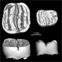
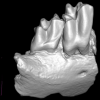
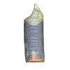
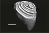
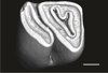
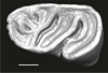
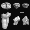
This contribution provides the raw files for the μCT-scan data and renderings of the three-dimensional digital models of two fossil teeth of a geomyin geomorph rodent (Caribeomys merzeraudi), discovered from lower Oligocene deposits of Puerto Rico, San Sebastian Formation (locality LACM Loc. 8060). These fossils were described, figured and discussed in the following publication: Marivaux et al. (2021), An unpredicted ancient colonization of the West Indies by North American rodents: dental evidence of a geomorph from the early Oligocene of Puerto Rico. Papers in Palaeontology. https://doi.org/10.1002/spp2.1388
Caribeomys merzeraudi LACM 162478 View specimen
|
M3#712Right lower dp4: isolated deciduous premolar. The specimen was scanned with a resolution of 5 µm using a μ-CT-scanning station EasyTom 150 / Rx Solutions (Montpellier RIO Imaging, ISE-M, Montpellier, France). AVIZO 7.1 (Visualization Sciences Group) software was used for visualization, segmentation, and 3D rendering. This isolated tooth was prepared within a “labelfield” module of AVIZO, using the segmentation threshold selection tool. Type: "3D_surfaces"doi: 10.18563/m3.sf.712 state:published |
Download 3D surface file |
|
M3#7145µm µCT data set . Right lower dp4: isolated deciduous premolar. The specimen was scanned with a resolution of 5 µm using a μ-CT-scanning station EasyTom 150 / Rx Solutions (Montpellier RIO Imaging, ISE-M, Montpellier, France). Type: "3D_CT"doi: 10.18563/m3.sf.714 state:published |
Download CT data |
Caribeomys merzeraudi LACM 162449 View specimen
|
M3#713Right lower molar (m1 or m2). The specimen was scanned with a resolution of 4.5 µm using a μ-CT-scanning station EasyTom 150 / Rx Solutions (Montpellier RIO Imaging, ISE-M, Montpellier, France). AVIZO 7.1 (Visualization Sciences Group) software was used for visualization, segmentation, and 3D rendering. This isolated tooth was prepared within a “labelfield” module of AVIZO, using the segmentation threshold selection tool. Type: "3D_surfaces"doi: 10.18563/m3.sf.713 state:published |
Download 3D surface file |
|
M3#715µCT data at 4.5µm Type: "3D_CT"doi: 10.18563/m3.sf.715 state:published |
Download CT data |
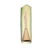
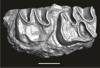
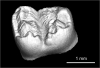
This contribution contains the 3D models of the fossil teeth of two chinchilloid caviomorph rodents (Borikenomys praecursor and Chinchilloidea gen. et sp. indet.) discovered from lower Oligocene deposits of Puerto Rico, San Sebastian Formation (locality LACM Loc. 8060). These fossils were described and figured in the following publication: Marivaux et al. (2020), Early Oligocene chinchilloid caviomorphs from Puerto Rico and the initial rodent colonization of the West Indies. Proceedings of the Royal Society B. http://dx.doi.org/10.1098/rspb.2019.2806
Borikenomys praecursor LACM 162447 View specimen
|
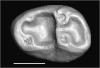
M3#638Right lower m3. This isolated tooth was scanned with a resolution of 6 µm using a μ-CT-scanning station EasyTom 150 / Rx Solutions (Montpellier RIO Imaging, ISE-M, Montpellier, France). AVIZO 7.1 (Visualization Sciences Group) software was used for visualization, segmentation, and 3D rendering. The specimen was prepared within a “labelfield” module of AVIZO, using the segmentation threshold selection tool. Type: "3D_surfaces"doi: 10.18563/m3.sf.638 state:published |
Download 3D surface file |
Borikenomys praecursor LACM 162446 View specimen
|
M3#639Fragment of lower molar (most of the mesial part). This isolated broken tooth was scanned with a resolution of 6 µm using a μ-CT-scanning station EasyTom 150 / Rx Solutions (Montpellier RIO Imaging, ISE-M, Montpellier, France). AVIZO 7.1 (Visualization Sciences Group) software was used for visualization, segmentation, and 3D rendering. The specimen was prepared within a “labelfield” module of AVIZO, using the segmentation threshold selection tool. Type: "3D_surfaces"doi: 10.18563/m3.sf.639 state:published |
Download 3D surface file |
indet indet LACM 162448 View specimen
|
M3#640Fragment of either an upper tooth (mesial laminae) or a lower tooth (distal laminae). The specimen was scanned with a resolution of 6 µm using a μ-CT-scanning station EasyTom 150 / Rx Solutions (Montpellier RIO Imaging, ISE-M, Montpellier, France). AVIZO 7.1 (Visualization Sciences Group) software was used for visualization, segmentation, and 3D rendering. This fragment of tooth was prepared within a “labelfield” module of AVIZO, using the segmentation threshold selection tool. Type: "3D_surfaces"doi: 10.18563/m3.sf.640 state:published |
Download 3D surface file |
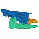
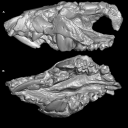
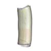
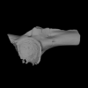
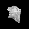
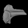
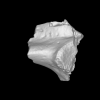
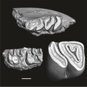
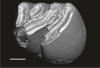
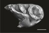
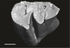
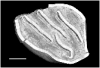
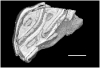
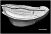
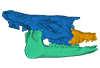
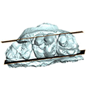
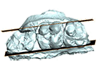
The present 3D Dataset contains two 3D models described in Tissier et al. (https://doi.org/10.1098/rsos.200633): the only known complete mandible of the early-branching rhinocerotoid Epiaceratherium magnum Uhlig, 1999, and a hypothetical reconstruction of the complete archetypic skull of Epiaceratherium Heissig, 1969, created by merging three cranial parts from three distinct Epiaceratherium species.
Epiaceratherium magnum NMB.O.B.928 View specimen
|
M3#5343D surface model of the mandible NMB.O.B.928 of Epiaceratherium magnum, with texture file. Type: "3D_surfaces"doi: 10.18563/m3.sf.534 state:published |
Download 3D surface file |
Epiaceratherium magnum NMB.O.B.928 + MJSN POI007–245 + NMB.I.O.43 View specimen
|
M3#535Archetypal reconstruction of the skull of Epiaceratherium, generated by 3D virtual association of the cranium of E. delemontense (MJSN POI007–245, in blue), mandible of E. magnum (NMB.O.B.928, green) and snout of E. bolcense (NMB.I.O.43, in orange). Type: "3D_surfaces"doi: 10.18563/m3.sf.535 state:published |
Download 3D surface file |
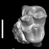
This contribution contains the 3D models of the fossil teeth of a small-bodied platyrrhine primate, Neosaimiri cf. fieldsi (Cebinae, Cebidae, Platyrrhini) discovered from Laventan deposits (late Middle Miocene) of Peruvian Amazonia, San Martín Department (TAR-31: Tarapoto/Juan Guerra vertebrate fossil-bearing locus n°31). These fossils were described and figured in the following publication: Marivaux et al. (2020), New record of Neosaimiri (Cebidae, Platyrrhini) from the late Middle Miocene of Peruvian Amazonia. Journal of Human Evolution. https://doi.org/10.1016/j.jhevol.2020.102835
Neosaimiri cf. fieldsi MUSM-3888 View specimen
|
M3#538MUSM-3888, right m3 of Neosaimiri cf. fieldsi. Type: "3D_surfaces"doi: 10.18563/m3.sf.538 state:published |
Download 3D surface file |
Neosaimiri cf. fieldsi MUSM-3890 View specimen
|
M3#540MUSM-3890, left dp2 of Neosaimiri cf. fieldsi. Type: "3D_surfaces"doi: 10.18563/m3.sf.540 state:published |
Download 3D surface file |
Neosaimiri cf. fieldsi MUSM-3895 View specimen
|
M3#541MUSM-3895, right DC1 of Neosaimiri cf. fieldsi. Type: "3D_surfaces"doi: 10.18563/m3.sf.541 state:published |
Download 3D surface file |
Neosaimiri cf. fieldsi MUSM-3891 View specimen
|
M3#542MUSM-3891, lingual part of a fragmentary right M1 or M2 of Neosaimiri cf. fieldsi. Type: "3D_surfaces"doi: 10.18563/m3.sf.542 state:published |
Download 3D surface file |
Neosaimiri cf. fieldsi MUSM-3892 View specimen
|
M3#543MUSM-3892, distobuccal part of a fragmentary right upper molar (metacone region) of Neosaimiri cf. fieldsi. Type: "3D_surfaces"doi: 10.18563/m3.sf.543 state:published |
Download 3D surface file |
Neosaimiri cf. fieldsi MUSM-3893 View specimen
|
M3#544MUSM-3893, buccal part of a fragmentary right P3 or P4 of Neosaimiri cf. fieldsi. Type: "3D_surfaces"doi: 10.18563/m3.sf.544 state:published |
Download 3D surface file |
Neosaimiri cf. fieldsi MUSM-3894 View specimen
|
M3#545MUSM-3894, lingual part of a fragmentary left P3 or P4 of Neosaimiri cf. fieldsi. Type: "3D_surfaces"doi: 10.18563/m3.sf.545 state:published |
Download 3D surface file |
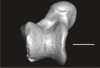
This contribution contains the 3D model of the fossil talus of a small-bodied anthropoid primate (Platyrrhini, Cebidae, Cebinae) discovered from lower Miocene deposits of Peruvian Amazonia (MD-61 locality, Upper Madre de Dios Basin). This fossil was described and figured in the following publication: Marivaux et al. (2012), A platyrrhine talus from the early Miocene of Peru (Amazonian Madre de Dios Sub-Andean Zone). Journal of Human Evolution. http://dx.doi.org/10.1016/j.jhevol.2012.07.005
Cebinae indet. sp. MUSM-2024 View specimen
|
M3#380Right talus 3D surface of a Miocene Cebinae indet. primate Type: "3D_surfaces"doi: 10.18563/m3.sf.380 state:published |
Download 3D surface file |
This contribution contains the 3D models of the isolated teeth attributed to stem representatives of the Cebuella and Cebus lineages (Cebuella sp. and Cebus sp.), described and figured in the following publication: Marivaux et al. (2016), Dental remains of cebid platyrrhines from the earliest late Miocene of Western Amazonia, Peru: macroevolutionary implications on the extant capuchin and marmoset lineages. American Journal of Physical Anthropology. http://dx.doi.org/10.1002/ajpa.23052
Cebus sp. MUSM-3243 View specimen
|
M3#2823D model of left lower m1 (lingual part) Type: "3D_surfaces"doi: 10.18563/m3.sf.282 state:published |
Download 3D surface file |
Cebuella sp. MUSM-3239 View specimen
|
M3#2833D model of left lower p4 Type: "3D_surfaces"doi: 10.18563/m3.sf.283 state:published |
Download 3D surface file |
Cebuella sp. MUSM-3240 View specimen
|
M3#2943D model of right upper P3 or P4 (buccal part) Type: "3D_surfaces"doi: 10.18563/m3.sf.294 state:published |
Download 3D surface file |
Cebuella sp. MUSM-3241 View specimen
|
M3#2953D model of right upper P2 Type: "3D_surfaces"doi: 10.18563/m3.sf.295 state:published |
Download 3D surface file |
Cebuella sp. MUSM-3242 View specimen
|
M3#2963D model of upper I2 Type: "3D_surfaces"doi: 10.18563/m3.sf.296 state:published |
Download 3D surface file |
This contribution contains the 3D models of the isolated teeth of Canaanimico amazonensis, a new stem platyrrhine primate, described and figured in the following publication: Marivaux et al. (2016), Neotropics provide insights into the emergence of New World monkeys: new dental evidence from the late Oligocene of Peruvian Amazonia. Journal of Human Evolution. http://dx.doi.org/10.1016/j.jhevol.2016.05.011
Canaanimico amazonensis MUSM-2499 View specimen
|
M3#2893D model of left upper M2 Type: "3D_surfaces"doi: 10.18563/m3.sf.289 state:published |
Download 3D surface file |
Canaanimico amazonensis MUSM-2500 View specimen
|
M3#2903D model of left upper M1 (lingual part) Type: "3D_surfaces"doi: 10.18563/m3.sf.290 state:published |
Download 3D surface file |
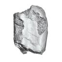
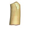
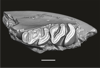
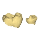
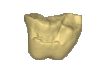
This contribution contains the 3D models described and figured in the following publication: Orliac M.J., Karadenizli L., Antoine P.-O., Sen S. 2015. Small suids (Mammalia, Artiodactyla) from the late Early Miocene of Turkey and a short overview of Early Miocene small suoids in the Old World. Paleontologia electronica 18(2): 18.2.30A: 1-48. https://doi.org/10.26879/547
?Nguruwe galaticum SMT-1 View specimen
|
M3#16fragment of palate with left broken M1-M3 Type: "3D_surfaces"doi: 10.18563/m3.sf16 state:published |
Download 3D surface file |
Page 1 of 1, showing 13 record(s) out of 13 total


