Explodable 3D Dog Skull for Veterinary Education
3D models of a Sheep and Goat Skull and Inner ear
3D models of Miocene vertebrates from Tavers
3D GM dataset of bird skeletal variation
Skeletal embryonic development in the catshark
Bony connexions of the petrosal bone of extant hippos
bony labyrinth (11) , inner ear (10) , Eocene (8) , South America (8) , Paleobiogeography (7) , skull (7) , phylogeny (6)
Lionel Hautier (23) , Maëva Judith Orliac (21) , Laurent Marivaux (16) , Rodolphe Tabuce (14) , Bastien Mennecart (13) , Pierre-Olivier Antoine (12) , Renaud Lebrun (11)

|
3D models related to the publication: An unpredicted ancient colonization of the West Indies by North American rodents: dental evidence of a geomorph from the early Oligocene of Puerto RicoLaurent Marivaux
Published online: 16/07/2021 |
|

|
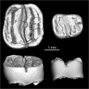
M3#713Right lower molar (m1 or m2). The specimen was scanned with a resolution of 4.5 µm using a μ-CT-scanning station EasyTom 150 / Rx Solutions (Montpellier RIO Imaging, ISE-M, Montpellier, France). AVIZO 7.1 (Visualization Sciences Group) software was used for visualization, segmentation, and 3D rendering. This isolated tooth was prepared within a “labelfield” module of AVIZO, using the segmentation threshold selection tool. Type: "3D_surfaces"doi: 10.18563/m3.sf.713 Data citation: Laurent Marivaux, Jorge Velez-Juarbe and Pierre-Olivier Antoine, 2021. M3#713. doi: 10.18563/m3.sf.713 state:published |
Download 3D surface file |

|
M3#715µCT data at 4.5µm Type: "3D_CT"doi: 10.18563/m3.sf.715 Data citation: Laurent Marivaux, Jorge Velez-Juarbe and Pierre-Olivier Antoine, 2021. M3#715. doi: 10.18563/m3.sf.715 state:published |
Download CT data |