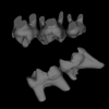
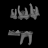
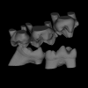
3D models of Cainotheriids Ossicular chain
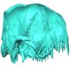
Explodable 3D Dog Skull for Veterinary Education
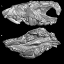
3D models of Kalakocetus, the earliest Cetacea
3D GM dataset of bird skeletal variation
Skeletal embryonic development in the catshark
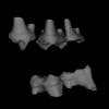
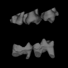
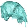
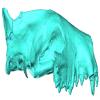
Bony connexions of the petrosal bone of extant hippos
bony labyrinth (14) , inner ear (11) , Eocene (11) , geometric morphometrics (10) , CT-scan (10) , Oligocene (9) , Micro-CT (9)
Lionel Hautier (25) , Maëva Judith Orliac (24) , Laurent Marivaux (18) , Renaud Lebrun (15) , Rodolphe Tabuce (15) , Pierre-Olivier Antoine (13) , Bastien Mennecart (13)
MorphoMuseuM, also referred to as M3, is a peer reviewed, online data journal that publishes 3D models of vertebrates, including models of type specimens, anatomy atlases, reconstruction of deformed or damaged specimens, and 3D datasets (see https://doi.org/10.1017/scs.2017.14 for details).
M3 comes along with a free software, MorphoDig, which contains a set of tools for editing, positioning, deforming, labelling, measuring and rendering sets of 3D surfaces.
All 3D data presented on this website are licensed under a Creative Commons Attribution-NonCommercial 4.0. International License. 
|
3D models related to the publication: A comprehensive evolutionary history of Peradectidae (Mammalia: Metatheria) revealed by a new dental morphology-based phylogenetic framework, with insights from late early Eocene French specimensKillian Gernelle
Published online: 16/07/2026 |
|
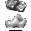
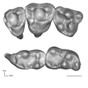
M3#1889right m3 (holotype) Type: "3D_surfaces"doi: 10.18563/m3.sf.1889 state:in_press |
Download 3D surface file |
Europeradectes williamsoni MNHN.F.PRE643 View specimen
|
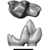
M3#1890left m2 Type: "3D_surfaces"doi: 10.18563/m3.sf.1890 state:in_press |
Download 3D surface file |
Europeradectes williamsoni MNHN.F.PRE272 View specimen
|
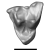
M3#1891right m1 Type: "3D_surfaces"doi: 10.18563/m3.sf.1891 state:in_press |
Download 3D surface file |
Europeradectes williamsoni MNHN.F.PMT167 View specimen
|
M3#1892left m4 Type: "3D_surfaces"doi: 10.18563/m3.sf.1892 state:in_press |
Download 3D surface file |
Europeradectes marivauxi MNHN.F.PMT169 View specimen
|
M3#1893left M2 Type: "3D_surfaces"doi: 10.18563/m3.sf.1893 state:in_press |
Download 3D surface file |
Europeradectes marivauxi MNHN.F.PMT166 View specimen
|
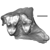
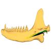
M3#1894partial right maxilla with M3-M4 (holotype) Type: "3D_surfaces"doi: 10.18563/m3.sf.1894 state:in_press |
Download 3D surface file |
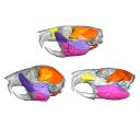
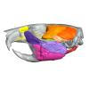
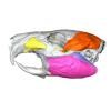
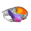
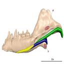
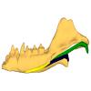
This contribution contains the 3D models described and figured in the following publication: Hautier L, Da Cunha L, Alves Filho M, Moison B, Besson L, Fabre P-H. 20XX. A 3D atlas of the trigeminal nerve and its relevance for comparative studies of the masticatory apparatus in rodents. Journal of Anatomy
Rattus norvegicus UM-ZOOL-3105 View specimen
|
M3#19363D reconstruction of the masticatory musculature, trigeminal nerve and skull of the brown rat Rattus norvegicus Type: "3D_surfaces"doi: 10.18563/m3.sf.1936 state:in_press |
Download 3D surface file |
Cavia porcellus UM-ZOOL-499V View specimen
|
M3#19353D reconstruction of the masticatory musculature, trigeminal nerve and skull of the Guinea pig Cavia porcellus Type: "3D_surfaces"doi: 10.18563/m3.sf.1935 state:in_press |
Download 3D surface file |
Sciurus vulgaris BOUM-2022.2.12 View specimen
|
M3#19373D reconstruction of the masticatory musculature, trigeminal nerve and skull of the red squirrel Sciurus vulgaris Type: "3D_surfaces"doi: 10.18563/m3.sf.1937 state:in_press |
Download 3D surface file |
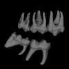
The present dataset contains the 3D models of the cheek teeth of eight raccoons analyzed in Koomen, S. E., Lang, A. J. & Martin, T. (2026). Tooth Function of the Northern Raccoon (Procyon lotor) and Adaptations to Omnivory in the Order Carnivora. Journal of Morphology.
Procyon lotor ZFMK-MAM-2013.0341 View specimen
|
M3#1838Cheek teeth of P. lotor specimen ZFMK-MAM-2013.0341 Type: "3D_surfaces"doi: 10.18563/m3.sf.1838 state:in_press |
Download 3D surface file |
Procyon lotor ZFMK-MAM-1993.0289 View specimen
|
M3#1839Cheek teeth of P. lotor specimen ZFMK-MAM-1993.0289 Type: "3D_surfaces"doi: 10.18563/m3.sf.1839 state:in_press |
Download 3D surface file |
Procyon lotor ZFMK-MAM-2016.0912 View specimen
|
M3#1840Cheek teeth of P. lotor specimen ZFMK-MAM-2016.0912 Type: "3D_surfaces"doi: 10.18563/m3.sf.1840 state:in_press |
Download 3D surface file |
Procyon lotor ZFMK-MAM-2016.0914 View specimen
|
M3#1841Cheek teeth of P. lotor specimen ZFMK-MAM-2016.0914 Type: "3D_surfaces"doi: 10.18563/m3.sf.1841 state:in_press |
Download 3D surface file |
Procyon lotor ZFMK-MAM-2016.0923 View specimen
|
M3#1842Cheek teeth of P. lotor specimen ZFMK-MAM-2016.0923 Type: "3D_surfaces"doi: 10.18563/m3.sf.1842 state:in_press |
Download 3D surface file |
Procyon lotor ZFMK-MAM-2016.0937 View specimen
|
M3#1843Cheek teeth of P. lotor specimen ZFMK-MAM-2016.0937 Type: "3D_surfaces"doi: 10.18563/m3.sf.1843 state:in_press |
Download 3D surface file |
Procyon lotor ZFMK-MAM-2016.0938 View specimen
|
M3#1844Cheek teeth of P. lotor specimen ZFMK-MAM-2016.0938 Type: "3D_surfaces"doi: 10.18563/m3.sf.1844 state:in_press |
Download 3D surface file |
Procyon lotor ZFMK-MAM-2016.0967 View specimen
|
M3#1845Cheek teeth of P. lotor specimen ZFMK-MAM-2016.0967 Type: "3D_surfaces"doi: 10.18563/m3.sf.1845 state:in_press |
Download 3D surface file |
|
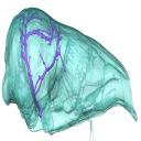
3D models related to the publication: Brain drain: exceptional pattern of calvarial venation in pangolins and its phylogenetic significance for FeraeGuillaume Billet
Published online: 26/03/2026 |
|
M3#1847cranium & intradiploic canals (sinuses & diploic veins) Type: "3D_surfaces"doi: 10.18563/m3.sf.1847 state:published |
Download 3D surface file |
Manis javanica NHM-UK 9.1.5.858 View specimen
|
M3#1848cranium & intradiploic canals (sinuses & diploic veins) Type: "3D_surfaces"doi: 10.18563/m3.sf.1848 state:published |
Download 3D surface file |
Felis silvestris UM-ZOOL-149N View specimen
|
M3#1849cranium & intradiploic canals (sinuses & diploic veins) Type: "3D_surfaces"doi: 10.18563/m3.sf.1849 state:published |
Download 3D surface file |
Erinaceus europaeus SMNS40759 View specimen
|
M3#1850cranium & intradiploic canals (sinuses & diploic veins) Type: "3D_surfaces"doi: 10.18563/m3.sf.1850 state:published |
Download 3D surface file |
Pterodon dasyuroides MNHN.F.Qu8301 View specimen
|
M3#1851cranium & intradiploic canals (sinuses & diploic veins) Type: "3D_surfaces"doi: 10.18563/m3.sf.1851 state:published |
Download 3D surface file |
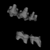
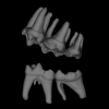
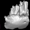
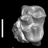
This contribution contains the 3D digital models of some fossil specimens of Wamradolops telloi Stutz and Pozodolops manuelorum Stutz (Metatheria: Polydolopimorphia), from several Palaeogene locations of Peruvian Amazonia. These taxa were described and analyzed in detail in the following publication: Stutz et al. (2026), Hidden diversity of Palaeogene metatherians: a new family of polydolopimorphian marsupials from Peruvian Amazonia. Zoological Journal of the Linnean Society. https://doi.org/10.1093/zoolinnean/zlag006.
Pozodolops manuelorum MUSM 4029 View specimen

|
M3#1874Pozodolops manuelorum, fragmentary left dentary with p3–m1 Type: "3D_surfaces"doi: 10.18563/m3.sf.1874 state:published |
Download 3D surface file |
Wamradolops telloi MUSM 4032 View specimen
|
M3#1875Wamradolops telloi, fragmentary right dentary with m1–m2 Type: "3D_surfaces"doi: 10.18563/m3.sf.1875 state:published |
Download 3D surface file |
Pozodolops manuelorum MUSM 4036 View specimen
|
M3#1876Pozodolops manuelorum, holotype, fragmentary right maxilla with M1 Type: "3D_surfaces"doi: 10.18563/m3.sf.1876 state:published |
Download 3D surface file |
Pozodolops manuelorum MUSM 4041 View specimen

|
M3#1877Pozodolops manuelorum, fragmentary right dentary with m1 Type: "3D_surfaces"doi: 10.18563/m3.sf.1877 state:published |
Download 3D surface file |
Pozodolops manuelorum MUSM 4058 View specimen

|
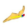
M3#1878Pozodolops manuelorum, fragmentary left P3 Type: "3D_surfaces"doi: 10.18563/m3.sf.1878 state:published |
Download 3D surface file |
Wamradolops telloi MUSM 4144 View specimen

|
M3#1879Wamradolops telloi, fragmentary left dentary with p2–m1 Type: "3D_surfaces"doi: 10.18563/m3.sf.1879 state:published |
Download 3D surface file |
Wamradolops telloi MUSM 4179 View specimen

|
M3#1880Wamradolops telloi, holotype, partial skull, with right P2–M4 and left I4–M3, plus boneless lower teeth below upper teeth, belonging to the same individual Type: "3D_surfaces"doi: 10.18563/m3.sf.1880 state:published |
Download 3D surface file |
Wamradolops telloi MUSM 4221 View specimen

|
M3#1881Wamradolops telloi, fragmentary of right dentary with p3–m2 Type: "3D_surfaces"doi: 10.18563/m3.sf.1881 state:published |
Download 3D surface file |
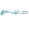
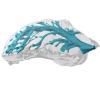
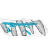
The present dataset contains 3D models used for illustration purposes in Schultz, J. A., Weaver, L. N., Jäger, K. R. K. & Grossnickle, D. M., 2026. Reexamining the evolutionary history of the mammalian medial pterygoid muscle. Evolution. The dataset includes 3D models based on micro-computed tomography (µCT) data of the postdentary area of the morganucodontan Morganucodon, the docodontan Docodon, the eutriconodontan Priacodon and the cladotherian Dryolestes. In addition, the dataset includes manually reconstructed schematic 3D models of the middle ear bones for the morganucodontan Morganucodon, the docodontan Docodon, the eutriconodontan Priacodon and schematic middle ear bones and a virtually rendered juvenile lower jaw of a juvenile monotreme Ornithorhynchus (based on illustrations of Zeller [1989]).
Morganucodon sp. NHM M84028 View specimen
|
M3#1883Lower jaw with manually reconstructed Meckel’s cartilage and schematic middle ear bones (Ectotympanic, Articular/part of Malleus, Surangular/ part of Malleus) Type: "3D_surfaces"doi: 10.18563/m3.sf.1883 state:published |
Download 3D surface file |
Priacodon fruitaensis LACM 120451 View specimen

|
M3#1884Lower jaw with manually reconstructed Meckel’s cartilage and schematic middle ear bones (Ectotympanic, Articular/part of Malleus, Surangular/part of Malleus) Type: "3D_surfaces"doi: 10.18563/m3.sf.1884 state:published |
Download 3D surface file |
Dryolestes leiriensis GuiMam 3-78 View specimen

|
M3#1888Lower jaw with manually reconstructed schematic Meckel’s cartilage Type: "3D_surfaces"doi: 10.18563/m3.sf.1888 state:published |
Download 3D surface file |
Docodon victor YPM10649, YPM13735, YPM11823 and YPM11826 View specimen
|
M3#1886Lower jaw with manually reconstructed Meckel’s cartilage and schematic middle ear bones (Ectotympanic, Articular/part of Malleus, Surangular/part of Malleus), for specification of jaw see Schultz et al. (2019) Type: "3D_surfaces"doi: 10.18563/m3.sf.1886 state:published |
Download 3D surface file |
Ornithorhynchus anatinus S32167 View specimen
|
M3#1887Lower jaw with manually reconstructed Meckel’s cartilage and schematic middle ear bones (Prearticular, Articular/part of the Malleus, Surangular/part of Malleus, Angular/Incus) Type: "3D_surfaces"doi: 10.18563/m3.sf.1887 state:published |
Download 3D surface file |
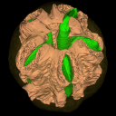
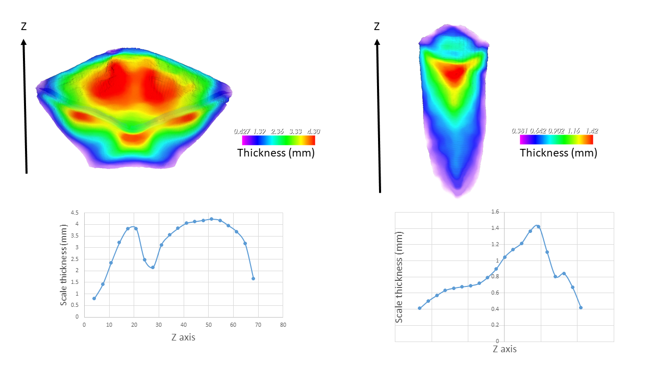
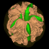
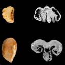
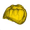
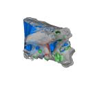
The present 3D Dataset contains the 3D models of two endocarps from the Oligocene of Baraval Quercy locality. These endocarps document new fossil genera within the Anacardiaceae family and illustrate the morphological diversity of this family during the Palaeogene. The CT-scan data were processed with ImageJ and Mimics Innovation Suite version 1.13 to reconstruct the specimens. Here we provide .stl files easy to read with the software Meshlab.
Palaeochoerospondias sheppeyensis UM-BAV-675 View specimen
|
M3#1895Surfaces of the endocarp, seeds and lacunae Type: "3D_surfaces"doi: 10.18563/m3.sf.1895 state:published |
Download 3D surface file |
Baravalosphaera operculata UM-BAV-689 View specimen
|
M3#1896Surfaces of the endocarp Type: "3D_surfaces"doi: 10.18563/m3.sf.1896 state:published |
Download 3D surface file |
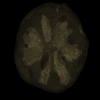
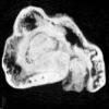
The present 3D dataset contains the 3D models of two fossil seeds of Vitaceae from the Quercy fossiliferous area, southwestern France. One seed comes from the Eocene locality of Fontoffre 2, and the other from the Oligocene locality of Baraval. These seeds document new fossil species within the Vitaceae family and illustrate the morphological diversity of this family during the Palaeogene. The CT scan data were processed with ImageJ and Mimics Innovation Suite version 1.13 to reconstruct the specimens. Here we provide .stl files that can be easily opened with the software MeshLab.
Vitis quercyensis UM-FTF2-167 View specimen
|
M3#1930Surface of the seed Type: "3D_surfaces"doi: 10.18563/m3.sf.1930 state:published |
Download 3D surface file |
|
M3#1933Seed : 8.43 μm µCT scan Type: "3D_CT"doi: 10.18563/m3.sf.1933 state:published |
Download CT data |
Vitis praerotundifolia UM-BAV-660 View specimen
|
M3#1931Seed : 8.43 μm µCT scan Type: "3D_CT"doi: 10.18563/m3.sf.1931 state:published |
Download CT data |
|
M3#1932Surface of the seed Type: "3D_surfaces"doi: 10.18563/m3.sf.1932 state:published |
Download 3D surface file |
The present 3D Dataset contains the 3D models of three specimens of Spinosauridae: two Cristatusaurus lapparenti Taquet & Russell, 1998 (MNHN.F.GDF365 and MNHN.F.GDF366) and one Spinosaurus maroccanus Russell, 1996 (MNHN.F.SAM124). These specimens are analyzed and discussed in Pittet F. 2026. Neurovascular system and dental renewal in the rostrum of Spinosauridae: new descriptions and implications on non-olfactive snout sensitivity of dinosaurs, Geodiversitas.
Cristatusaurus lapparenti MNHN.F.GDF365 View specimen
|
M3#1755Right and left premaxillae of MNHN.F.GDF365 (mature specimen of Cristatusaurus lapparenti): bony surface, teeth and neurovascular system Type: "3D_surfaces"doi: 10.18563/m3.sf.1755 state:published |
Download 3D surface file |
Spinosaurus maroccanus MNHN.F.SAM124 View specimen
|
M3#1756Premaxillae and maxillae of MNHN.F.SAM124 (Spinosaurus maroccanus): bony surface, teeth and neurovascular system Type: "3D_surfaces"doi: 10.18563/m3.sf.1756 state:published |
Download 3D surface file |
Cristatusaurus lapparenti MNHN.F.GDF366 View specimen
|
M3#1757Right and left premaxillae of MNHN.F.GDF366 (young specimen of Cristatusaurus lapparenti): bony surface, teeth and neurovascular system Type: "3D_surfaces"doi: 10.18563/m3.sf.1757 state:published |
Download 3D surface file |
|
M3#1758Right maxilla portion of MNHN.F.GDF366 (young specimen of Cristatusaurus lapparenti): bony surface, teeth and neurovascular system Type: "3D_surfaces"doi: 10.18563/m3.sf.1758 state:published |
Download 3D surface file |
The present 3D Dataset contains the 3D models analyzed in the following publication: Paulina-Carabajal, A., and Porfiri, J.D. 2026. Novel information on the braincase of Megaraptor namunhuaiquii (Dinosauria: Theropoda) using X-ray tomography: pneumaticity, paleoneurology and their paleobiological implications. Ameghiniana 63(1), 16-32
Megaraptor namunhuaiquii MUC-PV 595 View specimen
|
M3#18733D models of the braincase, lateral wall of the braincase, brain, right and left inner ears Type: "3D_surfaces"doi: 10.18563/m3.sf.1873 state:published |
Download 3D surface file |