

| Plane | Position | Flip |
| Show planes | Show edges |
0.0
M3#15
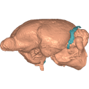
Labelled 3D model of the endocranial cast and sinuse of Microchoerus erinaceus.
Data citation:
Maëva J. Orliac , 2015. M3#15. doi: 10.18563/m3.sf15
Tag legend:
Endocast, Sinus
Model solid/transparent
Flags:
brain stem, capsuloparietal emissary vein canal, circular fissure, inferior petrosal sinus, IX, X, XI, lateral lobe of cerebellum, lateral sulcus, neocortex, olfactory bulb, optic chiasma, orbitotemporal canal, parafloccular lobe, piriform lobe, pituitary, sagital sinus, Sylvian fissure, temporal sulcus, transverse sinus, V3, vermis, VII, VIII, XII

|
The endocranial cast of Microchoerus erinaceus (Euprimates, Tarsiiformes).Maëva J. OrliacPublished online: 24/09/2015Keywords: endocast; Late Eocene; Omomyiformes; Primate https://doi.org/10.18563/m3.1.3.e4 Abstract This contribution contains the 3D model described and figured in the following publication: Ramdarshan A., Orliac M.J., 2015. Endocranial morphology of Microchoerus erinaceus (Euprimates, Tarsiiformes) and early evolution of the Euprimates brain. American Journal of Physical Anthropology. doi: 10.1002/ajpa.22868 See original publication M3 article infos Published in Volume 01, Issue 03 (2015) |
|