Holotype of Hamadasuchus rebouli
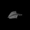
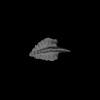
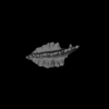
Inner ear morphology in wild vs laboratory mice
3D models of the endocranial anatomy of Voay robustus and comparative specimens
3D GM dataset of bird skeletal variation
Skeletal embryonic development in the catshark
Bony connexions of the petrosal bone of extant hippos
bony labyrinth (11) , inner ear (10) , South America (8) , Eocene (8) , skull (7) , brain (6) , Oligocene (6)
Maëva Judith Orliac (17) , Lionel Hautier (17) , Bastien Mennecart (12) , Laurent Marivaux (11) , Pierre-Olivier Antoine (11) , Leonardo Kerber (10) , Renaud Lebrun (9)
|
3D model related to the publication: The largest freshwater odontocete: a South Asian river dolphin relative from the Proto-AmazoniaAldo Benites-Palomino
Published online: 21/03/2024 |
|
M3#1394Holotype skull of Pebanista yacuruna MUSM 4017 Type: "3D_surfaces"doi: 10.18563/m3.sf.1394 state:in_press |
Download 3D surface file |
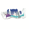
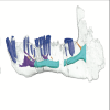
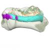
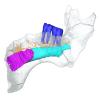
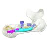
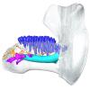
This contribution contains the 3D models described and figured in the following publication: Hautier L, Gomes Rodrigues H, Ferreira-Cardoso S, Emerling CA, Porcher M-L, Asher R, Portela Miguez R, Delsuc F. 2023. From teeth to pad: tooth loss and development of keratinous structures in sirenians. Proceedings of the Royal Society B. https://doi.org/10.1098/rspb.2023.1932
Dugong dugon 2005.51 View specimen

|
M3#1275Internal mandibular morphology. Orange = dorsal canaliculi; purple = mental branches; cyan = mandibular canal; dark blue = teeth; green = tooth alveoli. Type: "3D_surfaces"doi: 10.18563/m3.sf.1275 state:published |
Download 3D surface file |
Dugong dugon 2023.66 View specimen
|
M3#1274Internal mandibular morphology. Orange = dorsal canaliculi; purple = mental branches; cyan = mandibular canal; dark blue = teeth; green = tooth alveoli. Type: "3D_surfaces"doi: 10.18563/m3.sf.1274 state:published |
Download 3D surface file |
Dugong dugon 5386 View specimen
|
M3#1276Internal mandibular morphology. Orange = dorsal canaliculi; purple = mental branches; cyan = mandibular canal; dark blue = teeth; green = tooth alveoli. Type: "3D_surfaces"doi: 10.18563/m3.sf.1276 state:published |
Download 3D surface file |
Dugong dugon 1848.8.29.7/GERM 1027g View specimen
|
M3#1277Internal mandibular morphology. Orange = dorsal canaliculi; purple = mental branches; cyan = mandibular canal; dark blue = teeth. Type: "3D_surfaces"doi: 10.18563/m3.sf.1277 state:published |
Download 3D surface file |
Dugong dugon 1991.413 View specimen
|
M3#1278Internal mandibular morphology. Orange = dorsal canaliculi; purple = mental branches; cyan = mandibular canal; dark blue = teeth. Type: "3D_surfaces"doi: 10.18563/m3.sf.1278 state:published |
Download 3D surface file |
Dugong dugon 1991.427 View specimen

|
M3#1279Internal mandibular morphology. Orange = dorsal canaliculi; purple = mental branches; cyan = mandibular canal; dark blue = teeth. Type: "3D_surfaces"doi: 10.18563/m3.sf.1279 state:published |
Download 3D surface file |
Dugong dugon 2017-3-9 View specimen
|
M3#1280Internal mandibular morphology. Orange = dorsal canaliculi; purple = mental branches; cyan = mandibular canal; dark blue = teeth. Type: "3D_surfaces"doi: 10.18563/m3.sf.1280 state:published |
Download 3D surface file |
Eosiren lybica 1913-22 View specimen
|
M3#1281Internal mandibular morphology. Orange = dorsal canaliculi; purple = mental branches; cyan = mandibular canal; dark blue = teeth; green = tooth alveoli. Type: "3D_surfaces"doi: 10.18563/m3.sf.1281 state:published |
Download 3D surface file |
Halitherium taulannense RGHP C001 View specimen
|
M3#1282Internal mandibular morphology. Cyan = mandibular canal; dark blue = teeth. Type: "3D_surfaces"doi: 10.18563/m3.sf.1282 state:published |
Download 3D surface file |
Halitherium taulannense RGHP C009 View specimen
|
M3#1283Internal mandibular morphology. Orange = dorsal canaliculi; purple = mental branches; cyan = mandibular canal; dark blue = teeth; green = tooth alveoli. Type: "3D_surfaces"doi: 10.18563/m3.sf.1283 state:published |
Download 3D surface file |
Hydrodamalis gigas 1947.10.21.1 View specimen
|
M3#1284Internal mandibular morphology. Orange = dorsal canaliculi; purple = mental branches; cyan = mandibular canal Type: "3D_surfaces"doi: 10.18563/m3.sf.1284 state:published |
Download 3D surface file |
Hydrodamalis gigas C1021 View specimen
|
M3#1285Anterior part of the mandible Type: "3D_surfaces"doi: 10.18563/m3.sf.1285 state:published |
Download 3D surface file |

|
M3#1286Posterior part of the mandible Type: "3D_surfaces"doi: 10.18563/m3.sf.1286 state:published |
Download 3D surface file |
Hydrodamalis gigas 2023.67 View specimen
|
M3#1287Internal mandibular morphology. Orange = dorsal canaliculi; purple = mental branches; cyan = mandibular canal Type: "3D_surfaces"doi: 10.18563/m3.sf.1287 state:published |
Download 3D surface file |
Libysiren sickenbergi M.82429 View specimen
|
M3#1288Internal mandibular morphology. Orange = dorsal canaliculi; purple = mental branches; cyan = mandibular canal; dark blue = teeth; green = tooth alveoli. Type: "3D_surfaces"doi: 10.18563/m3.sf.1288 state:published |
Download 3D surface file |
Libysiren sickenbergi M.45675 View specimen
|
M3#1289Internal mandibular morphology. Orange = dorsal canaliculi; purple = mental branches; cyan = mandibular canal; dark blue = teeth; green = tooth alveoli. Type: "3D_surfaces"doi: 10.18563/m3.sf.1289 state:published |
Download 3D surface file |
Prorastomus sirenoides OR.448976 View specimen
|
M3#1290Internal morphology of the left mandible. Orange = dorsal canaliculi; purple = mental branches; cyan = mandibular canal; dark blue = teeth; green = tooth alveoli. Type: "3D_surfaces"doi: 10.18563/m3.sf.1290 state:published |
Download 3D surface file |
|
M3#1304Internal morphology of the right mandible. Orange = dorsal canaliculi; purple = mental branches; cyan = mandibular canal; dark blue = teeth. Type: "3D_surfaces"doi: 10.18563/m3.sf.1304 state:published |
Download 3D surface file |
Ribodon limbatus M.7073 View specimen
|
M3#1292Internal mandibular morphology. Orange = dorsal canaliculi; purple = mental branches; cyan = mandibular canal; dark blue = teeth; green = tooth alveoli. Type: "3D_surfaces"doi: 10.18563/m3.sf.1292 state:published |
Download 3D surface file |
Rytiodus capgrandi PAL2017-8-1 View specimen
|
M3#1293Internal mandibular morphology. Orange = dorsal canaliculi; purple = mental branches; cyan = mandibular canal; dark blue = teeth; green = tooth alveoli. Type: "3D_surfaces"doi: 10.18563/m3.sf.1293 state:published |
Download 3D surface file |
Trichechus inunguis 1868.12.19.2 View specimen
|
M3#1294Internal mandibular morphology. Orange = dorsal canaliculi; purple = mental branches; cyan = mandibular canal; dark blue = teeth; green = tooth alveoli. Type: "3D_surfaces"doi: 10.18563/m3.sf.1294 state:published |
Download 3D surface file |
Trichechus manatus 1843.3.10.12 View specimen
|
M3#1295Internal mandibular morphology. Orange = dorsal canaliculi; purple = mental branches; cyan = mandibular canal; dark blue = teeth. Type: "3D_surfaces"doi: 10.18563/m3.sf.1295 state:published |
Download 3D surface file |
Trichechus manatus 1864.6.5.1 View specimen
|
M3#1296Internal mandibular morphology. Orange = dorsal canaliculi; purple = mental branches; cyan = mandibular canal; dark blue = teeth; green = tooth alveoli. Type: "3D_surfaces"doi: 10.18563/m3.sf.1296 state:published |
Download 3D surface file |
Trichechus manatus 1950.1.23.1 View specimen
|
M3#1297Internal mandibular morphology. Orange = dorsal canaliculi; purple = mental branches; cyan = mandibular canal; dark blue = teeth; green = tooth alveoli. Type: "3D_surfaces"doi: 10.18563/m3.sf.1297 state:published |
Download 3D surface file |
Trichechus senegalensis 1885.6.30.2 View specimen
|
M3#1298Internal mandibular morphology. Orange = dorsal canaliculi; purple = mental branches; cyan = mandibular canal; dark blue = teeth; green = tooth alveoli. Type: "3D_surfaces"doi: 10.18563/m3.sf.1298 state:published |
Download 3D surface file |
Trichechus senegalensis 1894.7.25.8 View specimen
|
M3#1299Internal mandibular morphology. Orange = dorsal canaliculi; purple = mental branches; cyan = mandibular canal; dark blue = teeth. Type: "3D_surfaces"doi: 10.18563/m3.sf.1299 state:published |
Download 3D surface file |
Trichechus senegalensis V97 View specimen
|
M3#1302Mandibular internal morphology. Orange = dorsal canaliculi; purple = mental branches; cyan = mandibular canal; dark blue = teeth. Type: "3D_surfaces"doi: 10.18563/m3.sf.1302 state:published |
Download 3D surface file |
Trichechus sp. 65.4.28.9 View specimen
|
M3#1300Internal mandibular morphology. Orange = dorsal canaliculi; purple = mental branches; cyan = mandibular canal; dark blue = teeth; green = tooth alveoli. Type: "3D_surfaces"doi: 10.18563/m3.sf.1300 state:published |
Download 3D surface file |
Dugong dugon 1946.8.6.2 View specimen
|
M3#1301Mandibular internal morphology. Orange = dorsal canaliculi; purple = mental branches; cyan = mandibular canal; dark blue = teeth. Type: "3D_surfaces"doi: 10.18563/m3.sf.1301 state:published |
Download 3D surface file |
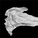
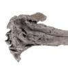
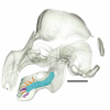
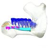
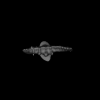
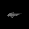
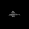
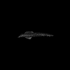
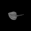
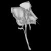
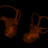
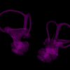
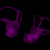
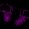
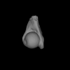
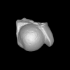
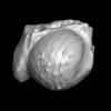
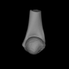
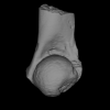
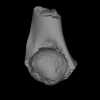
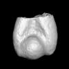
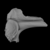
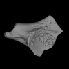
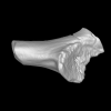
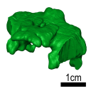
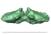
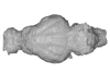
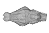
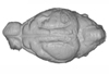
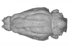
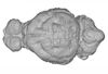
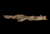
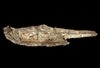
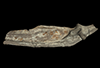
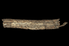
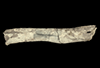
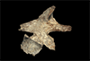
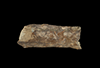
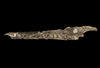
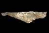
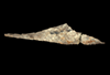
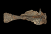
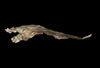
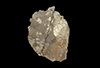
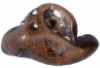
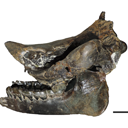
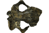
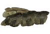
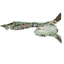
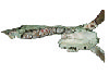
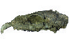
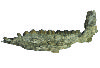
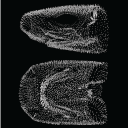
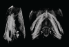
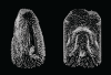
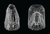
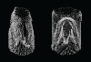
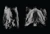
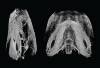
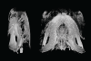
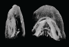
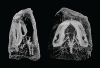
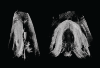
The holotype of Hamadasuchus rebouli Buffetaut 1994 from the Kem Kem beds of Morocco (Late Albian – Cenomanian) consists of a left dentary which is limited, fragmentary and reconstructed in some areas. To aid in assessing if the original diagnosis can be considered as valid, the specimen was CT scanned for the first time. This is especially important to resolve the taxonomic status of certain specimens that have been assigned to Hamadasuchus rebouli since then. The reconstructed structures in this contribution are in agreement with the original description, notably in terms of alveolar count; thus the original diagnosis of this taxon remains valid and some specimens are not referable to H. rebouli anymore.
Hamadasuchus rebouli MDE C001 View specimen

|
M3#1402Dentary and teeth Type: "3D_surfaces"doi: 10.18563/m3.sf.1402 state:in_press |
Download 3D surface file |

|
M3#1403Toothmarks Type: "3D_surfaces"doi: 10.18563/m3.sf.1403 state:in_press |
Download 3D surface file |
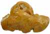
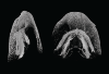
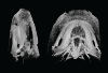
The present 3D Dataset contains the 3D models analyzed in Assemat et al. 2023: Shape diversity in conodont elements, a quantitative study using 3D topography. Marine Micropaleontology 184. https://doi.org/10.1016/j.marmicro.2023.102292
P1 elements represent dental components of the conodont apparatus that perform the final stage of food processing before ingestion. Consequently, quantifying the shape of P1 elements across the topographic indices of different conodont species becomes crucial for deciphering the diversity in feeding behavior within this group.
Bispathodus aculeatus UM CTB 082 View specimen
|
M3#1404P element Type: "3D_surfaces"doi: 10.18563/m3.sf.1404 state:in_press |
Download 3D surface file |
Bispathodus aculeatus UM CTB 083 View specimen

|
M3#1405P element Type: "3D_surfaces"doi: 10.18563/m3.sf.1405 state:in_press |
Download 3D surface file |
Bispathodus aculeatus UM CTB 086 View specimen
|
M3#1406P element Type: "3D_surfaces"doi: 10.18563/m3.sf.1406 state:in_press |
Download 3D surface file |
Bispathodus ultimus UM CTB 088 View specimen

|
M3#1407P element Type: "3D_surfaces"doi: 10.18563/m3.sf.1407 state:in_press |
Download 3D surface file |
Bispathodus aculeatus UM CTB 089 View specimen
|
M3#1408P element Type: "3D_surfaces"doi: 10.18563/m3.sf.1408 state:in_press |
Download 3D surface file |
Bispathodus costatus UM CTB 090 View specimen
|
M3#1409P element Type: "3D_surfaces"doi: 10.18563/m3.sf.1409 state:in_press |
Download 3D surface file |
Bispathodus ultimus UM CTB 092 View specimen
|
M3#1410P element Type: "3D_surfaces"doi: 10.18563/m3.sf.1410 state:in_press |
Download 3D surface file |
Bispathodus costatus UM CTB 093 View specimen
|
M3#1411P element Type: "3D_surfaces"doi: 10.18563/m3.sf.1411 state:in_press |
Download 3D surface file |
Bispathodus spinulicostatus UM CTB 094 View specimen
|
M3#1412P element Type: "3D_surfaces"doi: 10.18563/m3.sf.1412 state:in_press |
Download 3D surface file |
Bispathodus aculeatus UM CTB 096 View specimen

|
M3#1413P element Type: "3D_surfaces"doi: 10.18563/m3.sf.1413 state:in_press |
Download 3D surface file |
Bispathodus ultimus UM CTB 098 View specimen

|
M3#1414P element Type: "3D_surfaces"doi: 10.18563/m3.sf.1414 state:in_press |
Download 3D surface file |
Bispathodus costatus UM CTB 060 View specimen
|
M3#1415P element Type: "3D_surfaces"doi: 10.18563/m3.sf.1415 state:in_press |
Download 3D surface file |
Bispathodus spinulicostatus UM CTB 073 View specimen

|
M3#1416P element Type: "3D_surfaces"doi: 10.18563/m3.sf.1416 state:in_press |
Download 3D surface file |
Branmehla suprema UM CTB 049 View specimen

|
M3#1417P element Type: "3D_surfaces"doi: 10.18563/m3.sf.1417 state:in_press |
Download 3D surface file |
Branmehla inornata UM CTB 100 View specimen
|
M3#1418P element Type: "3D_surfaces"doi: 10.18563/m3.sf.1418 state:in_press |
Download 3D surface file |
Bispathodus stabilis (morphe 1) UM CTB 101 View specimen

|
M3#1419P element Type: "3D_surfaces"doi: 10.18563/m3.sf.1419 state:in_press |
Download 3D surface file |
Branmehla suprema UM CTB 102 View specimen
|
M3#1420P element Type: "3D_surfaces"doi: 10.18563/m3.sf.1420 state:in_press |
Download 3D surface file |
Branmehla suprema UM CTB 103 View specimen

|
M3#1421P element Type: "3D_surfaces"doi: 10.18563/m3.sf.1421 state:in_press |
Download 3D surface file |
Branmehla suprema UM CTB 104 View specimen

|
M3#1422P element Type: "3D_surfaces"doi: 10.18563/m3.sf.1422 state:in_press |
Download 3D surface file |
Branmehla suprema UM CTB 105 View specimen
|
M3#1423P element Type: "3D_surfaces"doi: 10.18563/m3.sf.1423 state:in_press |
Download 3D surface file |
Branmehla suprema UM CTB 106 View specimen
|
M3#1424P element Type: "3D_surfaces"doi: 10.18563/m3.sf.1424 state:in_press |
Download 3D surface file |
Branmehla suprema UM CTB 072 View specimen

|
M3#1425P element Type: "3D_surfaces"doi: 10.18563/m3.sf.1425 state:in_press |
Download 3D surface file |
Branmehla suprema UM CTB 107 View specimen
|
M3#1426P element Type: "3D_surfaces"doi: 10.18563/m3.sf.1426 state:in_press |
Download 3D surface file |
Branmehla suprema UM CTB 108 View specimen

|
M3#1427P element Type: "3D_surfaces"doi: 10.18563/m3.sf.1427 state:in_press |
Download 3D surface file |
Branmehla suprema UM CTB 109 View specimen
|
M3#1428P element Type: "3D_surfaces"doi: 10.18563/m3.sf.1428 state:in_press |
Download 3D surface file |
Bispathodus stabilis (morphe 1) UM CTB 110 View specimen

|
M3#1429P element Type: "3D_surfaces"doi: 10.18563/m3.sf.1429 state:in_press |
Download 3D surface file |
Palmatolepis gracilis UM CTB 112 View specimen
|
M3#1430P element Type: "3D_surfaces"doi: 10.18563/m3.sf.1430 state:in_press |
Download 3D surface file |
Palmatolepis gracilis UM CTB 061 View specimen

|
M3#1431P element Type: "3D_surfaces"doi: 10.18563/m3.sf.1431 state:in_press |
Download 3D surface file |
Palmatolepis gracilis UM CTB 115 View specimen
|
M3#1432P element Type: "3D_surfaces"doi: 10.18563/m3.sf.1432 state:in_press |
Download 3D surface file |
Palmatolepis gracilis UM CTB 116 View specimen
|
M3#1433P element Type: "3D_surfaces"doi: 10.18563/m3.sf.1433 state:in_press |
Download 3D surface file |
Palmatolepis gracilis UM CTB 117 View specimen
|
M3#1434P element Type: "3D_surfaces"doi: 10.18563/m3.sf.1434 state:in_press |
Download 3D surface file |
Palmatolepis gracilis UM CTB 062 View specimen

|
M3#1435P element Type: "3D_surfaces"doi: 10.18563/m3.sf.1435 state:in_press |
Download 3D surface file |
Palmatolepis gracilis UM CTB 118 View specimen

|
M3#1436P element Type: "3D_surfaces"doi: 10.18563/m3.sf.1436 state:in_press |
Download 3D surface file |
Palmatolepis gracilis UM CTB 119 View specimen
|
M3#1437P element Type: "3D_surfaces"doi: 10.18563/m3.sf.1437 state:in_press |
Download 3D surface file |
Palmatolepis gracilis UM CTB 120 View specimen
|
M3#1438P element Type: "3D_surfaces"doi: 10.18563/m3.sf.1438 state:in_press |
Download 3D surface file |
Polygnathus communis UM CTB 075 View specimen
|
M3#1439P element Type: "3D_surfaces"doi: 10.18563/m3.sf.1439 state:in_press |
Download 3D surface file |
Polygnathus communis UM CTB 121 View specimen
|
M3#1440P element Type: "3D_surfaces"doi: 10.18563/m3.sf.1440 state:in_press |
Download 3D surface file |
Polygnathus communis UM CTB 122 View specimen
|
M3#1441P element Type: "3D_surfaces"doi: 10.18563/m3.sf.1441 state:in_press |
Download 3D surface file |
Polygnathus communis UM CTB 123 View specimen
|
M3#1442P element Type: "3D_surfaces"doi: 10.18563/m3.sf.1442 state:in_press |
Download 3D surface file |
Polygnathus communis UM CTB 125 View specimen
|
M3#1443P element Type: "3D_surfaces"doi: 10.18563/m3.sf.1443 state:in_press |
Download 3D surface file |
Polygnathus communis UM CTB 126 View specimen
|
M3#1444P element Type: "3D_surfaces"doi: 10.18563/m3.sf.1444 state:in_press |
Download 3D surface file |
Polygnathus communis UM CTB 128 View specimen
|
M3#1445P element Type: "3D_surfaces"doi: 10.18563/m3.sf.1445 state:in_press |
Download 3D surface file |
Polygnathus communis UM CTB 130 View specimen
|
M3#1446P element Type: "3D_surfaces"doi: 10.18563/m3.sf.1446 state:in_press |
Download 3D surface file |
Polygnathus communis UM CTB 131 View specimen
|
M3#1447P element Type: "3D_surfaces"doi: 10.18563/m3.sf.1447 state:in_press |
Download 3D surface file |
Polygnathus communis UM CTB 132 View specimen

|
M3#1448P element Type: "3D_surfaces"doi: 10.18563/m3.sf.1448 state:in_press |
Download 3D surface file |
Polygnathus communis UM CTB 133 View specimen
|
M3#1449P element Type: "3D_surfaces"doi: 10.18563/m3.sf.1449 state:in_press |
Download 3D surface file |
Polygnathus symmetricus UM CTB 139 View specimen
|
M3#1450P element Type: "3D_surfaces"doi: 10.18563/m3.sf.1450 state:in_press |
Download 3D surface file |
Polygnathus symmetricus UM CTB 140 View specimen

|
M3#1451P element Type: "3D_surfaces"doi: 10.18563/m3.sf.1451 state:in_press |
Download 3D surface file |
Polygnathus symmetricus UM CTB 141 View specimen
|
M3#1452P element Type: "3D_surfaces"doi: 10.18563/m3.sf.1452 state:in_press |
Download 3D surface file |
Polygnathus symmetricus UM CTB 142 View specimen
|
M3#1453P element Type: "3D_surfaces"doi: 10.18563/m3.sf.1453 state:in_press |
Download 3D surface file |
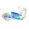
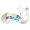
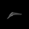
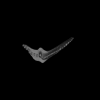
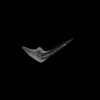
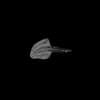
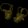
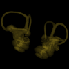
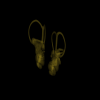
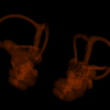
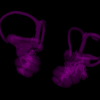
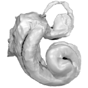
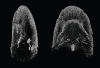
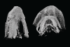
The present 3D Dataset contains 3D models of the cranium surface and of the bony labyrinth endocast of the stem bat Vielasia sigei. They are used by (Hand et al., 2023) to explore the phylogenetic position of this species, to infer its laryngeal echolocating capabilities, and to eventually discuss chiropteran evolution before the crown clade diversification.
Vielasia sigei UM VIE-250 View specimen
|
M3#1269External surface of the cranium Type: "3D_surfaces"doi: 10.18563/m3.sf.1269 state:published |
Download 3D surface file |
|
M3#1270Virtual endocast of the right bony labyrinth Type: "3D_surfaces"doi: 10.18563/m3.sf.1270 state:published |
Download 3D surface file |
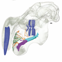
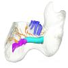
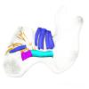
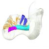
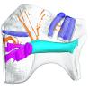
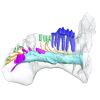
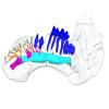
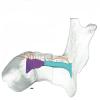
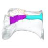
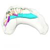
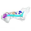
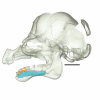
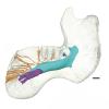
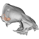
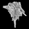
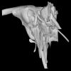
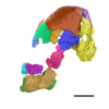
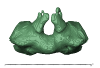
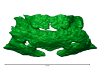
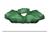
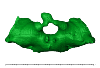
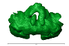
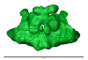
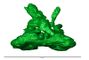
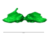
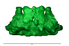
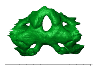
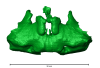
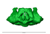
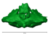
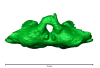
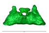
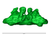
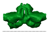
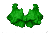
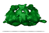
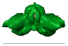
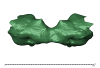
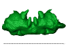
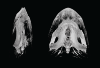
The present 3D Dataset contains the 3D models analyzed in: Perrichon et al., 2023. Neuroanatomy and pneumaticity of Voay robustus and its implications for crocodylid phylogeny and palaeoecology.
Crocodylus niloticus MHNL 50001387 View specimen
|
M3#1202Skull, inner ear, pharyngotympanic sinus and neurovascular system Type: "3D_surfaces"doi: 10.18563/m3.sf.1202 state:published |
Download 3D surface file |
Voay robustus MNHN F.1908-5 View specimen
|
M3#1203Skull, inner ear, pharyngotympanic sinus and neurovascular system Type: "3D_surfaces"doi: 10.18563/m3.sf.1203 state:published |
Download 3D surface file |
Voay robustus NHMUK PV R 36684 View specimen

|
M3#1204Skull, inner ear, pharyngotympanic sinus and neurovascular system Type: "3D_surfaces"doi: 10.18563/m3.sf.1204 state:published |
Download 3D surface file |
Voay robustus NHMUK PV R 36685 View specimen
|
M3#1205Skull, inner ear, pharyngotympanic sinus and neurovascular system Type: "3D_surfaces"doi: 10.18563/m3.sf.1205 state:published |
Download 3D surface file |
Osteolaemus tetraspis UCBLZ 2019-1-236 View specimen
|
M3#1208Skull, inner ear, pharyngotympanic sinus and neurovascular system Type: "3D_surfaces"doi: 10.18563/m3.sf.1208 state:published |
Download 3D surface file |
Mecistops sp. UM N89 View specimen
|
M3#1207Skull, inner ear, pharyngotympanic sinus and neurovascular system Type: "3D_surfaces"doi: 10.18563/m3.sf.1207 state:published |
Download 3D surface file |
Voay robustus NHMUK PV R 2204 View specimen

|
M3#1206Skull, inner ear, pharyngotympanic sinus, intertympanic sinus and neurovascular system Type: "3D_surfaces"doi: 10.18563/m3.sf.1206 state:published |
Download 3D surface file |
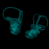
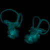
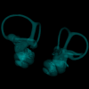
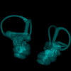
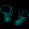
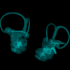
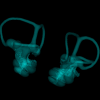
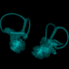
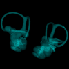
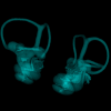
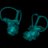
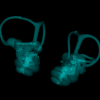
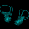
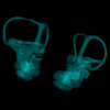
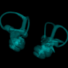
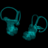
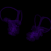
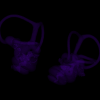
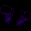
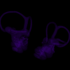
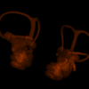
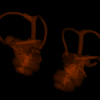
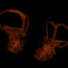
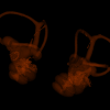
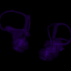
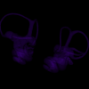
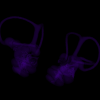
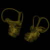
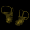
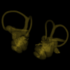
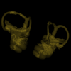
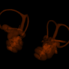
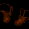
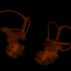
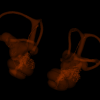
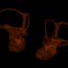
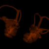
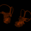
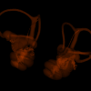
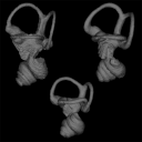
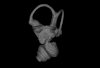
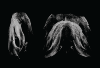
This contribution contains 3D models of left and right house mouse (Mus musculus domesticus) inner ears analyzed in Renaud et al. (2024). The studied mice belong to four groups: wild-trapped mice, wild-derived lab offspring, a typical laboratory strain (Swiss) and hybrids between wild-derived and Swiss mice. They have been analyzed to assess the impact of mobility reduction on inner ear morphology, including patterns of divergence, levels of inter-individual variance (disparity) and intra-individual variance (fluctuating asymmetry)
Mus musculus Tourch_7819 View specimen
|
M3#1366Two bony labyrinths Type: "3D_surfaces"doi: 10.18563/m3.sf.1366 state:in_press |
Download 3D surface file |
Mus musculus Tourch_7821 View specimen
|
M3#1367Two bony labyrinths Type: "3D_surfaces"doi: 10.18563/m3.sf.1367 state:in_press |
Download 3D surface file |
Mus musculus Tourch_7839 View specimen
|
M3#1368Two bony labyrinths Type: "3D_surfaces"doi: 10.18563/m3.sf.1368 state:in_press |
Download 3D surface file |
Mus musculus Tourch_7873 View specimen
|
M3#1369Two bony labyrinths Type: "3D_surfaces"doi: 10.18563/m3.sf.1369 state:in_press |
Download 3D surface file |
Mus musculus Tourch_7877 View specimen
|
M3#1370Two bony labyrinths Type: "3D_surfaces"doi: 10.18563/m3.sf.1370 state:in_press |
Download 3D surface file |
Mus musculus Tourch_7922 View specimen
|
M3#1371Two bony labyrinths Type: "3D_surfaces"doi: 10.18563/m3.sf.1371 state:in_press |
Download 3D surface file |
Mus musculus Tourch_7923 View specimen
|
M3#1372Two bony labyrinths Type: "3D_surfaces"doi: 10.18563/m3.sf.1372 state:in_press |
Download 3D surface file |
Mus musculus Tourch_7925 View specimen
|
M3#1373Two bony labyrinths Type: "3D_surfaces"doi: 10.18563/m3.sf.1373 state:in_press |
Download 3D surface file |
Mus musculus Tourch_7927 View specimen
|
M3#1374Two bony labyrinths Type: "3D_surfaces"doi: 10.18563/m3.sf.1374 state:in_press |
Download 3D surface file |
Mus musculus Tourch_7932 View specimen
|
M3#1375Two bony labyrinths Type: "3D_surfaces"doi: 10.18563/m3.sf.1375 state:in_press |
Download 3D surface file |
Mus musculus Bal02 View specimen
|
M3#1320Two bony labyrinths Type: "3D_surfaces"doi: 10.18563/m3.sf.1320 state:in_press |
Download 3D surface file |
Mus musculus Bal04 View specimen

|
M3#1321Two bony labyrinths Type: "3D_surfaces"doi: 10.18563/m3.sf.1321 state:in_press |
Download 3D surface file |
Mus musculus Bal06 View specimen
|
M3#1322Two bony labyrinths Type: "3D_surfaces"doi: 10.18563/m3.sf.1322 state:in_press |
Download 3D surface file |
Mus musculus Bal08 View specimen
|
M3#1323Two bony labyrinths Type: "3D_surfaces"doi: 10.18563/m3.sf.1323 state:in_press |
Download 3D surface file |
Mus musculus Bal11 View specimen
|
M3#1324Two bony labyrinths Type: "3D_surfaces"doi: 10.18563/m3.sf.1324 state:in_press |
Download 3D surface file |
Mus musculus Bal12 View specimen
|
M3#1325Two bony labyrinths Type: "3D_surfaces"doi: 10.18563/m3.sf.1325 state:in_press |
Download 3D surface file |
Mus musculus Bal15 View specimen
|
M3#1326Two bony labyrinths Type: "3D_surfaces"doi: 10.18563/m3.sf.1326 state:in_press |
Download 3D surface file |
Mus musculus Bal16 View specimen
|
M3#1327Two bony labyrinths Type: "3D_surfaces"doi: 10.18563/m3.sf.1327 state:in_press |
Download 3D surface file |
Mus musculus Bal17 View specimen
|
M3#1328Two bony labyrinths Type: "3D_surfaces"doi: 10.18563/m3.sf.1328 state:in_press |
Download 3D surface file |
Mus musculus Bal18 View specimen
|
M3#1329Two bony labyrinths Type: "3D_surfaces"doi: 10.18563/m3.sf.1329 state:in_press |
Download 3D surface file |
Mus musculus Bal19 View specimen
|
M3#1330Two bony labyrinths Type: "3D_surfaces"doi: 10.18563/m3.sf.1330 state:in_press |
Download 3D surface file |
Mus musculus Bal20 View specimen
|
M3#1331Two bony labyrinths Type: "3D_surfaces"doi: 10.18563/m3.sf.1331 state:in_press |
Download 3D surface file |
Mus musculus Bal21 View specimen
|
M3#1332Two bony labyrinths Type: "3D_surfaces"doi: 10.18563/m3.sf.1332 state:in_press |
Download 3D surface file |
Mus musculus Bal22 View specimen
|
M3#1333Two bony labyrinths Type: "3D_surfaces"doi: 10.18563/m3.sf.1333 state:in_press |
Download 3D surface file |
Mus musculus Bal23 View specimen
|
M3#1334Two bony labyrinths Type: "3D_surfaces"doi: 10.18563/m3.sf.1334 state:in_press |
Download 3D surface file |
Mus musculus Bal24 View specimen
|
M3#1335Two bony labyrinths Type: "3D_surfaces"doi: 10.18563/m3.sf.1335 state:in_press |
Download 3D surface file |
Mus musculus Bal25 View specimen
|
M3#1336Two bony labyrinths Type: "3D_surfaces"doi: 10.18563/m3.sf.1336 state:in_press |
Download 3D surface file |
Mus musculus Balan_LAB_035 View specimen
|
M3#1337Two bony labyrinths Type: "3D_surfaces"doi: 10.18563/m3.sf.1337 state:in_press |
Download 3D surface file |
Mus musculus Balan_LAB_046 View specimen
|
M3#1338Two bony labyrinths Type: "3D_surfaces"doi: 10.18563/m3.sf.1338 state:in_press |
Download 3D surface file |
Mus musculus Balan_LAB_054 View specimen
|
M3#1339Two bony labyrinths Type: "3D_surfaces"doi: 10.18563/m3.sf.1339 state:in_press |
Download 3D surface file |
Mus musculus Balan_LAB_056 View specimen
|
M3#1340Two bony labyrinths Type: "3D_surfaces"doi: 10.18563/m3.sf.1340 state:in_press |
Download 3D surface file |
Mus musculus Balan_LAB_082 View specimen
|
M3#1341Two bony labyrinths Type: "3D_surfaces"doi: 10.18563/m3.sf.1341 state:in_press |
Download 3D surface file |
Mus musculus Balan_LAB_086 View specimen

|
M3#1342Two bony labyrinths Type: "3D_surfaces"doi: 10.18563/m3.sf.1342 state:in_press |
Download 3D surface file |
Mus musculus Balan_LAB_092 View specimen
|
M3#1343Two bony labyrinths Type: "3D_surfaces"doi: 10.18563/m3.sf.1343 state:in_press |
Download 3D surface file |
Mus musculus Balan_LAB_319 View specimen
|
M3#1344Two bony labyrinths Type: "3D_surfaces"doi: 10.18563/m3.sf.1344 state:in_press |
Download 3D surface file |
Mus musculus Balan_LAB_325 View specimen
|
M3#1345Two bony labyrinths Type: "3D_surfaces"doi: 10.18563/m3.sf.1345 state:in_press |
Download 3D surface file |
Mus musculus Balan_LAB_329 View specimen
|
M3#1346Two bony labyrinths Type: "3D_surfaces"doi: 10.18563/m3.sf.1346 state:in_press |
Download 3D surface file |
Mus musculus Balan_LAB_330 View specimen
|
M3#1347Two bony labyrinths Type: "3D_surfaces"doi: 10.18563/m3.sf.1347 state:in_press |
Download 3D surface file |
Mus musculus Balan_LAB_F2b View specimen
|
M3#1348Two bony labyrinths Type: "3D_surfaces"doi: 10.18563/m3.sf.1348 state:in_press |
Download 3D surface file |
Mus musculus Balan_LAB_BB3weeks View specimen
|
M3#1349Two bony labyrinths Type: "3D_surfaces"doi: 10.18563/m3.sf.1349 state:in_press |
Download 3D surface file |
Mus musculus SW0ter View specimen
|
M3#1376Two bony labyrinths Type: "3D_surfaces"doi: 10.18563/m3.sf.1376 state:in_press |
Download 3D surface file |
Mus musculus SW343 View specimen
|
M3#1377Two bony labyrinths Type: "3D_surfaces"doi: 10.18563/m3.sf.1377 state:in_press |
Download 3D surface file |
Mus musculus SW1 View specimen
|
M3#1378Two bony labyrinths Type: "3D_surfaces"doi: 10.18563/m3.sf.1378 state:in_press |
Download 3D surface file |
Mus musculus SW2 View specimen
|
M3#1379Two bony labyrinths Type: "3D_surfaces"doi: 10.18563/m3.sf.1379 state:in_press |
Download 3D surface file |
Mus musculus BAL_F1_30x17_27j View specimen
|
M3#1350Two bony labyrinths Type: "3D_surfaces"doi: 10.18563/m3.sf.1350 state:in_press |
Download 3D surface file |
Mus musculus BAL_F1_167_48j View specimen
|
M3#1351Two bony labyrinths Type: "3D_surfaces"doi: 10.18563/m3.sf.1351 state:in_press |
Download 3D surface file |
Mus musculus BAL_F1_188_32j View specimen
|
M3#1352Two bony labyrinths Type: "3D_surfaces"doi: 10.18563/m3.sf.1352 state:in_press |
Download 3D surface file |
Mus musculus BAL_F1_192_28j View specimen
|
M3#1353Two bony labyrinths Type: "3D_surfaces"doi: 10.18563/m3.sf.1353 state:in_press |
Download 3D surface file |
Mus musculus BAL_F1_194_46j View specimen
|
M3#1354Two bony labyrinths Type: "3D_surfaces"doi: 10.18563/m3.sf.1354 state:in_press |
Download 3D surface file |
Mus musculus BAL_F1_196_44j View specimen
|
M3#1355Two bony labyrinths Type: "3D_surfaces"doi: 10.18563/m3.sf.1355 state:in_press |
Download 3D surface file |
Mus musculus BAL_F2_40x56_24j View specimen

|
M3#1356Two bony labyrinths Type: "3D_surfaces"doi: 10.18563/m3.sf.1356 state:in_press |
Download 3D surface file |
Mus musculus BAL_F2_47x61_22j View specimen
|
M3#1357Two bony labyrinths Type: "3D_surfaces"doi: 10.18563/m3.sf.1357 state:in_press |
Download 3D surface file |
Mus musculus Gardouch_3419 View specimen
|
M3#1358Two bony labyrinths Type: "3D_surfaces"doi: 10.18563/m3.sf.1358 state:in_press |
Download 3D surface file |
Mus musculus Gardouch_3432 View specimen
|
M3#1359Two bony labyrinths Type: "3D_surfaces"doi: 10.18563/m3.sf.1359 state:in_press |
Download 3D surface file |
Mus musculus Gardouch_3437 View specimen
|
M3#1360Two bony labyrinths Type: "3D_surfaces"doi: 10.18563/m3.sf.1360 state:in_press |
Download 3D surface file |
Mus musculus Gardouch_3439 View specimen
|
M3#1361Two bony labyrinths Type: "3D_surfaces"doi: 10.18563/m3.sf.1361 state:in_press |
Download 3D surface file |
Mus musculus Gardouch_3450 View specimen
|
M3#1362Two bony labyrinths Type: "3D_surfaces"doi: 10.18563/m3.sf.1362 state:in_press |
Download 3D surface file |
Mus musculus Gardouch_3453 View specimen
|
M3#1363Two bony labyrinths Type: "3D_surfaces"doi: 10.18563/m3.sf.1363 state:in_press |
Download 3D surface file |
Mus musculus Gardouch_3459 View specimen
|
M3#1364Two bony labyrinths Type: "3D_surfaces"doi: 10.18563/m3.sf.1364 state:in_press |
Download 3D surface file |
Mus musculus Gardouch_3462 View specimen
|
M3#1365Two bony labyrinths Type: "3D_surfaces"doi: 10.18563/m3.sf.1365 state:in_press |
Download 3D surface file |
Mus musculus SW5 View specimen
|
M3#1380Two bony labyrinths Type: "3D_surfaces"doi: 10.18563/m3.sf.1380 state:in_press |
Download 3D surface file |
Mus musculus SWF3 View specimen
|
M3#1381Two bony labyrinths Type: "3D_surfaces"doi: 10.18563/m3.sf.1381 state:in_press |
Download 3D surface file |
Mus musculus SW342 View specimen
|
M3#1382Two bony labyrinths Type: "3D_surfaces"doi: 10.18563/m3.sf.1382 state:in_press |
Download 3D surface file |
Mus musculus SW341 View specimen
|
M3#1383Two bony labyrinths Type: "3D_surfaces"doi: 10.18563/m3.sf.1383 state:in_press |
Download 3D surface file |
Mus musculus SW339 View specimen
|
M3#1384Two bony labyrinths Type: "3D_surfaces"doi: 10.18563/m3.sf.1384 state:in_press |
Download 3D surface file |
Mus musculus SWF4 View specimen
|
M3#1385Two bony labyrinths Type: "3D_surfaces"doi: 10.18563/m3.sf.1385 state:in_press |
Download 3D surface file |
Mus musculus SW0bis_350 View specimen
|
M3#1386Two bony labyrinths Type: "3D_surfaces"doi: 10.18563/m3.sf.1386 state:in_press |
Download 3D surface file |
Mus musculus SW0_348 View specimen
|
M3#1387Two bony labyrinths Type: "3D_surfaces"doi: 10.18563/m3.sf.1387 state:in_press |
Download 3D surface file |
Mus musculus SW347 View specimen
|
M3#1388Two bony labyrinths Type: "3D_surfaces"doi: 10.18563/m3.sf.1388 state:in_press |
Download 3D surface file |
Mus musculus SW345 View specimen
|
M3#1389Two bony labyrinths Type: "3D_surfaces"doi: 10.18563/m3.sf.1389 state:in_press |
Download 3D surface file |
Mus musculus hyb_125xSW_01 View specimen
|
M3#1390Two bony labyrinths Type: "3D_surfaces"doi: 10.18563/m3.sf.1390 state:in_press |
Download 3D surface file |
Mus musculus hyb_125xSW_02 View specimen
|
M3#1391Two bony labyrinths Type: "3D_surfaces"doi: 10.18563/m3.sf.1391 state:in_press |
Download 3D surface file |
Mus musculus hyb_SWx126_01 View specimen
|
M3#1392Two bony labyrinths Type: "3D_surfaces"doi: 10.18563/m3.sf.1392 state:in_press |
Download 3D surface file |
Mus musculus hyb_SWx126_02 View specimen
|
M3#1393Two bony labyrinths Type: "3D_surfaces"doi: 10.18563/m3.sf.1393 state:in_press |
Download 3D surface file |
The present contribution contains the 3D models of fossil humeri and ilia of anurans from various Eocene-Miocene deposits of Peruvian Amazonia. These fossils were described and figured in the following publication: Jansen et al. (2023), First Eocene–Miocene anuran fossils from Peruvian Amazonia: insights into Neotropical frog evolution and diversity. Papers in Palaeontology, The Palaeontological Association.
Indet. indet. MUSM 4746 View specimen
|
M3#1231Humeral fragment (distal end) Type: "3D_surfaces"doi: 10.18563/m3.sf.1231 state:published |
Download 3D surface file |
Indet. indet. MUSM 4747 View specimen
|
M3#1232Humeral fragment (distal end) Type: "3D_surfaces"doi: 10.18563/m3.sf.1232 state:published |
Download 3D surface file |
Indet. indet. MUSM 4748 View specimen
|
M3#1233Humeral fragment (distal end) Type: "3D_surfaces"doi: 10.18563/m3.sf.1233 state:published |
Download 3D surface file |
Indet. indet. MUSM 4755 View specimen
|
M3#1234Humeral fragment (distal end) Type: "3D_surfaces"doi: 10.18563/m3.sf.1234 state:published |
Download 3D surface file |
Indet. indet. MUSM 4756 View specimen
|
M3#1235Humeral fragment (distal end) Type: "3D_surfaces"doi: 10.18563/m3.sf.1235 state:published |
Download 3D surface file |
Indet. indet. MUSM 4757 View specimen

|
M3#1236Humeral fragment (distal end) Type: "3D_surfaces"doi: 10.18563/m3.sf.1236 state:published |
Download 3D surface file |
Indet. indet. MUSM 4761 View specimen
|
M3#1237Humeral fragment (distal end) Type: "3D_surfaces"doi: 10.18563/m3.sf.1237 state:published |
Download 3D surface file |
Indet. indet. MUSM 4763 View specimen
|
M3#1238Humeral fragment (distal end) Type: "3D_surfaces"doi: 10.18563/m3.sf.1238 state:published |
Download 3D surface file |
Indet. indet. MUSM 4765 View specimen
|
M3#1239Humeral fragment (distal end) Type: "3D_surfaces"doi: 10.18563/m3.sf.1239 state:published |
Download 3D surface file |
Indet. indet. MUSM 4766 View specimen
|
M3#1240Humeral fragment (distal end) Type: "3D_surfaces"doi: 10.18563/m3.sf.1240 state:published |
Download 3D surface file |
Indet. indet. MUSM 4775 View specimen
|
M3#1241Humeral fragment (distal end) Type: "3D_surfaces"doi: 10.18563/m3.sf.1241 state:published |
Download 3D surface file |
cf. Pipa sp. MUSM 4776 View specimen
|
M3#1242Humeral fragment (distal end) Type: "3D_surfaces"doi: 10.18563/m3.sf.1242 state:published |
Download 3D surface file |
Indet. indet. MUSM 4788 View specimen
|
M3#1243Ilial fragment Type: "3D_surfaces"doi: 10.18563/m3.sf.1243 state:published |
Download 3D surface file |
Indet. indet. MUSM 4789 View specimen
|
M3#1244Ilial fragment Type: "3D_surfaces"doi: 10.18563/m3.sf.1244 state:published |
Download 3D surface file |
Indet. indet. MUSM 4790 View specimen
|
M3#1245Ilial fragment Type: "3D_surfaces"doi: 10.18563/m3.sf.1245 state:published |
Download 3D surface file |
Indet. indet. MUSM 4792 View specimen
|
M3#1246Ilial fragment Type: "3D_surfaces"doi: 10.18563/m3.sf.1246 state:published |
Download 3D surface file |
Indet. indet. MUSM 4793 View specimen
|
M3#1247Ilial fragment Type: "3D_surfaces"doi: 10.18563/m3.sf.1247 state:published |
Download 3D surface file |
Indet. indet. MUSM 4794 View specimen
|
M3#1249Ilial fragment Type: "3D_surfaces"doi: 10.18563/m3.sf.1249 state:published |
Download 3D surface file |
Indet. indet. MUSM 4795 View specimen
|
M3#1250Ilial fragment Type: "3D_surfaces"doi: 10.18563/m3.sf.1250 state:published |
Download 3D surface file |
cf. Pipa sp. MUSM 4796 View specimen
|
M3#1251Ilial fragment Type: "3D_surfaces"doi: 10.18563/m3.sf.1251 state:published |
Download 3D surface file |
cf. Pipa sp. MUSM 4797 View specimen
|
M3#1252Ilial fragment Type: "3D_surfaces"doi: 10.18563/m3.sf.1252 state:published |
Download 3D surface file |

The present 3D Dataset contains the 3D model analyzed in the publication : On Roth’s “human fossil” from Baradero, Buenos Aires Province, Argentina: morphological and genetic analysis. The “human fossil” from Baradero, Buenos Aires Province, Argentina, is a collection of skeleton parts first recovered by Swiss paleontologist Santiago Roth and further studied by anthropologist Rudolf Martin. By the end of the 19th century and beginning of the 20th century it was considered as one of the oldest human skeletons from the southern cone. We studied the cranial anatomy and contextualized the ancient individual remains. We discuss the context of the finding, conducted an osteobiographical assessment and performed a 3D virtual reconstruction of the skull, using micro-CT-scans on selected skull fragments and the mandible. This was followed by the extraction of bone tissue and teeth samples for radiocarbon and genetic analyses, which brought only limited results due to poor preservation and possible contamination. We estimate that the individual from Baradero is a middle-aged adult male. We conclude that the revision of foundational collections with current methodological tools brings new insights and clarifies long held assumptions on the significance of samples that were recovered when archaeology was not yet professionalized.
Homo sapiens PIMUZ A/V 4217 View specimen
|
M3#11983D virtual reconstruction of the skull Type: "3D_surfaces"doi: 10.18563/m3.sf.1198 state:published |
Download 3D surface file |
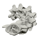
The present 3D Dataset contains the 3D models analyzed in Merten, L.J.F, Manafzadeh, A.R., Herbst, E.C., Amson, E., Tambusso, P.S., Arnold, P., Nyakatura, J.A., 2023. The functional significance of aberrant cervical counts in sloths: insights from automated exhaustive analysis of cervical range of motion. Proceedings of the Royal Society B. doi: 10.1098/rspb.2023.1592
Ailurus fulgens PMJ_Mam_6639 View specimen
|
M3#1260cervical vertebral series (7 vertebrae) Type: "3D_surfaces"doi: 10.18563/m3.sf.1260 state:published |
Download 3D surface file |
Bradypus variegatus ZMB_Mam_91345 View specimen
|
M3#1261cervical vertebral series (8 vertebrae) + first thoracic vertebra Type: "3D_surfaces"doi: 10.18563/m3.sf.1261 state:published |
Download 3D surface file |
Bradypus variegatus ZMB_Mam_35824 View specimen

|
M3#1262cervical vertebral series (8 vertebrae) + first & second thoracic vertebra Type: "3D_surfaces"doi: 10.18563/m3.sf.1262 state:published |
Download 3D surface file |
Choloepus didactylus ZMB_Mam_38388 View specimen
|
M3#1263cervical vertebral series (7 vertebrae) Type: "3D_surfaces"doi: 10.18563/m3.sf.1263 state:published |
Download 3D surface file |
Choloepus didactylus ZMB_Mam_102634 View specimen
|
M3#1264cervical vertebral series (6 vertebrae) + first thoracic vertebra Type: "3D_surfaces"doi: 10.18563/m3.sf.1264 state:published |
Download 3D surface file |
Tamandua tetradactyla ZMB_Mam_91288 View specimen
|
M3#1266cervical vertebral series (7 vertebrae) + first thoracic vertebra Type: "3D_surfaces"doi: 10.18563/m3.sf.1266 state:published |
Download 3D surface file |
Glossotherium robustum MNHN_n/n View specimen
|
M3#1267cervical vertebral series (7 vertebrae) + first thoracic vertebra Type: "3D_surfaces"doi: 10.18563/m3.sf.1267 state:published |
Download 3D surface file |
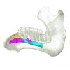
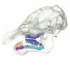
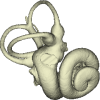
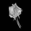
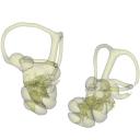
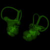
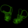
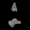
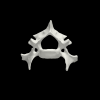
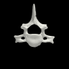
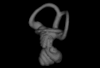
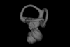
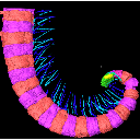
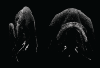
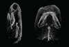
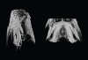
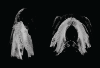
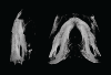
This contribution contains the three-dimensional models of the inner ear of the hetaxodontid rodents Amblyrhiza, Clidomys and Elasmodontomys from the West Indies. These specimens were analyzed and discussed in : The inner ear of caviomorph rodents: phylogenetic implications and application to extinct West Indian taxa.
Amblyrhiza inundata 11842 View specimen
|
M3#11543D surface of the left-oriented inner ear of Amblyrhiza. Type: "3D_surfaces"doi: 10.18563/m3.sf.1154 state:published |
Download 3D surface file |
Clidomys sp NA View specimen
|
M3#11553D surface of the left-oriented inner ear of Clidomys sp. Type: "3D_surfaces"doi: 10.18563/m3.sf.1155 state:published |
Download 3D surface file |
Elasmodontomys obliquus 17127 View specimen
|
M3#11563D surface of the left-oriented inner ear of Elasmodontomys obliquus. Type: "3D_surfaces"doi: 10.18563/m3.sf.1156 state:published |
Download 3D surface file |
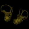
This contribution contains 3D models of upper molar rows of house mice (Mus musculus domesticus). The erupted part of the right row is presented for specimens belonging to four groups: wild-trapped mice, wild-derived lab offspring, a typical laboratory strain (Swiss) and hybrids between wild-derived and Swiss mice. These models are analyzed in the following publication: Savriama et al 2021: Wild versus lab house mice: Effects of age, diet, and genetics on molar geometry and topography. https://doi.org/10.1111/joa.13529
Mus musculus BW_03 View specimen

|
M3#736BW_03 Type: "3D_surfaces"doi: 10.18563/m3.sf.736 state:published |
Download 3D surface file |
Mus musculus BW_04 View specimen

|
M3#752BW_04 Type: "3D_surfaces"doi: 10.18563/m3.sf.752 state:published |
Download 3D surface file |
Mus musculus BW_06 View specimen

|
M3#753BW_06 Type: "3D_surfaces"doi: 10.18563/m3.sf.753 state:published |
Download 3D surface file |
Mus musculus BW_07 View specimen

|
M3#754BW_07 Type: "3D_surfaces"doi: 10.18563/m3.sf.754 state:published |
Download 3D surface file |
Mus musculus BW_08 View specimen

|
M3#755BW_08 Type: "3D_surfaces"doi: 10.18563/m3.sf.755 state:published |
Download 3D surface file |
Mus musculus BW_11 View specimen

|
M3#756BW_11 Type: "3D_surfaces"doi: 10.18563/m3.sf.756 state:published |
Download 3D surface file |
Mus musculus BW_12 View specimen

|
M3#757BW_12 Type: "3D_surfaces"doi: 10.18563/m3.sf.757 state:published |
Download 3D surface file |
Mus musculus Blab_035 View specimen

|
M3#758Blab_035 Type: "3D_surfaces"doi: 10.18563/m3.sf.758 state:published |
Download 3D surface file |
Mus musculus Blab_046 View specimen

|
M3#759Blab_046 Type: "3D_surfaces"doi: 10.18563/m3.sf.759 state:published |
Download 3D surface file |
Mus musculus Blab_054 View specimen

|
M3#760Blab_054 Type: "3D_surfaces"doi: 10.18563/m3.sf.760 state:published |
Download 3D surface file |
Mus musculus Blab_056 View specimen

|
M3#761Blab_056 Type: "3D_surfaces"doi: 10.18563/m3.sf.761 state:published |
Download 3D surface file |
Mus musculus Blab_082 View specimen

|
M3#762Blab_082 Type: "3D_surfaces"doi: 10.18563/m3.sf.762 state:published |
Download 3D surface file |
Mus musculus Blab_086 View specimen

|
M3#763Blab_086 Type: "3D_surfaces"doi: 10.18563/m3.sf.763 state:published |
Download 3D surface file |
Mus musculus Blab_092 View specimen

|
M3#764Blab_092 Type: "3D_surfaces"doi: 10.18563/m3.sf.764 state:published |
Download 3D surface file |
Mus musculus Blab_319 View specimen

|
M3#751Blab_319 Type: "3D_surfaces"doi: 10.18563/m3.sf.751 state:published |
Download 3D surface file |
Mus musculus Blab_325 View specimen

|
M3#750Blab_325 Type: "3D_surfaces"doi: 10.18563/m3.sf.750 state:published |
Download 3D surface file |
Mus musculus Blab_329 View specimen

|
M3#737Blab_329 Type: "3D_surfaces"doi: 10.18563/m3.sf.737 state:published |
Download 3D surface file |
Mus musculus Blab_330 View specimen

|
M3#738Blab_330 Type: "3D_surfaces"doi: 10.18563/m3.sf.738 state:published |
Download 3D surface file |
Mus musculus Blab_F2a View specimen

|
M3#739Blab_F2a Type: "3D_surfaces"doi: 10.18563/m3.sf.739 state:published |
Download 3D surface file |
Mus musculus Blab_F2b View specimen

|
M3#740Blab_F2b Type: "3D_surfaces"doi: 10.18563/m3.sf.740 state:published |
Download 3D surface file |
Mus musculus Blab_BB3w View specimen

|
M3#741Blab_BB3w Type: "3D_surfaces"doi: 10.18563/m3.sf.741 state:published |
Download 3D surface file |
Mus musculus hyb_BS01 View specimen

|
M3#742hyb_BS01 Type: "3D_surfaces"doi: 10.18563/m3.sf.742 state:published |
Download 3D surface file |
Mus musculus hyb_BS02 View specimen

|
M3#743hyb_BS02 Type: "3D_surfaces"doi: 10.18563/m3.sf.743 state:published |
Download 3D surface file |
Mus musculus hyb_SB01 View specimen

|
M3#744hyb_SB01 Type: "3D_surfaces"doi: 10.18563/m3.sf.744 state:published |
Download 3D surface file |
Mus musculus hyb_SB02 View specimen

|
M3#745hyb_SB02 Type: "3D_surfaces"doi: 10.18563/m3.sf.745 state:published |
Download 3D surface file |
Mus musculus SW_001 View specimen

|
M3#746SW_001 Type: "3D_surfaces"doi: 10.18563/m3.sf.746 state:published |
Download 3D surface file |
Mus musculus SW_002 View specimen
|
M3#747SW_002 Type: "3D_surfaces"doi: 10.18563/m3.sf.747 state:published |
Download 3D surface file |
Mus musculus SW_005 View specimen

|
M3#748SW_005 Type: "3D_surfaces"doi: 10.18563/m3.sf.748 state:published |
Download 3D surface file |
Mus musculus SW_0ter View specimen

|
M3#749SW_0ter Type: "3D_surfaces"doi: 10.18563/m3.sf.749 state:published |
Download 3D surface file |
Mus musculus SW_343 View specimen

|
M3#765SW_343 Type: "3D_surfaces"doi: 10.18563/m3.sf.765 state:published |
Download 3D surface file |
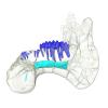
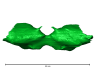
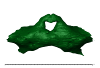
The present 3D Dataset contains the 3D models analyzed in: Perrichon et al. 2023. Ontogenetic variability of the intertympanic sinus distinguishes lineages within Crocodylia.
Mecistops sp. ag SVSTUA 022001 View specimen
|
M3#980Intertympanic sinus system (expressed in meters) Type: "3D_surfaces"doi: 10.18563/m3.sf.980 state:published |
Download 3D surface file |
Crocodylus niloticus ag SVSTUA 022002 View specimen
|
M3#981Intertympanic sinus system Type: "3D_surfaces"doi: 10.18563/m3.sf.981 state:published |
Download 3D surface file |
Mecistops sp. AMU Zoo 04721 View specimen
|
M3#982Intertympanic sinus system Type: "3D_surfaces"doi: 10.18563/m3.sf.982 state:published |
Download 3D surface file |
Crocodylus sp. MHNL QV14 View specimen
|
M3#983Intertympanic sinus system Type: "3D_surfaces"doi: 10.18563/m3.sf.983 state:published |
Download 3D surface file |
Crocodylus rhombifer MHNL 42006506 View specimen

|
M3#1008Intertympanic sinus system (incomplete) Type: "3D_surfaces"doi: 10.18563/m3.sf.1008 state:published |
Download 3D surface file |
Crocodylus rhombifer MHNL 42006507 View specimen
|
M3#1007Intertympanic sinus system (incomplete) Type: "3D_surfaces"doi: 10.18563/m3.sf.1007 state:published |
Download 3D surface file |
Crocodylus niloticus MHNL 50001387 View specimen
|
M3#1006Intertympanic sinus system Type: "3D_surfaces"doi: 10.18563/m3.sf.1006 state:published |
Download 3D surface file |
Crocodylus palustris MHNL 50001388 View specimen
|
M3#1004Intertympanic sinus system Type: "3D_surfaces"doi: 10.18563/m3.sf.1004 state:published |
Download 3D surface file |
Crocodylus porosus MHNL 50001389 View specimen
|
M3#1009Intertympanic sinus system Type: "3D_surfaces"doi: 10.18563/m3.sf.1009 state:published |
Download 3D surface file |
Mecistops sp. MHNL 50001393 View specimen
|
M3#1010Intertympanic sinus system Type: "3D_surfaces"doi: 10.18563/m3.sf.1010 state:published |
Download 3D surface file |
Crocodylus niloticus MHNL 50001397 View specimen
|
M3#1018Intertympanic sinus system Type: "3D_surfaces"doi: 10.18563/m3.sf.1018 state:published |
Download 3D surface file |
Crocodylus porosus MHNL 50001398 View specimen
|
M3#1017Intertympanic sinus system Type: "3D_surfaces"doi: 10.18563/m3.sf.1017 state:published |
Download 3D surface file |
Crocodylus niloticus MHNL 50001405 View specimen
|
M3#1016Intertympanic sinus system Type: "3D_surfaces"doi: 10.18563/m3.sf.1016 state:published |
Download 3D surface file |
Gavialis gangeticus MHNL 50001407 View specimen
|
M3#1015Intertympanic sinus system Type: "3D_surfaces"doi: 10.18563/m3.sf.1015 state:published |
Download 3D surface file |
Crocodylus niloticus MHNL 90001850 View specimen
|
M3#1014Intertympanic sinus system Type: "3D_surfaces"doi: 10.18563/m3.sf.1014 state:published |
Download 3D surface file |
Crocodylus niloticus MHNL 90001851 View specimen
|
M3#1013Intertympanic sinus system Type: "3D_surfaces"doi: 10.18563/m3.sf.1013 state:published |
Download 3D surface file |
Crocodylus niloticus MHNL 90001855 View specimen
|
M3#1012Intertympanic sinus system Type: "3D_surfaces"doi: 10.18563/m3.sf.1012 state:published |
Download 3D surface file |
Osteolaemus tetraspis MHNM.9095.0 View specimen
|
M3#1011Intertympanic sinus system Type: "3D_surfaces"doi: 10.18563/m3.sf.1011 state:published |
Download 3D surface file |
Voay robustus MNHN F.1908-5 View specimen
|
M3#1003Intertympanic sinus system Type: "3D_surfaces"doi: 10.18563/m3.sf.1003 state:published |
Download 3D surface file |
Crocodylus sp. MNHN-F.1908-5-2 View specimen
|
M3#1005Intertympanic sinus system Type: "3D_surfaces"doi: 10.18563/m3.sf.1005 state:published |
Download 3D surface file |
Osteolaemus tetraspis MZS Cro 040 View specimen
|
M3#1002Intertympanic sinus system Type: "3D_surfaces"doi: 10.18563/m3.sf.1002 state:published |
Download 3D surface file |
Crocodylus acutus MZS Cro 055 View specimen
|
M3#991Intertympanic sinus system Type: "3D_surfaces"doi: 10.18563/m3.sf.991 state:published |
Download 3D surface file |
Melanosuchus niger MZS Cro 073 View specimen
|
M3#989Intertympanic sinus system Type: "3D_surfaces"doi: 10.18563/m3.sf.989 state:published |
Download 3D surface file |
Mecistops sp. MZS Cro 083 View specimen

|
M3#990Intertympanic sinus system Type: "3D_surfaces"doi: 10.18563/m3.sf.990 state:published |
Download 3D surface file |
Tomistoma schlegelii MZS Cro 094 View specimen
|
M3#988Intertympanic sinus system Type: "3D_surfaces"doi: 10.18563/m3.sf.988 state:published |
Download 3D surface file |
Gavialis gangeticus NHMUK 1846.1.7.3 View specimen
|
M3#987Intertympanic sinus system Type: "3D_surfaces"doi: 10.18563/m3.sf.987 state:published |
Download 3D surface file |
Osteolaemus tetraspis NHMUK 1862.6.30.5 View specimen
|
M3#986Intertympanic sinus system Type: "3D_surfaces"doi: 10.18563/m3.sf.986 state:published |
Download 3D surface file |
Gavialis gangeticus NHMUK 1873 View specimen
|
M3#985intertympanic sinus system Type: "3D_surfaces"doi: 10.18563/m3.sf.985 state:published |
Download 3D surface file |
Tomistoma schlegelii NHMUK 1893.3.6.14 View specimen
|
M3#984Intertympanic sinus system Type: "3D_surfaces"doi: 10.18563/m3.sf.984 state:published |
Download 3D surface file |
Mecistops sp. NHMUK 1924.5.10.1 View specimen
|
M3#992Intertympanic sinus system Type: "3D_surfaces"doi: 10.18563/m3.sf.992 state:published |
Download 3D surface file |
Voay robustus NHMUK PV R 36684 View specimen
|
M3#993Intertympanic sinus system Type: "3D_surfaces"doi: 10.18563/m3.sf.993 state:published |
Download 3D surface file |
Voay robustus NHMUK PV R 36685 View specimen
|
M3#1001Intertympanic sinus system Type: "3D_surfaces"doi: 10.18563/m3.sf.1001 state:published |
Download 3D surface file |
Crocodylus niloticus UCBL FSL 532077 View specimen

|
M3#1000Intertympanic sinus system Type: "3D_surfaces"doi: 10.18563/m3.sf.1000 state:published |
Download 3D surface file |
Crocodylus porosus/siamensis UCBLZ 2019-1-237 View specimen
|
M3#999Intertympanic sinus system Type: "3D_surfaces"doi: 10.18563/m3.sf.999 state:published |
Download 3D surface file |
Osteolaemus tetraspis UCBLZ 2019-1-236 View specimen
|
M3#998Intertympanic sinus system Type: "3D_surfaces"doi: 10.18563/m3.sf.998 state:published |
Download 3D surface file |
Alligator mississipiensis UCBLZ WB35 View specimen
|
M3#997Intertympanic sinus system Type: "3D_surfaces"doi: 10.18563/m3.sf.997 state:published |
Download 3D surface file |
Crocodylus siamensis UCBLZ WB41 View specimen
|
M3#996Intertympanic sinus system Type: "3D_surfaces"doi: 10.18563/m3.sf.996 state:published |
Download 3D surface file |
Tomistoma schlegelii UM 1097 View specimen
|
M3#995Intertympanic sinus system Type: "3D_surfaces"doi: 10.18563/m3.sf.995 state:published |
Download 3D surface file |
Crocodylus niloticus UM 2001-1756-1-434 NR View specimen
|
M3#994Intertympanic sinus system Type: "3D_surfaces"doi: 10.18563/m3.sf.994 state:published |
Download 3D surface file |
Mecistops sp. UM N89 View specimen
|
M3#1019Intertympanic sinus system Type: "3D_surfaces"doi: 10.18563/m3.sf.1019 state:published |
Download 3D surface file |
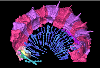
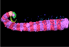
The present 3D Dataset contains the 3D models analyzed in the publication Fossils from the Montceau-les-Mines Lagerstätte (305 Ma) shed light on the anatomy, ecology and phylogeny of Carboniferous millipedes. Authors: Lheritier Mickael, Perroux Maëva, Vannier Jean, Escarguel Gilles, Wesener Thomas, Moritz Leif, Chabard Dominique, Adrien Jerome and Perrier Vincent. Journal of Systematics Palaeontology. https://doi.org/10.1080/14772019.2023.2169891
Amynilyspes fatimae MNHN.F.SOT.2134 View specimen

|
M3#1073Nearly complete specimen. Type: "3D_surfaces"doi: 10.18563/m3.sf.1073 state:published |
Download 3D surface file |
Amynilyspes fatimae MNHN.F.SOT.14983 View specimen
|
M3#1074Nearly complete specimen. Type: "3D_surfaces"doi: 10.18563/m3.sf.1074 state:published |
Download 3D surface file |
Amynilyspes fatimae MNHN.F.SOT.2129 View specimen
|
M3#1075Nearly complete specimen. Type: "3D_surfaces"doi: 10.18563/m3.sf.1075 state:published |
Download 3D surface file |
Blanzilius parriati MNHN.F.SOT.2114A View specimen
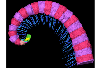
|
M3#1076Front part. Type: "3D_surfaces"doi: 10.18563/m3.sf.1076 state:published |
Download 3D surface file |
Blanzilius parriati MNHN.F.SOT.5148 View specimen
|
M3#1077Front part. Type: "3D_surfaces"doi: 10.18563/m3.sf.1077 state:published |
Download 3D surface file |
Blanzilius parriati MNHN.F.SOT.2113 View specimen
|
M3#1078Fragment with legs, sternites and possible tracheal openings. Type: "3D_surfaces"doi: 10.18563/m3.sf.1078 state:published |
Download 3D surface file |
Blanzilius parriati MNHN.F.SOT.81522 View specimen
|
M3#1079Nealry complete specimen. Type: "3D_surfaces"doi: 10.18563/m3.sf.1079 state:published |
Download 3D surface file |
The present 3D Dataset contains the 3D models illustrated and described in the chapter “Paleoneurology of Artiodactyla, an overview of the evolution of the artiodactyl brain” (Orliac et al. 2022) published in "Paleoneurology of amniotes: new directions in the study of fossil endocasts", edited by Dozo, Paulina-Carabajal, Macrini and Walsh.
Homacodon vagans AMNH 12695 View specimen
|
M3#1063Endocranial cast Type: "3D_surfaces"doi: 10.18563/m3.sf.1063 state:published |
Download 3D surface file |
Helohyus sp. AMNH 13079 View specimen
|
M3#1064Endocranial cast Type: "3D_surfaces"doi: 10.18563/m3.sf.1064 state:published |
Download 3D surface file |
Leptauchenia sp. AMNH 45508 View specimen
|
M3#1065endocranial cast Type: "3D_surfaces"doi: 10.18563/m3.sf.1065 state:published |
Download 3D surface file |
Agriochoerus sp. AMNH 95330 View specimen
|
M3#1067endocranial cast Type: "3D_surfaces"doi: 10.18563/m3.sf.1067 state:published |
Download 3D surface file |
Mouillacitherium elegans UM ACQ 6625 View specimen
|
M3#1068endocranial cast Type: "3D_surfaces"doi: 10.18563/m3.sf.1068 state:published |
Download 3D surface file |
Caenomeryx filholi UM PDS 2570 View specimen
|
M3#1069endocranial cast Type: "3D_surfaces"doi: 10.18563/m3.sf.1069 state:published |
Download 3D surface file |
Dichobune leporina MNHN.F.QU16586 View specimen
|
M3#1070endocranial cast Type: "3D_surfaces"doi: 10.18563/m3.sf.1070 state:published |
Download 3D surface file |
Anoplotherium sp. not numbered View specimen
|
M3#1071endocranial cast Type: "3D_surfaces"doi: 10.18563/m3.sf.1071 state:published |
Download 3D surface file |
The democratization of 3D techniques in recent years provides exciting new opportunities for the study of complex fossils. In the present contribution, we provide a virtual reconstruction of a partial, disarticulated metriorhynchid (Metriorhynchidae, Thalattosuchia, Crocodylomorpha) skull from the Late Jurassic of northwestern Switzerland. This virtual reconstruction was used to produce high quality scientific illustrations of the whole skull for descriptive purposes. The reconstructed skull also served for the estimation of the total body length of the specimen and to propose a life reconstruction of the animal in its paleoenvironment. In an effort for transparency, we review the sources that were consulted for the life reconstruction and explain the choices that we had to make.
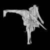
Torvoneustes jurensis BSY008-465 View specimen
|
M3#1037Left dentary (3 meshes) Type: "3D_surfaces"doi: 10.18563/m3.sf.1037 state:published |
Download 3D surface file |
|
M3#1038Right dentary (3 meshes) Type: "3D_surfaces"doi: 10.18563/m3.sf.1038 state:published |
Download 3D surface file |
|
M3#1039Left ramus (2 meshes) Type: "3D_surfaces"doi: 10.18563/m3.sf.1039 state:published |
Download 3D surface file |
|
M3#1040Right ramus (3 meshes) Type: "3D_surfaces"doi: 10.18563/m3.sf.1040 state:published |
Download 3D surface file |
|
M3#1041Left splenial (2 meshes) Type: "3D_surfaces"doi: 10.18563/m3.sf.1041 state:published |
Download 3D surface file |
|
M3#1042Right splenial (2 meshes) Type: "3D_surfaces"doi: 10.18563/m3.sf.1042 state:published |
Download 3D surface file |
|
M3#1043Frontal and left prefrontal Type: "3D_surfaces"doi: 10.18563/m3.sf.1043 state:published |
Download 3D surface file |
|
M3#1044Left maxilla (4 meshes) Type: "3D_surfaces"doi: 10.18563/m3.sf.1044 state:published |
Download 3D surface file |
|
M3#1045Right maxilla Type: "3D_surfaces"doi: 10.18563/m3.sf.1045 state:published |
Download 3D surface file |
|
M3#1046Left nasal Type: "3D_surfaces"doi: 10.18563/m3.sf.1046 state:published |
Download 3D surface file |
|
M3#1047Right nasal Type: "3D_surfaces"doi: 10.18563/m3.sf.1047 state:published |
Download 3D surface file |
|
M3#1048Parietal Type: "3D_surfaces"doi: 10.18563/m3.sf.1048 state:published |
Download 3D surface file |
|
M3#1049Right postorbital Type: "3D_surfaces"doi: 10.18563/m3.sf.1049 state:published |
Download 3D surface file |
|
M3#1050Right prefrontal Type: "3D_surfaces"doi: 10.18563/m3.sf.1050 state:published |
Download 3D surface file |
|
M3#1051Right premaxilla Type: "3D_surfaces"doi: 10.18563/m3.sf.1051 state:published |
Download 3D surface file |
|
M3#1052Left squamosal Type: "3D_surfaces"doi: 10.18563/m3.sf.1052 state:published |
Download 3D surface file |
|
M3#1053Right squamosal Type: "3D_surfaces"doi: 10.18563/m3.sf.1053 state:published |
Download 3D surface file |

|
M3#1054Reconstruction of the mandible Type: "3D_surfaces"doi: 10.18563/m3.sf.1054 state:published |
Download 3D surface file |

|
M3#1055Reconstruction of the cranium Type: "3D_surfaces"doi: 10.18563/m3.sf.1055 state:published |
Download 3D surface file |
This contribution contains the 3D models described and figured in the following publication: Aguirre-Fernández G, Jost J, and Hilfiker S. 2022. First records of extinct kentriodontid and squalodelphinid dolphins from the Upper Marine Molasse (Burdigalian age) of Switzerland and a reappraisal of the Swiss cetacean fauna.
Kentriodon sp. NMBE 5023944 View specimen
|
M3#8583D models of left periotic and bony labyrinth of NMBE 5023944 (Kentriodon sp.) Type: "3D_surfaces"doi: 10.18563/m3.sf.858 state:published |
Download 3D surface file |
Kentriodon sp. NMBE 5023945 View specimen
|
M3#8593D models of right periotic and bony labyrinth of NMBE 5023945 (Kentriodontidae indet.) Type: "3D_surfaces"doi: 10.18563/m3.sf.859 state:published |
Download 3D surface file |
Kentriodon sp. NMBE 5023946 View specimen
|
M3#8603D models of left periotic and bony labyrinth of NMBE 5023946 (Kentriodon sp.) Type: "3D_surfaces"doi: 10.18563/m3.sf.860 state:published |
Download 3D surface file |
Kentriodon sp. NMBE 5036436 View specimen
|
M3#8613D models of right periotic and bony labyrinth of NMBE 5036436 (Kentriodontidae indet.) Type: "3D_surfaces"doi: 10.18563/m3.sf.861 state:published |
Download 3D surface file |
indet. indet. NMBE 5023942 View specimen
|
M3#8623D models of right periotic and bony labyrinth of NMBE 5023942 (Squalodelphinidae indet.) Type: "3D_surfaces"doi: 10.18563/m3.sf.862 state:published |
Download 3D surface file |
indet. indet. NMBE 5023943 View specimen
|
M3#8633D models of left periotic and bony labyrinth of NMBE 5023943 (Squalodelphinidae indet.) Type: "3D_surfaces"doi: 10.18563/m3.sf.863 state:published |
Download 3D surface file |
indet. indet. NMBE 5036437 View specimen
|
M3#8643D models of left periotic and bony labyrinth of NMBE 5036437 (Physeteridae indet.) Type: "3D_surfaces"doi: 10.18563/m3.sf.864 state:published |
Download 3D surface file |
This contribution contains the 3D models described and figured in the following publication: Tissier et al. (in prep.).
Sellamynodon zimborensis UBB MPS 15795 View specimen
|
M3#297Incomplete skull with left M3. Type: "3D_surfaces"doi: 10.18563/m3.sf.297 state:published |
Download 3D surface file |
Sellamynodon zimborensis UBB MPS 15795 View specimen

|
M3#298Mandible with complete molar and premolar rows, lacking symphysis. Type: "3D_surfaces"doi: 10.18563/m3.sf.298 state:published |
Download 3D surface file |
Amynodontopsis aff. bodei UBB MPS V545 View specimen
|
M3#299Maxillary fragment with M1-3. Type: "3D_surfaces"doi: 10.18563/m3.sf.299 state:published |
Download 3D surface file |
Amynodontopsis aff. bodei UBB MPS V546 View specimen
|
M3#300Unworn m1/2 on mandible fragment. Type: "3D_surfaces"doi: 10.18563/m3.sf.300 state:published |
Download 3D surface file |
This contribution contains the 3D models described and figured in: New remains of Neotropical bunodont litopterns and the systematics of Megadolodinae (Mammalia: Litopterna). Geodiversitas.
Megadolodus molariformes VPPLT 974 View specimen
|
M3#1020Partial mandible with the symphysis and left body, bearing the alveoli of ?i2, right and left ?i3, alveolus of right c and p1, roots of left p1, and the left p2–m3 of Megadolodus molariformes (Litopterna, Mammalia) Type: "3D_surfaces"doi: 10.18563/m3.sf.1020 state:published |
Download 3D surface file |
Neodolodus colombianus VPPLT 1696 View specimen
|
M3#1021Almost complete skull with left and right ?I2 and P1–M3 Type: "3D_surfaces"doi: 10.18563/m3.sf.1021 state:published |
Download 3D surface file |
|
M3#1022Partial mandible with complete right and left dentition except for left ?i2 Type: "3D_surfaces"doi: 10.18563/m3.sf.1022 state:published |
Download 3D surface file |
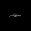
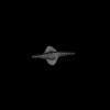
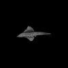
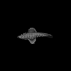
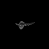
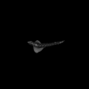
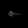
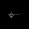
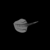
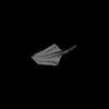
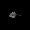
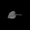
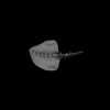
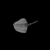
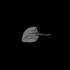
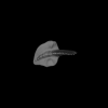
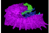
The present dataset contains the 3D models analyzed in Berio, F., Bayle, Y., Baum, D., Goudemand, N., and Debiais-Thibaud, M. 2022. Hide and seek shark teeth in Random Forests: Machine learning applied to Scyliorhinus canicula. It contains the head surfaces of 56 North Atlantic and Mediterranean small-spotted catsharks Scyliorhinus canicula, from which tooth surfaces were further extracted to perform geometric morphometrics and machine learning.
Scyliorhinus canicula 081118A View specimen
|
M3#941Head of a 10.6 cm long Scyliorhinus canicula female from a North Atlantic population. Type: "3D_surfaces"doi: 10.18563/m3.sf.941 state:published |
Download 3D surface file |
Scyliorhinus canicula 081118B View specimen
|
M3#942Head of a 11.0 cm long Scyliorhinus canicula female from a North Atlantic population. Type: "3D_surfaces"doi: 10.18563/m3.sf.942 state:published |
Download 3D surface file |
Scyliorhinus canicula 200118I View specimen
|
M3#959Head of a 45.0 cm long Scyliorhinus canicula female from a Mediterranean population. Type: "3D_surfaces"doi: 10.18563/m3.sf.959 state:published |
Download 3D surface file |
Scyliorhinus canicula 200118H View specimen
|
M3#958Head of a 47.0 cm long Scyliorhinus canicula female from a Mediterranean population. Type: "3D_surfaces"doi: 10.18563/m3.sf.958 state:published |
Download 3D surface file |
Scyliorhinus canicula 200118G View specimen
|
M3#957Head of a 40.0 cm long Scyliorhinus canicula female from a Mediterranean population. Type: "3D_surfaces"doi: 10.18563/m3.sf.957 state:published |
Download 3D surface file |
Scyliorhinus canicula 081118C View specimen
|
M3#940Head of a 11.2 cm long Scyliorhinus canicula female from a North Atlantic population. Type: "3D_surfaces"doi: 10.18563/m3.sf.940 state:published |
Download 3D surface file |
Scyliorhinus canicula 081118D View specimen
|
M3#939Head of a 10.2 cm long Scyliorhinus canicula female from a North Atlantic population. Type: "3D_surfaces"doi: 10.18563/m3.sf.939 state:published |
Download 3D surface file |
Scyliorhinus canicula 081118E View specimen
|
M3#938Head of a 12.0 cm long Scyliorhinus canicula male from a North Atlantic population. Type: "3D_surfaces"doi: 10.18563/m3.sf.938 state:published |
Download 3D surface file |
Scyliorhinus canicula 081118F View specimen
|
M3#937Head of a 10.7 cm long Scyliorhinus canicula male from a North Atlantic population. Type: "3D_surfaces"doi: 10.18563/m3.sf.937 state:published |
Download 3D surface file |
Scyliorhinus canicula 081118G View specimen
|
M3#936Head of a 10.8 cm long Scyliorhinus canicula male from a North Atlantic population. Type: "3D_surfaces"doi: 10.18563/m3.sf.936 state:published |
Download 3D surface file |
Scyliorhinus canicula 200118F View specimen
|
M3#935Head of a 41.5 cm long Scyliorhinus canicula female from a Mediterranean population. Type: "3D_surfaces"doi: 10.18563/m3.sf.935 state:published |
Download 3D surface file |
Scyliorhinus canicula 200118E View specimen
|
M3#934Head of a 40.0 cm long Scyliorhinus canicula female from a Mediterranean population. Type: "3D_surfaces"doi: 10.18563/m3.sf.934 state:published |
Download 3D surface file |
Scyliorhinus canicula 200118D View specimen
|
M3#933Head of a 42.0 cm long Scyliorhinus canicula male from a Mediterranean population. Type: "3D_surfaces"doi: 10.18563/m3.sf.933 state:published |
Download 3D surface file |
Scyliorhinus canicula 200118C View specimen
|
M3#943Head of a 41.0 cm long Scyliorhinus canicula male from a Mediterranean population. Type: "3D_surfaces"doi: 10.18563/m3.sf.943 state:published |
Download 3D surface file |
Scyliorhinus canicula 200118B View specimen
|
M3#945Head of a 44.0 cm long Scyliorhinus canicula male from a Mediterranean population. Type: "3D_surfaces"doi: 10.18563/m3.sf.945 state:published |
Download 3D surface file |
Scyliorhinus canicula 200118A View specimen
|
M3#944Head of a 46.0 cm long Scyliorhinus canicula male from a Mediterranean population. Type: "3D_surfaces"doi: 10.18563/m3.sf.944 state:published |
Download 3D surface file |
Scyliorhinus canicula 030418A View specimen
|
M3#956Head of a 13.9 cm long Scyliorhinus canicula female from a North Atlantic population. Type: "3D_surfaces"doi: 10.18563/m3.sf.956 state:published |
Download 3D surface file |
Scyliorhinus canicula 030418B View specimen
|
M3#955Head of a 13.6 cm long Scyliorhinus canicula female from a North Atlantic population. Type: "3D_surfaces"doi: 10.18563/m3.sf.955 state:published |
Download 3D surface file |
Scyliorhinus canicula 030418C View specimen
|
M3#954Head of a 13.4 cm long Scyliorhinus canicula male from a North Atlantic population. Type: "3D_surfaces"doi: 10.18563/m3.sf.954 state:published |
Download 3D surface file |
Scyliorhinus canicula 030418D View specimen
|
M3#953Head of a 13.2 cm long Scyliorhinus canicula male from a North Atlantic population. Type: "3D_surfaces"doi: 10.18563/m3.sf.953 state:published |
Download 3D surface file |
Scyliorhinus canicula 071118A View specimen
|
M3#952Head of a 36.0 cm long Scyliorhinus canicula female from a North Atlantic population. Type: "3D_surfaces"doi: 10.18563/m3.sf.952 state:published |
Download 3D surface file |
Scyliorhinus canicula 071118B View specimen
|
M3#951Head of a 33.0 cm long Scyliorhinus canicula female from a North Atlantic population. Type: "3D_surfaces"doi: 10.18563/m3.sf.951 state:published |
Download 3D surface file |
Scyliorhinus canicula 071118C View specimen
|
M3#950Head of a 32.0 cm long Scyliorhinus canicula female from a North Atlantic population. Type: "3D_surfaces"doi: 10.18563/m3.sf.950 state:published |
Download 3D surface file |
Scyliorhinus canicula 071118D View specimen
|
M3#949Head of a 35.0 cm long Scyliorhinus canicula male from a North Atlantic population. Type: "3D_surfaces"doi: 10.18563/m3.sf.949 state:published |
Download 3D surface file |
Scyliorhinus canicula 071118E View specimen
|
M3#948Head of a 35.0 cm long Scyliorhinus canicula male from a North Atlantic population. Type: "3D_surfaces"doi: 10.18563/m3.sf.948 state:published |
Download 3D surface file |
Scyliorhinus canicula 071118F View specimen
|
M3#947Head of a 33.0 cm long Scyliorhinus canicula male from a North Atlantic population. Type: "3D_surfaces"doi: 10.18563/m3.sf.947 state:published |
Download 3D surface file |
Scyliorhinus canicula 121118G View specimen
|
M3#946Head of a 36.0 cm long Scyliorhinus canicula female from a North Atlantic population. Type: "3D_surfaces"doi: 10.18563/m3.sf.946 state:published |
Download 3D surface file |
Scyliorhinus canicula 121118H View specimen
|
M3#932Head of a 35.0 cm long Scyliorhinus canicula female from a North Atlantic population. Type: "3D_surfaces"doi: 10.18563/m3.sf.932 state:published |
Download 3D surface file |
Scyliorhinus canicula 121118I View specimen
|
M3#931Head of a 33.0 cm long Scyliorhinus canicula male from a North Atlantic population. Type: "3D_surfaces"doi: 10.18563/m3.sf.931 state:published |
Download 3D surface file |
Scyliorhinus canicula 121118J View specimen
|
M3#917Head of a 36.0 cm long Scyliorhinus canicula male from a North Atlantic population. Type: "3D_surfaces"doi: 10.18563/m3.sf.917 state:published |
Download 3D surface file |
Scyliorhinus canicula 180118A View specimen
|
M3#916Head of a 57.0 cm long Scyliorhinus canicula female from a North Atlantic population. Type: "3D_surfaces"doi: 10.18563/m3.sf.916 state:published |
Download 3D surface file |
Scyliorhinus canicula 180118B View specimen
|
M3#915Head of a 58.0 cm long Scyliorhinus canicula female from a North Atlantic population. Type: "3D_surfaces"doi: 10.18563/m3.sf.915 state:published |
Download 3D surface file |
Scyliorhinus canicula 180118C View specimen
|
M3#911Head of a 58.5 cm long Scyliorhinus canicula female from a North Atlantic population. Type: "3D_surfaces"doi: 10.18563/m3.sf.911 state:published |
Download 3D surface file |
Scyliorhinus canicula 180118D View specimen
|
M3#914Head of a 56.0 cm long Scyliorhinus canicula male from a North Atlantic population. Type: "3D_surfaces"doi: 10.18563/m3.sf.914 state:published |
Download 3D surface file |
Scyliorhinus canicula 180118E View specimen
|
M3#913Head of a 58.0 cm long Scyliorhinus canicula male from a North Atlantic population. Type: "3D_surfaces"doi: 10.18563/m3.sf.913 state:published |
Download 3D surface file |
Scyliorhinus canicula 180118F View specimen
|
M3#912Head of a 59.0 cm long Scyliorhinus canicula male from a North Atlantic population. Type: "3D_surfaces"doi: 10.18563/m3.sf.912 state:published |
Download 3D surface file |
Scyliorhinus canicula 270918A View specimen
|
M3#910Head of a 56.0 cm long Scyliorhinus canicula male from a North Atlantic population. Type: "3D_surfaces"doi: 10.18563/m3.sf.910 state:published |
Download 3D surface file |
Scyliorhinus canicula 270918B View specimen
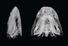
|
M3#908Head of a 59.5 cm long Scyliorhinus canicula male from a North Atlantic population. Type: "3D_surfaces"doi: 10.18563/m3.sf.908 state:published |
Download 3D surface file |
Scyliorhinus canicula 270918C View specimen
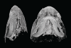
|
M3#909Head of a 63.0 cm long Scyliorhinus canicula female from a North Atlantic population. Type: "3D_surfaces"doi: 10.18563/m3.sf.909 state:published |
Download 3D surface file |
Scyliorhinus canicula 270918D View specimen
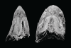
|
M3#907Head of a 64.0 cm long Scyliorhinus canicula female from a North Atlantic population. Type: "3D_surfaces"doi: 10.18563/m3.sf.907 state:published |
Download 3D surface file |
Scyliorhinus canicula 12111931 View specimen
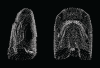
|
M3#905Head of a 9.5 cm long Scyliorhinus canicula male from a Mediterranean population. Type: "3D_surfaces"doi: 10.18563/m3.sf.905 state:published |
Download 3D surface file |
Scyliorhinus canicula 12111933 View specimen
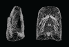
|
M3#906Head of a 9.5 cm long Scyliorhinus canicula female from a Mediterranean population. Type: "3D_surfaces"doi: 10.18563/m3.sf.906 state:published |
Download 3D surface file |
Scyliorhinus canicula 190118A View specimen
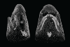
|
M3#918Head of a 8.8 cm long Scyliorhinus canicula female from a Mediterranean population. Type: "3D_surfaces"doi: 10.18563/m3.sf.918 state:published |
Download 3D surface file |
Scyliorhinus canicula 190118C View specimen
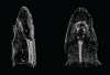
|
M3#930Head of a 9.0 cm long Scyliorhinus canicula female from a Mediterranean population. Type: "3D_surfaces"doi: 10.18563/m3.sf.930 state:published |
Download 3D surface file |
Scyliorhinus canicula 190118D View specimen
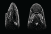
|
M3#929Head of a 8.9 cm long Scyliorhinus canicula male from a Mediterranean population. Type: "3D_surfaces"doi: 10.18563/m3.sf.929 state:published |
Download 3D surface file |
Scyliorhinus canicula 190118F View specimen
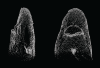
|
M3#928Head of a 9.1 cm long Scyliorhinus canicula male from a Mediterranean population. Type: "3D_surfaces"doi: 10.18563/m3.sf.928 state:published |
Download 3D surface file |
Scyliorhinus canicula 060718A View specimen
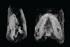
|
M3#927Head of a 25.5 cm long Scyliorhinus canicula male from a Mediterranean population. Type: "3D_surfaces"doi: 10.18563/m3.sf.927 state:published |
Download 3D surface file |
Scyliorhinus canicula 060718B View specimen
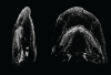
|
M3#926Head of a 23.0 cm long Scyliorhinus canicula female from a Mediterranean population. Type: "3D_surfaces"doi: 10.18563/m3.sf.926 state:published |
Download 3D surface file |
Scyliorhinus canicula 060718C View specimen
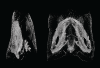
|
M3#925Head of a 28.0 cm long Scyliorhinus canicula male from a Mediterranean population. Type: "3D_surfaces"doi: 10.18563/m3.sf.925 state:published |
Download 3D surface file |
Scyliorhinus canicula 060718D View specimen
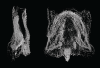
|
M3#924Head of a 21.0 cm long Scyliorhinus canicula male from a Mediterranean population. Type: "3D_surfaces"doi: 10.18563/m3.sf.924 state:published |
Download 3D surface file |
Scyliorhinus canicula 060718E View specimen
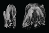
|
M3#923Head of a 23.5 cm long Scyliorhinus canicula male from a Mediterranean population. Type: "3D_surfaces"doi: 10.18563/m3.sf.923 state:published |
Download 3D surface file |
Scyliorhinus canicula 060718F View specimen
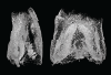
|
M3#922Head of a 22.5 cm long Scyliorhinus canicula female from a Mediterranean population. Type: "3D_surfaces"doi: 10.18563/m3.sf.922 state:published |
Download 3D surface file |
Scyliorhinus canicula 121218A View specimen
|
M3#921Head of a 31.0 cm long Scyliorhinus canicula female from a Mediterranean population. Type: "3D_surfaces"doi: 10.18563/m3.sf.921 state:published |
Download 3D surface file |
Scyliorhinus canicula 121218B View specimen
|
M3#920Head of a 31.0 cm long Scyliorhinus canicula female from a Mediterranean population. Type: "3D_surfaces"doi: 10.18563/m3.sf.920 state:published |
Download 3D surface file |
Scyliorhinus canicula 121218C View specimen
|
M3#919Head of a 31.0 cm long Scyliorhinus canicula female from a Mediterranean population. Type: "3D_surfaces"doi: 10.18563/m3.sf.919 state:published |
Download 3D surface file |
Scyliorhinus canicula 121218D View specimen
|
M3#904Head of a 31.0 cm long Scyliorhinus canicula male from a Mediterranean population. Type: "3D_surfaces"doi: 10.18563/m3.sf.904 state:published |
Download 3D surface file |
