

| Plane | Position | Flip |
| Show planes | Show edges |
0.0
M3#128
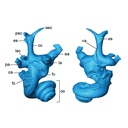
Left bony labyrinth of an adult Bos taurus
Data citation:
Loïc Costeur and Bastien Mennecart
, 2016. M3#128. doi: 10.18563/m3.sf.128
Tag legend:
Cochlear aqueduct, Labyrinth, Vestibular aqueduct/Endolymphatic sac
Model solid/transparent
Flags:
anterior semicircular canal (asc), asc ampulla, cochlea, cochlear aqueduct, common crus, endolymphatic sac, fenestra cochleae, fenestra vestibuli, lateral semicircular canal (lsc), lsc ampulla, posterior semicircular canal (psc), psc ampulla, vestibular aqueduct

|
3D models related to the publication: Prenatal growth stages show the development of the ruminant bony labyrinth and petrosal bone.Loïc Costeur and Bastien MennecartPublished online: 19/10/2016Keywords: bony labyrinth; foetus; ossification timing; phylogeny; Ruminantia https://doi.org/10.18563/m3.2.2.e3 Abstract The present 3D Dataset contains the 3D models analyzed in Costeur L., Mennecart B., Müller B., Schulz G., 2016. Prenatal growth stages show the development of the ruminant bony labyrinth and petrosal bone. Journal of Anatomy. https://doi.org/10.1111/joa.12549 See original publication M3 article infos Published in Volume 02, Issue 02 (2017) |
|