

| Plane | Position | Flip |
| Show planes | Show edges |
Measured length
0.0
0.0
M3#45
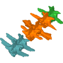
1st and 2nd lumbar vertebrae, and 5th lumbar and sacral vertebrae
Data citation:
Julia Molnar , Stephanie E. Pierce
, Bhart-Anjan Bhullar
, Alan Turner
and John Hutchinson
, 2015. M3#45. doi: 10.18563/m3.sf45
Tag legend:
Dorsal vertebrae, Lumbar vertebrae, Sacral vertebrae
Model solid/transparent

|
3D models related to the publication: Morphological and functional changes in the vertebral column with increasing aquatic adaptation in crocodylomorphsJulia Molnar, Stephanie E. Pierce, Bhart-Anjan Bhullar, Alan Turner and John HutchinsonPublished online: 06/11/2015Keywords: archosaur; axial skeleton; Vertebrae https://doi.org/10.18563/m3.1.3.e5 Abstract This contribution contains the 3D models described and figured in the following publication: Molnar, JL, Pierce, SE, Bhullar, B-A, Turner, AH, Hutchinson, JR (accepted). Morphological and functional changes in the crocodylomorph vertebral column with increasing aquatic adaptation. Royal Society Open Science. See original publication M3 article infos Published in Volume 01, Issue 03 (2015) |
|