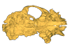
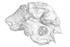
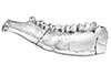
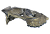
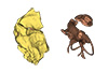
Holotype of Hamadasuchus rebouli
3D model of the holotype specimen of Pebanista yacuruna
3D models of Eocene–Miocene anuran fossils from Peruvian Amazonia
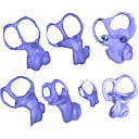
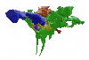
3D GM dataset of bird skeletal variation
Skeletal embryonic development in the catshark
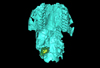
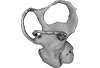
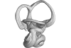
Bony connexions of the petrosal bone of extant hippos
bony labyrinth (11) , inner ear (10) , South America (8) , Eocene (8) , skull (7) , brain (6) , Oligocene (6)
Maëva Judith Orliac (17) , Lionel Hautier (17) , Bastien Mennecart (12) , Laurent Marivaux (11) , Pierre-Olivier Antoine (11) , Leonardo Kerber (10) , Renaud Lebrun (9)

|
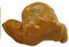
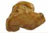
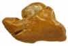
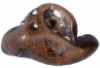
3D models related to the publication: Micromeryx? eiselei - a new moschid species from Steinheim am Albuch, Germany, and the first comprehensive description of moschid cranial material from the Miocene of Central EuropeManuela Aiglstorfer
Published online: 16/10/2017 |
|
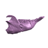
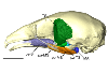
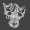
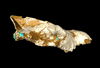
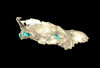
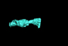
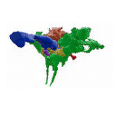
M3#284The 3 D surfaces comprises the skull, petrosal, and bony labyrinth of NMB Sth.833, the holotype of Micromeryx? eiselei Type: "3D_surfaces"doi: 10.18563/m3.sf.284 state:published |
Download 3D surface file |
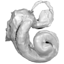
Here, the semicircular canals of the most aquatic seal, the rare Antarctic Ross Seal (Ommatophoca rossii), are presented for the first time, along with representatives of every species in the Lobodontini: the leopard seal (Hydrurga leptonyx), Weddell seal (Leptonychotes weddellii), and crabeater seal (Lobodon carcinophagus). Because encounters with wild Ross seal are rare, and few specimens are available in collections worldwide, this dataset increases accessibility to a rare species. For further comparison, we present the bony labyrinths of other carnivorans, the elephant seal (Mirounga leonina), harbor seal (Phoca vitulina), walrus (Odobenus rosmarus), South American sea lion (Otaria byronia).
Odobenus rosmarus MVZ 125566 View specimen
|
M3#173Surface of the semicircular canals and cochlea of the walrus, Odobenus rosmarus Type: "3D_surfaces"doi: 10.18563/m3.sf.173 state:published |
Download 3D surface file |
Phoca vitulina UZNH 17973 View specimen
|
M3#174Endocast surface of the semicircular canals and cochlea of the harbor seal, Phoca vitulina. Type: "3D_surfaces"doi: 10.18563/m3.sf.174 state:published |
Download 3D surface file |
Hydrurga leptonyx MLP 14.IV.48.11 View specimen
|
M3#285Endocast surface of the semicircular canals and cochlea of the leopard seal, Hydrurga leptonyx. Type: "3D_surfaces"doi: 10.18563/m3.sf.285 state:published |
Download 3D surface file |
Leptonychotes weddellii IAA 02-13 View specimen
|
M3#288Endocast surface of the semicircular canals and cochlea of the Weddell seal Leptonychotes weddellii. Type: "3D_surfaces"doi: 10.18563/m3.sf.288 state:published |
Download 3D surface file |
Lobodon carcinophagus IAA 530 View specimen
|
M3#286Endocast surface of the semicircular canals and cochlea of the crabeater seal, Lobodon carcinophagus. Type: "3D_surfaces"doi: 10.18563/m3.sf.286 state:published |
Download 3D surface file |
Ommatophoca rossii MACN 48259 View specimen
|
M3#176Endocast surface of the semicircular canals and cochlea of the Ross seal Ommatophoca rossii. Type: "3D_surfaces"doi: 10.18563/m3.sf.176 state:published |
Download 3D surface file |
Mirounga leonina IAA 03-5 View specimen
|
M3#287Right endocast surface of the semicircular canals and cochlea of the elephant seal, Mirounga leonina. Type: "3D_surfaces"doi: 10.18563/m3.sf.287 state:published |
Download 3D surface file |
This contribution contains the 3D model described and figured in the following publication: Martin, T., Averianov, A. O., Schultz, J. A., & Schwermann, A. H. (2023). A stem therian mammal from the Lower Cretaceous of Germany. Journal of Vertebrate Paleontology, e2224848.
Spelaeomolitor speratus WMNM P99101 View specimen

|
M3#12573D_model_Spelaeomolitor_lower_molar Type: "3D_surfaces"doi: 10.18563/m3.sf.1257 state:published |
Download 3D surface file |
|
M3#1258CT imagestack (jpgs) and info data sheet (pca file) in one zip folder Type: "3D_CT"doi: 10.18563/m3.sf.1258 state:published |
Download CT data |
The present 3D Dataset contains the 3D models analyzed in 3D Finite Element Analysis and Geometric Morphometrics of Sloths (Xenarthra, Folivora) Mandibles Show Insights on the Dietary Specializations of Fossil Taxa. Journal of South American Earth Sciences. https://doi.org/10.1016/j.jsames.2023.104445
Mylodon darwinii CAV 379 View specimen
|
M3#1159Right hemimandible Type: "3D_surfaces"doi: 10.18563/m3.sf.1159 state:published |
Download 3D surface file |
Scelidotherium leptocephalum MNHN-M 137,722 View specimen
|
M3#1160Mandible Type: "3D_surfaces"doi: 10.18563/m3.sf.1160 state:published |
Download 3D surface file |
Glossotherium robustum MNHN-M 914 View specimen
|
M3#1161Mandible Type: "3D_surfaces"doi: 10.18563/m3.sf.1161 state:published |
Download 3D surface file |
Lestodon armatus MPAC 899 View specimen
|
M3#1162Mandible Type: "3D_surfaces"doi: 10.18563/m3.sf.1162 state:published |
Download 3D surface file |
Valgipes bucklandi NHMD.Z.M.K. 1/1845:3540 View specimen
|
M3#1163Mandible Type: "3D_surfaces"doi: 10.18563/m3.sf.1163 state:published |
Download 3D surface file |
The present 3D Dataset contains the 3D model of the skin of Allosaurus described in Hendrickx, C. et al. in press. Morphology and distribution of scales, dermal ossifications, and other non-feather integumentary structures in non-avialan theropod dinosaurs. Biological Reviews.
Allosaurus jimmadseni UMNH VP C481 View specimen
|
M3#902The material consists of a 3D reconstruction of the counterpart of a 30 cm2 patch of skin impression associated with the anterior dorsal ribs/pectoral region of the specimen of Allosaurus jimmadseni UMNH VP C481. The skin shows a semi-uniform basement of 1-2 mm diameter pebbles with a smaller number of slightly larger (up to 3 mm) ovoid scales. The irregular shape, distribution, and overall small size of these larger scales suggest that they are not classifiable as feature scales but rather as variations in the basement scales. Type: "3D_surfaces"doi: 10.18563/m3.sf.902 state:published |
Download 3D surface file |
This contribution contains the 3D models described and figured in the following publication: Aguirre-Fernández G, Jost J, and Hilfiker S. 2022. First records of extinct kentriodontid and squalodelphinid dolphins from the Upper Marine Molasse (Burdigalian age) of Switzerland and a reappraisal of the Swiss cetacean fauna.
Kentriodon sp. NMBE 5023944 View specimen
|
M3#8583D models of left periotic and bony labyrinth of NMBE 5023944 (Kentriodon sp.) Type: "3D_surfaces"doi: 10.18563/m3.sf.858 state:published |
Download 3D surface file |
Kentriodon sp. NMBE 5023945 View specimen
|
M3#8593D models of right periotic and bony labyrinth of NMBE 5023945 (Kentriodontidae indet.) Type: "3D_surfaces"doi: 10.18563/m3.sf.859 state:published |
Download 3D surface file |
Kentriodon sp. NMBE 5023946 View specimen
|
M3#8603D models of left periotic and bony labyrinth of NMBE 5023946 (Kentriodon sp.) Type: "3D_surfaces"doi: 10.18563/m3.sf.860 state:published |
Download 3D surface file |
Kentriodon sp. NMBE 5036436 View specimen
|
M3#8613D models of right periotic and bony labyrinth of NMBE 5036436 (Kentriodontidae indet.) Type: "3D_surfaces"doi: 10.18563/m3.sf.861 state:published |
Download 3D surface file |
indet. indet. NMBE 5023942 View specimen
|
M3#8623D models of right periotic and bony labyrinth of NMBE 5023942 (Squalodelphinidae indet.) Type: "3D_surfaces"doi: 10.18563/m3.sf.862 state:published |
Download 3D surface file |
indet. indet. NMBE 5023943 View specimen
|
M3#8633D models of left periotic and bony labyrinth of NMBE 5023943 (Squalodelphinidae indet.) Type: "3D_surfaces"doi: 10.18563/m3.sf.863 state:published |
Download 3D surface file |
indet. indet. NMBE 5036437 View specimen
|
M3#8643D models of left periotic and bony labyrinth of NMBE 5036437 (Physeteridae indet.) Type: "3D_surfaces"doi: 10.18563/m3.sf.864 state:published |
Download 3D surface file |
The present 3D Dataset contains the 3D models described in “Comparative masticatory myology in anteaters and its implications for interpreting morphological convergence in myrmecophagous placentals”.
Cyclopes didactylus M1571_JAG View specimen
|
M3#522Skull, mandible, and muscles of Cyclopes didactylus Type: "3D_surfaces"doi: 10.18563/m3.sf.522 state:published |
Download 3D surface file |
Tamandua tetradactyla M3075_JAG View specimen
|
M3#524Skull, left mandibles, and muscles of Tamandua tetradactyla. Type: "3D_surfaces"doi: 10.18563/m3.sf.524 state:published |
Download 3D surface file |
Myrmecophaga tridactyla M3023_JAG View specimen
|
M3#523Skull, left mandible and muscles of Myrmecophaga tridactyla. Type: "3D_surfaces"doi: 10.18563/m3.sf.523 state:published |
Download 3D surface file |
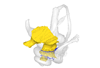
The present 3D Dataset contains the 3D models described and figured in the following publication: Grohé C., Bonis L. de, Chaimanee Y., Chavasseau O., Rugbumrung M., Yamee C., Suraprasit K., Gibert C., Surault J., Blondel C., Jaeger J.-J. 2020. The late middle Miocene Mae Moh Basin of northern Thailand: the richest Neogene assemblage of Carnivora from Southeast Asia and a paleobiogeographic analysis of Miocene Asian carnivorans. American Museum Novitates. http://digitallibrary.amnh.org/handle/2246/7223
Siamogale bounosa MM-54 View specimen
|
M3#5053D model of the skull of Siamogale bounosa The zip file contains: - the 3D surface in PLY - the orientation files in .pos and .ori - the project in .ntw Type: "3D_surfaces"doi: 10.18563/m3.sf.505 state:published |
Download 3D surface file |
Vishnuonyx maemohensis MM-78 View specimen
|
M3#5063D model of the skull of Vishnuonyx maemohensis The zip file contains: - the 3D surface in PLY - the orientation files in .pos and .ori - the project in .ntw Type: "3D_surfaces"doi: 10.18563/m3.sf.506 state:published |
Download 3D surface file |
|
M3#5073D model of the reconstructed upper teeth of Vishnuonyx maemohensis The zip file contains: - the 3D surface in PLY - the orientation files in .pos and .ori - the project in .ntw Type: "3D_surfaces"doi: 10.18563/m3.sf.507 state:published |
Download 3D surface file |
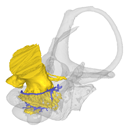
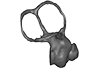
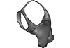
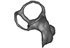
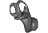
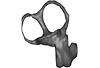
This contribution contains the 3D models described and figured in the publication entitled "The petrosal and bony labyrinth of Diplobune minor, an enigmatic Artiodactyla from the Oligocene of Western Europe" by Orliac, Araújo, and Lihoreau published in Journal of Morphology (Orliac et al. 2017) https://doi.org/10.1002/jmor.20702.
Diplobune minor UM ITD 1079 View specimen
|
M3#138right bony labyrinth of Diplobune minor from Itardies, France Type: "3D_surfaces"doi: 10.18563/m3.sf.138 state:published |
Download 3D surface file |
|
M3#139right isolated petrosal of Diplobune minor from Itardies, France Type: "3D_surfaces"doi: 10.18563/m3.sf.139 state:published |
Download 3D surface file |
Diplobune minor UM ITD 1080 View specimen
|
M3#140left bony labyrinth of Diplobune minor from Itardies, France Type: "3D_surfaces"doi: 10.18563/m3.sf.140 state:published |
Download 3D surface file |
|
M3#141left isolated petrosal of Diplobune minor from Itardies, France Type: "3D_surfaces"doi: 10.18563/m3.sf.141 state:published |
Download 3D surface file |
Diplobune minor UM ITD 1081 View specimen
|
M3#142right bony labyrinth and associated nerves and veins of Diplobune minor from Itardies, France Type: "3D_surfaces"doi: 10.18563/m3.sf.142 state:published |
Download 3D surface file |
|
M3#143right isolated petrosal of Diplobune minor from Itardies, France Type: "3D_surfaces"doi: 10.18563/m3.sf.143 state:published |
Download 3D surface file |
Diplobune minor UM ITD 1083 View specimen
|
M3#144left bony labyrinth of Diplobune minor from Itardies, France Type: "3D_surfaces"doi: 10.18563/m3.sf.144 state:published |
Download 3D surface file |
|
M3#145left petrosal of Diplobune minor from Itardies, France Type: "3D_surfaces"doi: 10.18563/m3.sf.145 state:published |
Download 3D surface file |
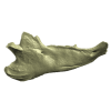
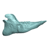
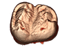
This contribution contains the 3D models described and figured in the following publication: Bonis et al. 2023. A new large pantherine and a sabre-toothed cat (Mammalia, Carnivora, Felidae) from the late Miocene hominoid-bearing Khorat sand pits, Nakhon Ratchasima Province, northeastern Thailand. The Science of Nature 110(5):42. https://doi.org/10.1007/s00114-023-01867-4
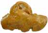
Pachypanthera piriyai CUF-KR-1 View specimen
|
M3#1209Holotype of Pachypanthera piriyai, a left hemi-mandible with alveoli for i1-i3 and canine, roots of p3, p4 and partially broken off m1 crown. Type: "3D_surfaces"doi: 10.18563/m3.sf.1209 state:published |
Download 3D surface file |
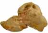
Pachypanthera piriyai CUF-KR-2 View specimen
|
M3#1210Paratype of Pachypanthera piriyai, a right hemi-maxilla with P3-P4, alveoli of C and M1, root of P2 Type: "3D_surfaces"doi: 10.18563/m3.sf.1210 state:published |
Download 3D surface file |
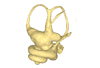
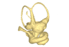
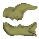
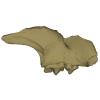
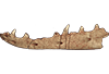
The present 3D Dataset contains the 3D models of a skull and lower jaw of the holotype of Santagnathus mariensis, described in “Old fossil findings in the Upper Triassic rocks of southern Brazil improve diversity of traversodontid cynodonts (Therapsida, Cynodontia)”
Santagnathus mariensis UFRGS-PV-1419-T View specimen
|
M3#1157Skull Type: "3D_surfaces"doi: 10.18563/m3.sf.1157 state:published |
Download 3D surface file |
|
M3#1158Lower jaw Type: "3D_surfaces"doi: 10.18563/m3.sf.1158 state:published |
Download 3D surface file |
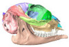
The present 3D Dataset contains the 3D models analyzed in Keppeler, H., Schultz, J. A., Ruf, I., & Martin, T., 2023. Cranial anatomy of Hypisodus minimus (Artiodactyla: Ruminantia) from the Oligocene Brule Formation of North America. Palaeontographica Abteilung A.
Hypisodus minimus SMNK-PAL 27212 View specimen
|
M3#1031CT image stack of a skull of Hypisodus minimus. Also includes a lumbar vertebra and a probable proximal phalanx of digit III or IV. Type: "3D_CT"doi: 10.18563/m3.sf.1031 state:published |
Download CT data |
|
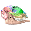
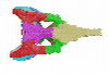
M3#10363D surface models of a skull of Hypisodus minimus (SMNK-PAL27212). The data includes a surface model for: basisphenoid, tympanic bullae, ethmoid (lamina perpendicularis), frontals, jugal (left), jugal (right), lacrimals, lower dentition, mandibles, mastoid processes, maxillaries, maxilloturbinals, nasals, occipital, palatine, parietals, petrosals, presphenoid, squamosals, turbinates, upper dentition, and the vomer. Type: "3D_surfaces"doi: 10.18563/m3.sf.1036 state:published |
Download 3D surface file |
Hypisodus minimus SMNK-PAL 27213 View specimen
|
M3#1033CT image stack of a skull of Hypisodus minimus. Also shows numerous postcranial material including an atlas articulated with the occipital bone, the distal part of a left humerus articulated to radius and ulna, a part of a femur, a part of a tibia and fibula, unidentifiable tarsal bones, parts of the metatarsals II, III, IV and V and their phalanges, a proximal phalanx of digit III or IV, a middle phalanx of digit III or IV, a possible patella and calcaneus, as well as numerous unidentifiable broken bony fragments. Type: "3D_CT"doi: 10.18563/m3.sf.1033 state:published |
Download CT data |
|
M3#10353D surface models of a skull of Hypisodus minimus (SMNK-PAL27213). The data includes a surface model for: atlas, basisphenoid, tympanic bullae, nasals, occipital, the petrosals, and the inner ear. Type: "3D_surfaces"doi: 10.18563/m3.sf.1035 state:published |
Download 3D surface file |
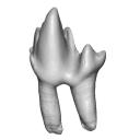
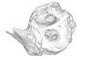
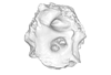
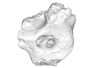
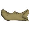
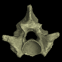
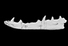
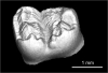
The present 3D Dataset contains the 3D models of the holotype mandible and referred fragmented skull of the new species Amphimoschus xishuiensis analyzed in the article Li, Y.-K., Mennecart, B., Aiglstorfer, M., Ni, X.-J., Li, Q., Deng, T. 2021. The early evolution of cranial appendages in Bovoidea revealed by new species of Amphimoschus (Mammalia: Ruminantia) from China. Zoological Journal of the Linnean Society https://doi.org/10.1093/zoolinnean/zlab053
Amphimoschus xishuiensis IVPP V 25521.1 View specimen
|
M3#803the holotype, a right hemimandible with tooth row p2 to m3 Type: "3D_surfaces"doi: 10.18563/m3.sf.803 state:published |
Download 3D surface file |
Amphimoschus xishuiensis IVPP V 25521.2 View specimen
|
M3#804referred material, anterior part of a skull with right P4-M3 and left P3-M2 Type: "3D_surfaces"doi: 10.18563/m3.sf.804 state:published |
Download 3D surface file |
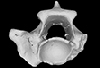
The present 3D Dataset contains the 3D models analyzed in the following publication: Georgalis, G. L., and T. M. Scheyer. A new species of Palaeopython (Serpentes) and other extinct squamates from the Eocene of Dielsdorf (Zurich, Switzerland). Swiss Journal of Geosciences (in press). https://doi.org/10.1007/s00015-019-00341-6
Palaeopython helveticus PIMUZ A/III 631 View specimen
|
M3#399ZIP file containing .ply of vertebra PIMUZ A/III 631 from Palaeopython helveticus n. sp. Type: "3D_surfaces"doi: 10.18563/m3.sf.399 state:published |
Download 3D surface file |
|
M3#403dataset of snake vertebra PIMUZ A/III 631 Type: "3D_CT"doi: 10.18563/m3.sf.403 state:published |
Download CT data |
Palaeopython helveticus PIMUZ A/III 634 View specimen
|
M3#400ZIP file containing .ply of vertebra PIMUZ A/III 634 from Palaeopython helveticus n. sp. (holotype) Type: "3D_surfaces"doi: 10.18563/m3.sf.400 state:published |
Download 3D surface file |
|
M3#404dataset of snake vertbra PIMUZ A/III 634 (holotype) Type: "3D_CT"doi: 10.18563/m3.sf.404 state:published |
Download CT data |
Palaeopython helveticus PIMUZ A/III 636 View specimen
|
M3#401ZIP file containing .ply of vertebra PIMUZ A/III 636 from Palaeopython helveticus n. sp. Type: "3D_surfaces"doi: 10.18563/m3.sf.401 state:published |
Download 3D surface file |
|
M3#406dataset of snake vertebra PIMUZ A/III 636 Type: "3D_CT"doi: 10.18563/m3.sf.406 state:published |
Download CT data |
Palaeovaranus sp. PIMUZ A/III 234 View specimen
|
M3#402ZIP file containing .ply of dentary PIMUZ A/III 234 of Palaeovaranus sp. Type: "3D_surfaces"doi: 10.18563/m3.sf.402 state:published |
Download 3D surface file |
|
M3#405dataset of dentary of Palaeovaranus sp. (PIMUZ A/III 234) Type: "3D_CT"doi: 10.18563/m3.sf.405 state:published |
Download CT data |
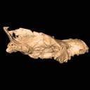
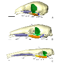
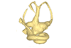
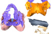
The present 3D Dataset contains the 3D model analyzed in the article : Dubied et al. (2021), Endocranium and ecology of Eurotherium theriodis, a European hyaenodont mammal from the Lutetian. Acta Palaeontologica Polonica 2021, https://doi.org/10.4202/app.00771.2020
Eurotherium theriodis NMB.Em12 View specimen
|
M3#381NMB.Em12 unprepared specimen Type: "3D_surfaces"doi: 10.18563/m3.sf.381 state:published |
Download 3D surface file |
|
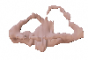
M3#382NMB.Em12 cranium Type: "3D_surfaces"doi: 10.18563/m3.sf.382 state:published |
Download 3D surface file |
|
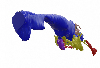
M3#383NMB.Em12 endocast Type: "3D_surfaces"doi: 10.18563/m3.sf.383 state:published |
Download 3D surface file |
This contribution contains the 3D models described and figured in: The Neogene record of northern South American native ungulates. Smithsonian Contributions to Paleobiology. Doi: 10.5479/si.1943-6688.101
Hilarcotherium miyou IGMp 881327 View specimen
|
M3#318Right upper M2 Type: "3D_surfaces"doi: 10.18563/m3.sf.318 state:published |
Download 3D surface file |
Hilarcotherium miyou MUN-STRI 34216 View specimen
|
M3#319Right upper P4 Type: "3D_surfaces"doi: 10.18563/m3.sf.319 state:published |
Download 3D surface file |
|
M3#320Right upper M2 Type: "3D_surfaces"doi: 10.18563/m3.sf.320 state:published |
Download 3D surface file |
Falcontoxodon aguilerai AMU-CURS 585 View specimen
|
M3#321Maxilla with left M3-P2 and right I2 Type: "3D_surfaces"doi: 10.18563/m3.sf.321 state:published |
Download 3D surface file |
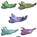
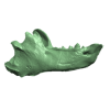
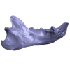
This contribution contains the 3D models described and figured in the following publication: Tissier et al. (in prep.).
Sellamynodon zimborensis UBB MPS 15795 View specimen
|
M3#297Incomplete skull with left M3. Type: "3D_surfaces"doi: 10.18563/m3.sf.297 state:published |
Download 3D surface file |
Sellamynodon zimborensis UBB MPS 15795 View specimen

|
M3#298Mandible with complete molar and premolar rows, lacking symphysis. Type: "3D_surfaces"doi: 10.18563/m3.sf.298 state:published |
Download 3D surface file |
Amynodontopsis aff. bodei UBB MPS V545 View specimen
|
M3#299Maxillary fragment with M1-3. Type: "3D_surfaces"doi: 10.18563/m3.sf.299 state:published |
Download 3D surface file |
Amynodontopsis aff. bodei UBB MPS V546 View specimen
|
M3#300Unworn m1/2 on mandible fragment. Type: "3D_surfaces"doi: 10.18563/m3.sf.300 state:published |
Download 3D surface file |
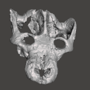
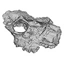
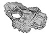
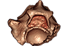
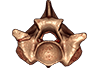
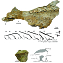
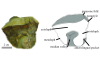
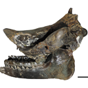
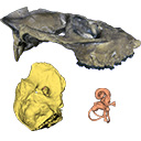
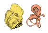
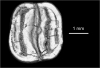
The present 3D Dataset contains the 3D models analyzed in Pochat-Cottilloux Y., Rinder N., Perrichon G., Adrien J., Amiot R., Hua S. & Martin J. E. (2023). The neuroanatomy and pneumaticity of Hamadasuchus from the Cretaceous of Morocco and its significance for the paleoecology of Peirosauridae and other altirostral crocodylomorphs. Journal of Anatomy, https://doi.org/10.1111/joa.13887
Hamadasuchus sp. UCBL-FSL 532408 View specimen
|
M3#10943D volume reconstruction of the braincase osteology Type: "3D_surfaces"doi: 10.18563/m3.sf.1094 state:published |
Download 3D surface file |
|
M3#10963D volume reconstruction of the endocast Type: "3D_surfaces"doi: 10.18563/m3.sf.1096 state:published |
Download 3D surface file |
|
M3#10973D volume reconstruction of the labyrinths Type: "3D_surfaces"doi: 10.18563/m3.sf.1097 state:published |
Download 3D surface file |
|
M3#10983D volume reconstruction of the pneumatic cavities Type: "3D_surfaces"doi: 10.18563/m3.sf.1098 state:published |
Download 3D surface file |
The present 3D Dataset contains the 3D models analyzed in Mennecart B., Métais G., Costeur L., Ginsburg L, and Rössner G. 2021, Reassessment of the enigmatic ruminant Miocene genus Amphimoschus Bourgeois, 1873 (Mammalia, Artiodactyla, Pecora). PlosOne. https://doi.org/10.1371/journal.pone.0244661
Amphimoschus ponteleviensis MNHN.F.AR3266 View specimen
|
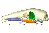
M3#701Surface scan of the cast of the skull of Amphimoschus ponteleviensis MNHN.F.AR3266 from Artenay (France) Type: "3D_surfaces"doi: 10.18563/m3.sf.701 state:published |
Download 3D surface file |
|
M3#702Right petrosal bone and bony labyrinth of the skull MNHN.F.AR3266 from Artenay (France) Type: "3D_surfaces"doi: 10.18563/m3.sf.702 state:published |
Download 3D surface file |
Amphimoschus ponteleviensis SMNS40693 View specimen
|
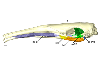
M3#704Left petrosal bone and bony labyrinth of the skull SMNS40693 from Langenau 1 (Germany) Type: "3D_surfaces"doi: 10.18563/m3.sf.704 state:published |
Download 3D surface file |
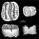
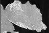
This contribution provides the raw files for the μCT-scan data and renderings of the three-dimensional digital models of two fossil teeth of a geomyin geomorph rodent (Caribeomys merzeraudi), discovered from lower Oligocene deposits of Puerto Rico, San Sebastian Formation (locality LACM Loc. 8060). These fossils were described, figured and discussed in the following publication: Marivaux et al. (2021), An unpredicted ancient colonization of the West Indies by North American rodents: dental evidence of a geomorph from the early Oligocene of Puerto Rico. Papers in Palaeontology. https://doi.org/10.1002/spp2.1388
Caribeomys merzeraudi LACM 162478 View specimen
|
M3#712Right lower dp4: isolated deciduous premolar. The specimen was scanned with a resolution of 5 µm using a μ-CT-scanning station EasyTom 150 / Rx Solutions (Montpellier RIO Imaging, ISE-M, Montpellier, France). AVIZO 7.1 (Visualization Sciences Group) software was used for visualization, segmentation, and 3D rendering. This isolated tooth was prepared within a “labelfield” module of AVIZO, using the segmentation threshold selection tool. Type: "3D_surfaces"doi: 10.18563/m3.sf.712 state:published |
Download 3D surface file |
|
M3#7145µm µCT data set . Right lower dp4: isolated deciduous premolar. The specimen was scanned with a resolution of 5 µm using a μ-CT-scanning station EasyTom 150 / Rx Solutions (Montpellier RIO Imaging, ISE-M, Montpellier, France). Type: "3D_CT"doi: 10.18563/m3.sf.714 state:published |
Download CT data |
Caribeomys merzeraudi LACM 162449 View specimen
|
M3#713Right lower molar (m1 or m2). The specimen was scanned with a resolution of 4.5 µm using a μ-CT-scanning station EasyTom 150 / Rx Solutions (Montpellier RIO Imaging, ISE-M, Montpellier, France). AVIZO 7.1 (Visualization Sciences Group) software was used for visualization, segmentation, and 3D rendering. This isolated tooth was prepared within a “labelfield” module of AVIZO, using the segmentation threshold selection tool. Type: "3D_surfaces"doi: 10.18563/m3.sf.713 state:published |
Download 3D surface file |
|
M3#715µCT data at 4.5µm Type: "3D_CT"doi: 10.18563/m3.sf.715 state:published |
Download CT data |


