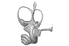
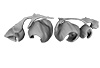
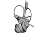
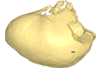
Holotype of Hamadasuchus rebouli
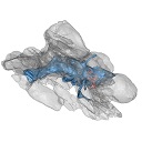
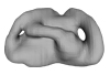
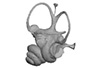
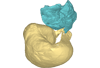
3D model of the holotype specimen of Pebanista yacuruna
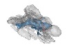
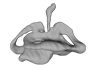
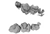
3D models of Eocene–Miocene anuran fossils from Peruvian Amazonia
3D GM dataset of bird skeletal variation
Skeletal embryonic development in the catshark
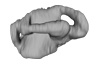
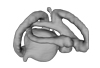
Bony connexions of the petrosal bone of extant hippos
bony labyrinth (11) , inner ear (10) , Eocene (8) , South America (8) , skull (7) , brain (6) , Oligocene (6)
Maëva Judith Orliac (17) , Lionel Hautier (17) , Bastien Mennecart (12) , Laurent Marivaux (11) , Pierre-Olivier Antoine (11) , Leonardo Kerber (10) , Renaud Lebrun (9)
|
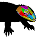
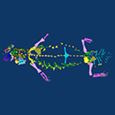
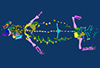
3D models related to the publication: Brief comment on the brain and inner ear of Giganotosaurus carolinii (Dinosauria: Theropoda) based on CT scans.Mauro N. Nieto
Published online: 01/04/2020 |
|
M3#504The current file contents 3D models of the braincase, brain, left and right inner ears Type: "3D_surfaces"doi: 10.18563/m3.sf.504 state:published |
Download 3D surface file |
The present 3D Dataset contains the 3D models analyzed in the publication ‘Ontogenetic development of the otic region in the new model organism, Leucoraja erinacea (Chondrichthyes; Rajidae)’, https://doi.org/10.1017/S1755691018000993
Leucoraja erinacea 2018.9.26.1 View specimen
|
M3#3673D model of the right skeletal labyrinth of the adult specimen of Leucoraja erincea. T Type: "3D_surfaces"doi: 10.18563/m3.sf.367 state:published |
Download 3D surface file |
Leucoraja erinacea 2018.9.25.2 View specimen
|
M3#3683D model of the right skeletal labyrinth of the stage 34 specimen of Leucoraja erincea. Type: "3D_surfaces"doi: 10.18563/m3.sf.368 state:published |
Download 3D surface file |
Leucoraja erinacea 2018.9.25.3 View specimen

|
M3#3693D model of the right skeletal labyrinth of the stage 32 specimen of Leucoraja erinacea. Type: "3D_surfaces"doi: 10.18563/m3.sf.369 state:published |
Download 3D surface file |
|
M3#3723D model of the right membranous system of stage 32 of Leucoraja erincea. Type: "3D_surfaces"doi: 10.18563/m3.sf.372 state:published |
Download 3D surface file |
Leucoraja erinacea 2018.9.25.4 View specimen
|
M3#3703D model of the right skeletal labyrinth of the stage 31 specimen of Leucoraja erinacea. Type: "3D_surfaces"doi: 10.18563/m3.sf.370 state:published |
Download 3D surface file |
Leucoraja erinacea 2018.9.26.5 View specimen

|
M3#3763D model of the right skeletal labyrinth of the stage 29 specimen of Leucoraja erinacea. Type: "3D_surfaces"doi: 10.18563/m3.sf.376 state:published |
Download 3D surface file |
The present 3D Dataset contains the 3D models analyzed in Costeur L., Mennecart B., Müller B., Schulz G., 2016. Prenatal growth stages show the development of the ruminant bony labyrinth and petrosal bone. Journal of Anatomy. https://doi.org/10.1111/joa.12549
Bos taurus NMB3038 View specimen
|
M3#124Right bony labyrinth of a Bos taurus foetus (gestational age 115 days) Type: "3D_surfaces"doi: 10.18563/m3.sf.124 state:published |
Download 3D surface file |
Bos taurus NMB3367 View specimen
|
M3#125Right bony labyrinth of a Bos taurus foetus (gestational age 165 days) Type: "3D_surfaces"doi: 10.18563/m3.sf.125 state:published |
Download 3D surface file |
Bos taurus NMB3365 View specimen
|
M3#126Right bony labyrinth of a Bos taurus foetus (gestational age 210 days) Type: "3D_surfaces"doi: 10.18563/m3.sf.126 state:published |
Download 3D surface file |
Bos taurus NMB2855 View specimen
|
M3#127Right bony labyrinth of a Bos taurus foetus (gestational age 260 days) Type: "3D_surfaces"doi: 10.18563/m3.sf.127 state:published |
Download 3D surface file |
Bos taurus NMB1037 View specimen
|
M3#128Left bony labyrinth of an adult Bos taurus Type: "3D_surfaces"doi: 10.18563/m3.sf.128 state:published |
Download 3D surface file |
The present 3D Dataset contains the 3D models analyzed in: Abel P., Pommery Y., Ford D. P., Koyabu D., Werneburg I. 2022. Skull sutures and cranial mechanics in the Permian reptile Captorhinus aguti and the evolution of the temporal region in early amniotes. Frontiers in Ecology and Evolution. https://doi.org/10.3389/fevo.2022.841784
Captorhinus aguti OMNH 44816 View specimen

|
M3#965Segmented cranial bone surfaces of OMNH 44816 Type: "3D_surfaces"doi: 10.18563/m3.sf.965 state:published |
Download 3D surface file |
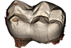
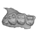
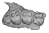
This contribution contains the 3D models described and figured in the following publication: Hautier L, Tabuce R, Kassegne KE, Amoudji YZ, Mourlam M, Orliac M, Quillévéré F, Charruault A-L, Johnson AKC, Guinot G. 2021. New middle Eocene proboscidean from Togo illuminates the early evolution of the elephantiform-like dental pattern.
Dagbatitherium tassyi ULDG-DAG1 View specimen
|
M3#7693D model of a molar of Dagbatitherium tassyi. Type: "3D_surfaces"doi: 10.18563/m3.sf.769 state:published |
Download 3D surface file |
|
M3#771µCT scan of a molar of Dagbatitherium tassyi. Type: "3D_CT"doi: 10.18563/m3.sf.771 state:published |
Download CT data |
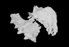
The present contribution contains the 3D virtual restoration of a Pliocene Lutrine right femur of Tobène, Senegal, described and figured in Lihoreau et al. (2021) : "A fossil terrestrial fauna from Tobène (Senegal) provides a unique early Pliocene window in Western Africa ". https://doi.org/10.1016/j.gr.2021.06.013
Indet indet SN-Tob-12-02 View specimen
|
M3#441Virtual restoration of SN-Tob-12-02 Type: "3D_surfaces"doi: 10.18563/m3.sf.441 state:published |
Download 3D surface file |
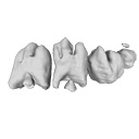
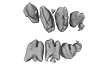
The present 3D Dataset contains the 3D models analyzed in ”Morphological features of tooth development and replacement in the rabbit Oryctolagus cuniculus”, Archives of Oral Biology, https://doi.org/10.1016/j.archoralbio.2019.104576
Oryctogalus cuniculus E14 View specimen
|
M3#390Right cheek teeth, Left and right incisors at 14 dpf Type: "3D_surfaces"doi: 10.18563/m3.sf.390 state:published |
Download 3D surface file |
Oryctogalus cuniculus E16 View specimen

|
M3#391Left cheek teeth, Left and right incisors at 16 dpf Type: "3D_surfaces"doi: 10.18563/m3.sf.391 state:published |
Download 3D surface file |
Oryctogalus cuniculus E18 View specimen
|
M3#392Left cheek teeth and incisors at 18 dpf Type: "3D_surfaces"doi: 10.18563/m3.sf.392 state:published |
Download 3D surface file |
Oryctogalus cuniculus E20 View specimen
|
M3#393Left cheek teeth and incisors at 20 dpf Type: "3D_surfaces"doi: 10.18563/m3.sf.393 state:published |
Download 3D surface file |
Oryctogalus cuniculus E22 View specimen
|
M3#394Left lower cheek teeth and incisors, right upper cheek teeth and incisors at 22 dpf Type: "3D_surfaces"doi: 10.18563/m3.sf.394 state:published |
Download 3D surface file |
Oryctogalus cuniculus E24 View specimen
|
M3#395Left cheek teeth and incisors at 24 dpf Type: "3D_surfaces"doi: 10.18563/m3.sf.395 state:published |
Download 3D surface file |
Oryctogalus cuniculus E28 View specimen
|
M3#396Right cheek teeth and incisors at 28 dpf Type: "3D_surfaces"doi: 10.18563/m3.sf.396 state:published |
Download 3D surface file |
Oryctogalus cuniculus E26 View specimen
|
M3#397Right cheek teeth and incisors at 26 dpf Type: "3D_surfaces"doi: 10.18563/m3.sf.397 state:published |
Download 3D surface file |
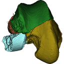
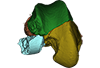
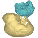
This contribution contains the 3D model described and figured in the following publication: Dubied, M., Mennecart, B. and Solé, F. 2019. The cranium of Proviverra typica (Mammalia, Hyaenodonta) and its impact on hyaenodont phylogeny and endocranial evolution. Palaeontology. https://doi.org/10.1111/pala.12437
Proviverra typica NMB Em18 View specimen
|
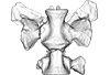
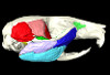
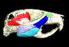
M3#355The file contain the cranium (yellow) and the endocast (blue) of the facial part and the brain case part of the type specimen of Proviverra typica (NMB Em18). Type: "3D_surfaces"doi: 10.18563/m3.sf.355 state:published |
Download 3D surface file |
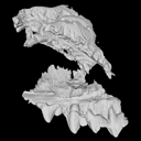
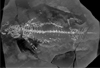
The present 3D Dataset contains the 3D model analyzed in the following publication: Solé et al. (2018), Niche partitioning of the European carnivorous mammals during the paleogene. Palaios. https://doi.org/10.2110/palo.2018.022
Hyaenodon leptorhynchus FSL848325 View specimen
|
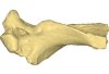
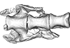
M3#336The specimen FSL848325 is separated in two fragments: the anterior part bears the incisors, the deciduous and permanent canines, while the posterior part bears the right P3, P4, M1 and M2. The P2 is isolated. When combined, the cranium length is approximatively 10.5 cm long. The anterior part is 6.9 cm long and 2.15 cm wide (taken at the level of the P1). The posterior part is 4.8 cm long. The anterior part of the cranium is very narrow. Type: "3D_surfaces"doi: 10.18563/m3.sf.336 state:published |
Download 3D surface file |
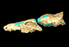
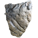
This contribution contains the 3D model of the holotype of Chambius kasserinensis, the basalmost ‘elephant-shrew’ figured in the following publication: New remains of Chambius kasserinensis from the Eocene of Tunisia and evaluation of proposed affinities for Macroscelidea (Mammalia, Afrotheria). https://doi.org/10.1080/08912963.2017.1297433
Chambius kasserinensis CBI-1-06 View specimen
|
M3#1463D model of the holotype maxilla of Chambius kasserinensis. The 3D surface was extracted manually from the limestone matrix within AVIZO 9.2 Type: "3D_surfaces"doi: 10.18563/m3.sf.146 state:published |
Download 3D surface file |
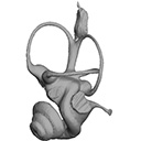
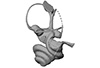
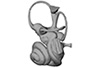
The present 3D Dataset contains the 3D models analyzed in the article Mennecart, B., and L. Costeur. 2016. A Dorcatherium (Mammalia, Ruminantia, Middle Miocene) petrosal bone and the tragulid ear region. Journal of Vertebrate Paleontology 36(6), 1211665(1)-1211665(7). DOI: 10.1080/02724634.2016.1211665.
Tragulus javanicus 10028 View specimen
|
M3#1193D surface of the left bony labyrinth of Tragulus javanicus NMB 10028 Type: "3D_surfaces"doi: 10.18563/m3.sf.119 state:published |
Download 3D surface file |
Moschiola meminna C.2453 View specimen
|
M3#1203D surface of the left bony labyrinth of Moschiola meminna NMB C.2453 Type: "3D_surfaces"doi: 10.18563/m3.sf.120 state:published |
Download 3D surface file |
Hyemoschus aquaticus C.1930 View specimen
|
M3#1223D surface of the right bony labyrinth of Hyemoschus aquaticus NMB C.1930 Type: "3D_surfaces"doi: 10.18563/m3.sf.122 state:published |
Download 3D surface file |
Dorcatherium crassum San.15053 View specimen
|
M3#1233D surface of the right bony labyrinth of Dorcatherium crassum NMB San.15053 Type: "3D_surfaces"doi: 10.18563/m3.sf.123 state:published |
Download 3D surface file |
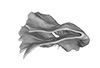
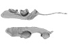
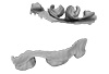
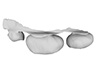
This contribution contains the 3D models described and figured in the following publication: Mourlam, M., Orliac, M. J. (2017), Protocetid (Cetacea, Artiodactyla) bullae and petrosals from the Middle Eocene locality of Kpogamé, Togo: new insights into the early history of cetacean hearing. Journal of Systematic Palaeontology https://doi.org/10.1080/14772019.2017.1328378
?Carolinacetus indet. UM KPG-M 164 View specimen
|
M3#132left petrosal of ?Carolinacetus sp. from the locality of Kpogamé, Togo Type: "3D_surfaces"doi: 10.18563/m3.sf.132 state:published |
Download 3D surface file |
indet. indet. UM KPG-M 73 View specimen

|
M3#133labelled surface of the left petrosal Type: "3D_surfaces"doi: 10.18563/m3.sf.133 state:published |
Download 3D surface file |
|
M3#134left bullaof Protocetidae indeterminate from Kpogamé, Togo Type: "3D_surfaces"doi: 10.18563/m3.sf.134 state:published |
Download 3D surface file |
|
M3#135petrotympanic complex of Protocetidae indeterminate from Kpogamé, Togo Type: "3D_surfaces"doi: 10.18563/m3.sf.135 state:published |
Download 3D surface file |
?Carolinacetus indet. UM KPG-M 33 View specimen

|
M3#136left auditory bulla of a juvenile specimen of ?Carolinacetus sp. from Kpogamé, Togo Type: "3D_surfaces"doi: 10.18563/m3.sf.136 state:published |
Download 3D surface file |
Togocetus traversei UM KPG-M 80 View specimen

|
M3#137fragmentary right auditory bulla of Togocetus traversei from Kpogamé, Togo Type: "3D_surfaces"doi: 10.18563/m3.sf.137 state:published |
Download 3D surface file |
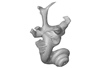
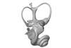
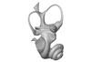
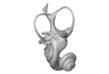
The present 3D Dataset contains the 3D models analyzed in: Toyoda S et al., 2015, Morphogenesis of the inner ear at different stages of normal human development. The Anatomical Record. doi : 10.1002/ar.23268
Homo sapiens KC-CS17IER29248 View specimen

|
M3#36Computationally reconstructed membranous labyrinth of a human embryo (KC-CS17IER29248) at Carnegie Stage 17 (Crown Rump Length= 7mm). Type: "3D_surfaces"doi: 10.18563/m3.sf36 state:published |
Download 3D surface file |
Homo sapiens KC-CS18IER17746 View specimen

|
M3#37Computationally reconstructed membranous labyrinth of a human embryo (KC-CS18IER17746) at Carnegie Stage 18 (Crown Rump Length= 12mm). Type: "3D_surfaces"doi: 10.18563/m3.sf37 state:published |
Download 3D surface file |
Homo sapiens KC-CS19IER16127 View specimen

|
M3#38Computationally reconstructed membranous labyrinth of a human embryo (KC-CS19IER16127) at Carnegie Stage 19 (Crown Rump Length= 13mm). Type: "3D_surfaces"doi: 10.18563/m3.sf38 state:published |
Download 3D surface file |
Homo sapiens KC-CS20IER20268 View specimen

|
M3#39Computationally reconstructed membranous labyrinth of a human embryo (KC-CS20IER20268) at Carnegie Stage 20 (Crown Rump Length= 13.7mm). Type: "3D_surfaces"doi: 10.18563/m3.sf39 state:published |
Download 3D surface file |
Homo sapiens KC-CS21IER28066 View specimen

|
M3#40Computationally reconstructed membranous labyrinth of a human embryo (KC-CS21IER28066) at Carnegie Stage 21 (Crown Rump Length= 16.7mm). Type: "3D_surfaces"doi: 10.18563/m3.sf40 state:published |
Download 3D surface file |
Homo sapiens KC-CS22IER35233 View specimen

|
M3#41Computationally reconstructed membranous labyrinth of a human embryo (KC-CS22IER35233) at Carnegie Stage 22 (Crown Rump Length= 22mm). Type: "3D_surfaces"doi: 10.18563/m3.sf41 state:published |
Download 3D surface file |
Homo sapiens KC-CS23IER15919 View specimen

|
M3#42Computationally reconstructed membranous labyrinth of a human embryo (KC-CS23IER15919) at Carnegie Stage 23 (Crown Rump Length= 32.3mm). Type: "3D_surfaces"doi: 10.18563/m3.sf42 state:published |
Download 3D surface file |
Homo sapiens KC-FIER52730 View specimen

|
M3#43Computationally reconstructed human membranous labyrinth in post embryonic phase (KC-FIER52730). Crown Rump Length: 43.5mm. Type: "3D_surfaces"doi: 10.18563/m3.sf43 state:published |
Download 3D surface file |
This contribution contains the 3D model described and figured in the following publication: Crochet, J.-Y., Hautier, L., Lehmann, T., 2015. A pangolin (Manidae, Pholidota, Mammalia) from the French Quercy phosphorites (Pech du Fraysse, Saint-Projet, Tarn-et-Garonne, late Oligocene, MP 28). Palaeovertebrata 39(2)-e4. doi: 10.18563/pv.39.2.e4
Necromanis franconica UM PFY 4051 View specimen
|
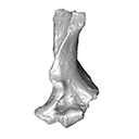
M3#12A partial left humerus from Pech du Fraysse (Saint-Projet, Tarn-et-Garonne, France), MP 28 (late Oligocene) Type: "3D_surfaces"doi: 10.18563/m3.sf12 state:published |
Download 3D surface file |
The present 3D Dataset contains the 3D model analyzed in Hendrickx, C. and Bell, P. R. 2021. The scaly skin of the abelisaurid Carnotaurus sastrei (Theropoda: Ceratosauria) from the Upper Cretaceous of Patagonia. Cretaceous Research. https://doi.org/10.1016/j.cretres.2021.104994
Carnotaurus sastrei MACN 894 View specimen
|
M3#8023D reconstruction of the biggest patch of skin (~1200 cm2) from the anterior tail region of the holotype of Carnotaurus, which is the largest single patch of squamous integument available for any saurischian. The skin consists of medium to large (up to 65 mm in diameter) conical feature scales surrounded by a network of low and small (< 14 mm) irregular basement scales separated by narrow interstitial tissue. Type: "3D_surfaces"doi: 10.18563/m3.sf.802 state:published |
Download 3D surface file |
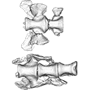
The present 3D Dataset contains the 3D models of the sacral vertebrae analyzed in “Sacral co-ossification in dinosaurs: The oldest record of fused sacral vertebrae in Dinosauria and the diversity of sacral co-ossification patterns in the group”.
Buriolestes schultzi CAPPA/UFSM 0035 View specimen
|
M3#705Sacral vertebrae of Buriolestes schultzi Type: "3D_surfaces"doi: 10.18563/m3.sf.705 state:published |
Download 3D surface file |
indet indet CAPPA/UFSM 0228 View specimen
|
M3#706Sacral vertebrae of a saurischian dinosaur indet. Type: "3D_surfaces"doi: 10.18563/m3.sf.706 state:published |
Download 3D surface file |
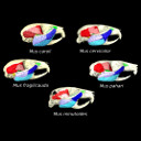
The present 3D Dataset contains the 3D models analyzed in the article entitled "One skull to rule them all? Descriptive and comparative anatomy of the masticatory apparatus in five mice species based on traditional and digital dissections" (Ginot et al. 2018, Journal of Morphology, https://doi.org/10.1002/jmor.20845).
Mus cervicolor R7314 View specimen
|
M3#343.ply surfaces of the skull and masticatory muscles of Mus cervicolor. Created with MorphoDig, .pos and .ntw files also included. Scans were obtained thanks to the Institut des Sciences de l'Evolution de Montpellier MRI platform. Type: "3D_surfaces"doi: 10.18563/m3.sf.343 state:published |
Download 3D surface file |
Mus caroli R7264 View specimen
|
M3#344.ply surfaces of the skull and masticatory muscles of Mus caroli. Created with MorphoDig, .pos and .ntw files also included. Scans were obtained thanks to the Institut des Sciences de l'Evolution de Montpellier MRI platform. Type: "3D_surfaces"doi: 10.18563/m3.sf.344 state:published |
Download 3D surface file |
Mus fragilicauda R7260 View specimen
|
M3#345.ply surfaces of the skull and masticatory muscles of Mus fragilicauda. Created with MorphoDig, .pos and .ntw files also included. Scans were obtained thanks to the Institut des Sciences de l'Evolution de Montpellier MRI platform. Type: "3D_surfaces"doi: 10.18563/m3.sf.345 state:published |
Download 3D surface file |
Mus pahari R7226 View specimen
|
M3#346.ply surfaces of the skull and masticatory muscles of Mus pahari. Created with MorphoDig, .pos and .ntw files also included. Scans were obtained thanks to the Institut des Sciences de l'Evolution de Montpellier MRI platform. Type: "3D_surfaces"doi: 10.18563/m3.sf.346 state:published |
Download 3D surface file |
Mus minutoides minutoides-1 View specimen
|
M3#347.ply surfaces of the skull and masticatory muscles of Mus minutoides. Created with MorphoDig, .pos and .ntw files also included. Scans were obtained thanks to the Institut des Sciences de l'Evolution de Montpellier MRI platform. Type: "3D_surfaces"doi: 10.18563/m3.sf.347 state:published |
Download 3D surface file |
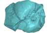
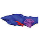
The present contribution contains the 3D model and dataset analyzed in the following publication: Scheyer, T. M., J. M. Neenan, T. Bodogan, H. Furrer, C. Obrist, and M. Plamondon. 2017. A new, exceptionally preserved juvenile specimen of Eusaurosphargis dalsassoi (Diapsida) and implications for Mesozoic marine diapsid phylogeny. Scientific Reports, https://doi.org/10.1038/s41598-017-04514-x .
Eusaurosphargis dalsassoi PIMUZ A/III 4380 View specimen
|
M3#17994 extracted surfaces of skeletal elements of PIMUZ A/III 4380 Type: "3D_surfaces"doi: 10.18563/m3.sf.179 state:published |
Download 3D surface file |
|
M3#180Accompanying CT scan dataset Type: "3D_CT"doi: 10.18563/m3.sf.180 state:published |
Download CT data |
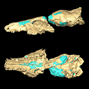
The present publication contains the µCT dataset and the 3D models analyzed in the following publication: Mautner, A.-K., A. E. Latimer, U. Fritz, and T. M. Scheyer. An updated description of the osteology of the pancake tortoise Malacochersus tornieri (Testudines: Testudinidae) with special focus on intraspecific variation. Journal of Morphology. https://doi.org/10.1002/jmor.20640
Malacochersus tornieri ZM 100.102 View specimen

|
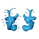
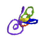
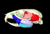
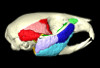
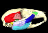
M3#129Virtual brain and inner ear endocast of Malacochersus tornieri (ZM 100.102; Zoological Museum of The University of Zurich). This virtual model is accompanied by the 3D dataset. Blue, endocranium; red, blood vessels; purple, semicircular canals; yellow, cranial nerves. Type: "3D_surfaces"doi: 10.18563/m3.sf.129 state:published |
Download 3D surface file |
|
M3#1303D dataset of skull of Malacochersus tornieri (ZM 100.102) Type: "3D_CT"doi: 10.18563/m3.sf.130 state:published |
Download CT data |
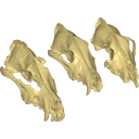
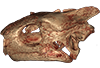
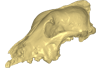
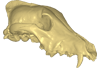
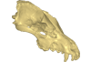
Archaeozoological studies are increasingly using new methods and approaches to explore questions about domestication. Here, we provide 3D models of three archaeological Canis lupus skulls from Belgium originating from the sites of Goyet (31,680±250BP; 31,890+240/-220BP), Trou des Nutons (21,810±90BP) and Trou Balleux (postglacial). Since their identification as either wolves or early dogs is still debated, we present these models as additional tools for further investigating their evolutionary history and the history of dog domestication.
Canis lupus Goyet 2860 View specimen
|
M3#213D surface model of the cranium of the Late Pleistocene Canis lupus "Goyet 2860" from the Royal Belgian Institute of Natural Sciences. Type: "3D_surfaces"doi: 10.18563/m3.sf21 state:published |
Download 3D surface file |
Canis lupus Trou Balleux no-nr View specimen
|
M3#223D surface model of the cranium of the Late Pleistocene Canis lupus "Trou Balleux no-nr" from the University of Liège, Belgium Type: "3D_surfaces"doi: 10.18563/m3.sf22 state:published |
Download 3D surface file |
Canis lupus Trou des Nutons 2559-1 View specimen
|
M3#233D surface model of the cranium of the Late Pleistocene Canis lupus "Trou des Nutons 2559-1" from the Royal Belgian Institute of Natural Sciences. Type: "3D_surfaces"doi: 10.18563/m3.sf23 state:published |
Download 3D surface file |


