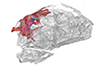
Holotype of Hamadasuchus rebouli
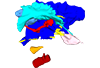
3D model of the holotype specimen of Pebanista yacuruna
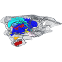
3D models of the endocranial anatomy of Voay robustus and comparative specimens
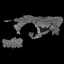
3D GM dataset of bird skeletal variation
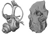
Skeletal embryonic development in the catshark
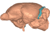
Bony connexions of the petrosal bone of extant hippos
bony labyrinth (11) , inner ear (10) , Eocene (8) , South America (8) , skull (7) , Oligocene (6) , phylogeny (6)
Maëva Judith Orliac (17) , Lionel Hautier (17) , Bastien Mennecart (12) , Laurent Marivaux (11) , Pierre-Olivier Antoine (11) , Leonardo Kerber (10) , Renaud Lebrun (9)
|
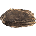
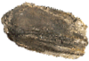
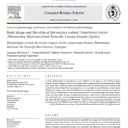
Holotype specimen of Donrussellia magna, an adapiform primate from the early Eocene (MP7) of Southern FranceAnusha Ramdarshan, Marc Godinot
Published online: 18/06/2015 |

|
M3#173D surface file model of UM PAT 17 (type specimen of Donrussellia magna), which is a well preserved left lower jaw with p4-m3. The teeth (and roots) were manually segmented. Type: "3D_surfaces"doi: 10.18563/m3.sf17 state:published |
Download 3D surface file |
|
M3#18CT Scan Data of Donrussellia magna UM PAT 17. Voxel size (in µm): 36µm (isotropic voxels). Dimensions in x,y,z : 594 pixels, 294 pixels, 1038 pixels. Image type : 8-bit voxels. Image format : raw data format (no header). Type: "3D_CT"doi: 10.18563/m3.sf18 state:published |
Download CT data |
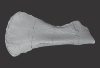
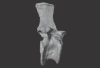
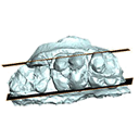
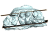
The present 3D Dataset contains the 3D models of an ilium, a vertebra, and a partial scapula of Prestosuchus sp. that were analyzed in “New Loricata remains from the Pinheiros-Chiniquá Sequence (Middle-Upper Triassic), southern Brazil”.
Prestosuchus sp. UFSM11603 View specimen
|
M3#1080Surface scan of a right ilium of Prestosuchus sp. with a 0.4 mm resolution. Type: "3D_surfaces"doi: 10.18563/m3.sf.1080 state:published |
Download 3D surface file |
Prestosuchus sp. UFSM11233 View specimen
|
M3#1081Surface scan of a partial right scapula of Prestosuchus sp. with a 0.4mm resolution. Type: "3D_surfaces"doi: 10.18563/m3.sf.1081 state:published |
Download 3D surface file |
Prestosuchus sp. UFSM11602a View specimen
|
M3#1082Surface scan of a anterior dorsal vertebra of Prestosuchus sp. with a 0.2 mm resolution. Type: "3D_surfaces"doi: 10.18563/m3.sf.1082 state:published |
Download 3D surface file |
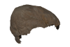
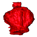
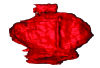
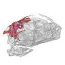
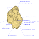
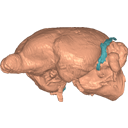
This contribution contains the 3D model of an endocranial cast analyzed in “A 10 ka intentionally deformed human skull from Northeast Asia”. There are many studies on the morphological characteristics of intentional cranial deformation (ICD), but few related 3D models were published. Here, we present the surface model of an intentionally deformed 10 ka human cranium for further research on ICD practice. The 3D model of the endocranial cast of this ICD cranium was discovered near Harbin City, Province Heilongjiang, Northeast China. The fossil preserved only the frontal, parietal, and occipital bones. To complete the endocast model of the specimen, we printed a 3D model and used modeling clay to reconstruct the missing part based on the general form of the modern human endocast morphology.
Homo sapiens IVPP-PA1616 View specimen
|
M3#972The frontal region of the endocast is flattened, probably formed by the constant pressure on the frontal bone during growth. There is a well-developed frontal crest on the endocranial surface. The endocast widens posteriorly from the frontal lobe. The widest point of the endocast is at the lateral border of the parietal lobe. The lower parietal areas display a marked lateral expansion. The overall shape of the endocast is asymmetrical, with the left side of the parietal lobe being more laterally expanded than the right side. Like the frontal lobe, the occipital lobe is also anteroposteriorly flattened. Type: "3D_surfaces"doi: 10.18563/m3.sf.972 state:published |
Download 3D surface file |
|
M3#976The original endocranial cast model (with texture) of IVPP-PA1616. It shows the original structures of the specimen, and was not altered in any way. Type: "3D_surfaces"doi: 10.18563/m3.sf.976 state:published |
Download 3D surface file |
The present 3D Dataset contains the 3D model of the brain endocast of Neoepiblema acreensis analyzed in “Small within the largest: Brain size and anatomy of the extinct Neoepiblema acreensis, a giant rodent from the Neotropics”. The 3D model was generated using CT-Scanning and techniques of virtual reconstruction.
Neoepiblema acreensis UFAC 4515 View specimen
|
M3#502Brain endocast of Neoepiblema acreensis Type: "3D_surfaces"doi: 10.18563/m3.sf.502 state:published |
Download 3D surface file |
The present 3D Dataset contains the 3D models analyzed in the following publication: Paulina-Carabajal, A., Ezcurra, M., Novas, F., 2019. New information on the braincase and endocranial morphology of the Late Triassic neotheropod Zupaysaurus rougieri using Computed Tomography data. Journal of Vertebrate Paleontology. https://doi.org/10.1080/02724634.2019.1630421
Zupaysaurus rougieri PULR 076 View specimen
|
M3#424The Zip contains 3 files, which correspond to: PULR_076-M1: Zupaysaurus rougieri skull, braincase and cranial endocast PULR_076-M2: Zupaysaurus rougieri braincase PULR_076-M1: Zupaysaurus rougieri brain and inner ear Type: "3D_surfaces"doi: 10.18563/m3.sf.424 state:published |
Download 3D surface file |
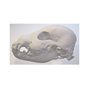
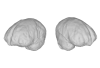
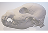
This contribution comprises the 3D models of three wolf pup skulls, which were used for the publication by Geiger et al. 2017 on Neomorphosis and heterochrony of skull shape in dog domestication.
Canis lupus CLL2 View specimen
|
M3#3123d model of a wolf pup skull Type: "3D_surfaces"doi: 10.18563/m3.sf.312 state:published |
Download 3D surface file |
Canis lupus CLL4 View specimen
|
M3#3133d model of a wolf pup skull Type: "3D_surfaces"doi: 10.18563/m3.sf.313 state:published |
Download 3D surface file |
Canis lupus CLL5 View specimen
|
M3#3143d model of a wolf pup skull Type: "3D_surfaces"doi: 10.18563/m3.sf.314 state:published |
Download 3D surface file |
This contribution contains the 3D model described and figured in the following publication: Albino, A., Carrillo-Briceño, J. D. & Neenan, J. M. 2016. An enigmatic aquatic snake from the Cenomanian of northern South America. PeerJ 4:e2027 http://dx.doi.org/10.7717/peerj.2027
Lunaophis aquaticus MCNC-1827-F View specimen
|
M3#116Articulated precloacal vertebrae of Lunaophis aquaticus Type: "3D_surfaces"doi: 10.18563/m3.sf.116 state:published |
Download 3D surface file |
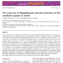
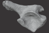
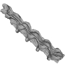
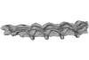
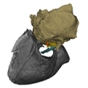
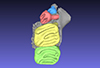
This contribution contains the 3D model described and figured in the following publication: Billet G., Germain D., Ruf I., Muizon C. de, Hautier L. 2013. The inner ear of Megatherium and the evolution of the vestibular system in sloths. Journal of Anatomy 123:557-567, DOI: 10.1111/joa.12114.
Megatherium americanum MNHN.F.PAM276 View specimen

|
M3#14This model corresponds to a virtually reconstructed bony labyrinth of the right inner ear of the skull MNHN-F-PAM 276, attributed to the extinct giant ground sloth Megatherium americanum. The fossil comes from Pleistocene deposits at Rio Salado (Prov. Buenos Aires, Argentina). The bony labyrinth of Megatherium shows semicircular canals that are proportionally much larger than in the modern two-toed and three-toed sloths. The cochlea in Megatherium shows 2.5 turns, which is a rather high value within Xenarthra. Overall, the shape of the bony labyrinth of Megatherium resembles more that of extant armadillos than that of its extant sloth relatives. Type: "3D_surfaces"doi: 10.18563/m3.sf14 state:published |
Download 3D surface file |
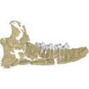
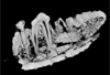
This project presents the 3D models of two isolated petrosals from the Oligocene locality of Pech de Fraysse (Quercy, France) here attributed to the genus Prodremotherium Filhol, 1877. Our aim is to describe the petrosal morphology of this Oligocene “early ruminant” as only few data are available in the literature for Oligocene taxa.
Prodremotherium sp. UM PFY 4053 View specimen

|
M3#7Labelled 3D model of right isolated petrosal of Prodremotherium sp. from Pech de Fraysse (Quercy, MP 28) Type: "3D_surfaces"doi: 10.18563/m3.sf7 state:published |
Download 3D surface file |
Prodremotherium sp. UM PFY 4054 View specimen

|
M3#8Labelled 3D model of right isolated petrosal of Prodremotherium sp. from Pech de Fraysse (Quercy, MP 28) Type: "3D_surfaces"doi: 10.18563/m3.sf8 state:published |
Download 3D surface file |
This contribution includes the 3D models of the reconstructed ossicular chain of the cainotheriid Caenomeryx filholi from the late Oligocene locality of Pech Desse (MP28, Quercy, France) described and figured in the publication of Assemat et al. (2020). It represents the oldest ossicular chain reconstruction for a Paleogene terrestrial artiodactyl species.
Caenomeryx filholi UM PDS 3353 View specimen
|
M3#508reconstruction of the middle ear with petrosal, bulla, stapes, incus, malleus Type: "3D_surfaces"doi: 10.18563/m3.sf.508 state:published |
Download 3D surface file |
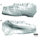
The present 3D Dataset contains the 3D model of a left dentary with m1-m3 analyzed in “A new fossil of Tayassuidae (Mammalia: Certartiodactyla) from the Pleistocene of northern Brazil”. The 3D model was generated using a laser scanning.
cf. Pecari tajacu UFSM 11606 View specimen
|
M3#498Left dentary with m1-m3 Type: "3D_surfaces"doi: 10.18563/m3.sf.498 state:published |
Download 3D surface file |
This contribution contains the 3D model of the holotype of Simplomys hugi, the new dormouse species from the locality of Glovelier described and figured in the following publication: New data on the Miocene dormouse Simplomys García-Paredes, 2009 from the peri-alpin basins of Switzerland and Germany: palaeodiversity of a rare genus in Central Europe. https://doi.org/10.1007/s12549-018-0339-y
Simplomys hugi MJSN-GLM017-0001 View specimen
|
M3#385the left maxilla with four teeth ( DP4, P4, M1 and M2) Type: "3D_surfaces"doi: 10.18563/m3.sf.385 state:published |
Download 3D surface file |
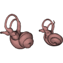
The present 3D Dataset contains the 3D models analyzed in Velazco P. M., Grohé C. 2017. Comparative anatomy of the bony labyrinth of the bats Platalina genovensium (Phyllostomidae, Lonchophyllinae) and Tomopeas ravus (Molossidae, Tomopeatinae). Biotempo 14(2).
Platalina genovensium 278520 View specimen

|
M3#276Right bony labyrinth surface positioned (.PLY) Labels associated (.FLG) Type: "3D_surfaces"doi: 10.18563/m3.sf.276 state:published |
Download 3D surface file |
Tomopeas ravus 278525 View specimen

|
M3#277Right bony labyrinth surface (.PLY) Labels associated (.FLG) Type: "3D_surfaces"doi: 10.18563/m3.sf.277 state:published |
Download 3D surface file |
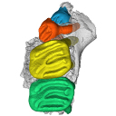
The present 3D Dataset contains the 3D models analyzed in: "a giant dapediid from the Late Triassic of Switzerland and insights into neopterygian phylogeny", Royal Society Open Science, https://doi.org/10.1098/rsos.180497
Scopulipiscis saxciput PIMUZ A/I 3026 View specimen
|
M3#1773D surfaces of the skull and endocranial spaces inside neurocranium, including the aortic canal, braincase, fossa bridgei, lateral cranial canal, nerves and other passageways, notochord, posterior myodome, and right semicircular canals. Type: "3D_surfaces"doi: 10.18563/m3.sf.177 state:published |
Download 3D surface file |

|
M3#178Scan of the neurocranium of PIMUZ A/I 3026 Type: "3D_CT"doi: 10.18563/m3.sf.178 state:published |
Download CT data |
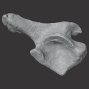
This contribution contains the 3D models described and figured in the following publication: Mennecart B., de Perthuis Ad., Rössner G.E., Guzmán J.A., de Perthuis Au., Costeur L. The first French tragulid skull (Mammalia, Ruminantia, Tragulidae) and associated tragulid remains from the Middle Miocene of Contres (Loir-et-Cher, France). Comptes Rendus Palévol. https://doi.org/10.1016/j.crpv.2017.08.004
Dorcatherium crassum NMB Fa.213.abg View specimen
|
M3#181The 3D surface files of the specimen NMB Fa.213 are the reconstructions of the main skull fragments, the right petrosal bone, and the left bony labyrinth. Type: "3D_surfaces"doi: 10.18563/m3.sf.181 state:published |
Download 3D surface file |
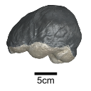
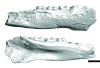
The presented dataset contains the 3D surface scan of the holotype of Birgeria americana, a partial skull described and depicted in: Romano, C., Jenks, J.F., Jattiot, R., Scheyer, T.M., Bylund, K.G. & Bucher, H. 2017. Marine Early Triassic Actinopterygii from Elko County (Nevada, USA): implications for the Smithian equatorial vertebrate eclipse. Journal of Paleontology. https://doi.org/10.1017/jpa.2017.36 .
Birgeria americana NMMNH P-66225 View specimen
|
M3#175NMMNH P-66225 is from upper lower Smithian to lower upper Smithian beds (Thaynes Group). The collecting site is located about 2.75 km south-southeast of the Winecup Ranch, east-central Elko County, Nevada, USA. P-66225 is a partial skull preserved within a large limestone nodule, with its right side exposed. It preserves the portion between the cleithrum posteriorly, and the level of the hind margin of the orbital opening anteriorly. The fossil has a length of 26 cm. Type: "3D_surfaces"doi: 10.18563/m3.sf.175 state:published |
Download 3D surface file |
This contribution contains the 3D models described and figured in the following publication: Orliac M.J., Karadenizli L., Antoine P.-O., Sen S. 2015. Small suids (Mammalia, Artiodactyla) from the late Early Miocene of Turkey and a short overview of Early Miocene small suoids in the Old World. Paleontologia electronica 18(2): 18.2.30A: 1-48. https://doi.org/10.26879/547
?Nguruwe galaticum SMT-1 View specimen
|
M3#16fragment of palate with left broken M1-M3 Type: "3D_surfaces"doi: 10.18563/m3.sf16 state:published |
Download 3D surface file |
This contribution contains the 3D reconstruction of Canariomys bravoi, described and figured in the following publication: Michaux J., Hautier L., Hutterer R., Lebrun R., Guy F., García-Talavera F., 2012 : Body shape and life style of the extinct rodent Canariomys bravoi (Mammalia, Murinae) from Tenerife, Canary Islands (Spain). Comptes Rendus Palevol 11 (7), 485-494. DOI: 10.1016/j.crpv.2012.06.004
Canariomys bravoi TFMCV872-873 View specimen

|
M3#6This file contains the 3D reconstruction of Canariomys bravoi, described and figured in the following publication: Michaux J., Hautier L., Hutterer R., Lebrun R., Guy F., García-Talavera F., 2012 : Body shape and life style of the extinct rodent Canariomys bravoi (Mammalia, Murinae) from Tenerife, Canary Islands (Spain). Comptes Rendus Palevol 11 (7), 485-494. Type: "3D_surfaces"doi: 10.18563/m3.sf6 state:published |
Download 3D surface file |
This contribution provides for the first time the 3D model of the type specimen of Molassitherium delemontense (Mammalia, Rhinocerotidae) described in the following publication: Becker et al. (2013), Journal of Systematic Palaeontology, Vol. 11, Issue 8, 947–972, https://doi.org/10.1080/14772019.2012.699007. Conservation issues of the specimen and solutions using 3D model and 3D prints are detailed.
Molassitherium delemontense MJSN POI007–245 View specimen
|
M3#384Skull of Molassitherium delemontense Becker and Antoine, 2013 (in Becker et al. 2013): holotype Type: "3D_surfaces"doi: 10.18563/m3.sf.384 state:published |
Download 3D surface file |
This contribution contains the 3D model described and figured in the following publication: Ramdarshan A., Orliac M.J., 2015. Endocranial morphology of Microchoerus erinaceus (Euprimates, Tarsiiformes) and early evolution of the Euprimates brain. American Journal of Physical Anthropology. doi: 10.1002/ajpa.22868
Microchoerus erinaceus UM-PRR1771 View specimen
|
M3#15Labelled 3D model of the endocranial cast and sinuse of Microchoerus erinaceus. Type: "3D_surfaces"doi: 10.18563/m3.sf15 state:published |
Download 3D surface file |
|
M3#130350µm voxel size µCT scan of the cranium of UM PRR1771 Type: "3D_CT"doi: 10.18563/m3.sf.1303 state:published |
Download CT data |


