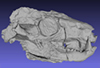
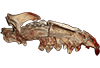
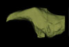
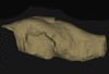
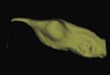
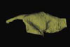
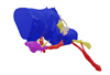
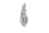
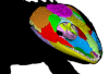
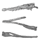
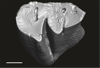
Holotype of Hamadasuchus rebouli
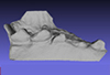
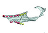
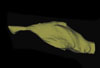
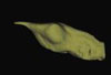
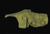
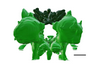
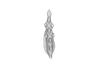
3D model of the holotype specimen of Pebanista yacuruna
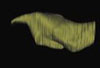
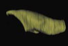
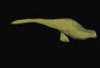
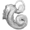
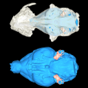
3D models of Eocene–Miocene anuran fossils from Peruvian Amazonia
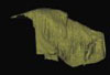
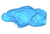
3D GM dataset of bird skeletal variation
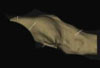
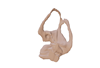
Skeletal embryonic development in the catshark
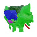
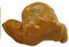
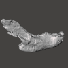
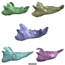
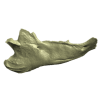
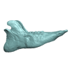
Bony connexions of the petrosal bone of extant hippos
bony labyrinth (11) , inner ear (10) , South America (8) , Eocene (8) , skull (7) , Paleobiogeography (6) , brain (6)
Lionel Hautier (17) , Maëva Judith Orliac (17) , Bastien Mennecart (12) , Pierre-Olivier Antoine (11) , Laurent Marivaux (11) , Leonardo Kerber (10) , Renaud Lebrun (9)
|
3D models related to the publication: Virtual brain endocast of Antifer (Mammalia: Cervidae), an extinct large cervid from South AmericaEmmanuelle Fontoura
Published online: 21/08/2020 |
|
M3#550Brain endocast Type: "3D_surfaces"doi: 10.18563/m3.sf.550 state:published |
Download 3D surface file |
Antifer ensenadensis MCN-PV 943 View specimen
|
M3#551Brain endocast Type: "3D_surfaces"doi: 10.18563/m3.sf.551 state:published |
Download 3D surface file |
The present 3D Dataset contains the 3D models analyzed in Hendrickx, C., Gaetano, L. C., Choiniere, J., Mocke, H. and Abdala, F. in press. A new traversodontid cynodont with a peculiar postcanine dentition from the Middle/Late Triassic of Namibia and dental evolution in basal gomphodonts. Journal of Systematic Palaeontology.
Etjoia dentitransitus GSN F1591 View specimen
|
M3#557Surface model derived from µCT data of the holotype of Etjoia dentitransitus Type: "3D_surfaces"doi: 10.18563/m3.sf.557 state:published |
Download 3D surface file |
|
M3#558Photogrammetric 3D surface model of the postcanines of the Holotype of Etjoia dentitransitus Type: "3D_surfaces"doi: 10.18563/m3.sf.558 state:published |
Download 3D surface file |
|
M3#559Photogrammetric 3D surface model of the Holotype of Etjoia dentitransitus Type: "3D_surfaces"doi: 10.18563/m3.sf.559 state:published |
Download 3D surface file |
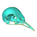
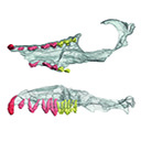
Macroevolution is integral to understanding the patterns of the diversification of life. As the life sciences increasingly use big data approaches, large multivariate datasets are required to test fundamental macroevolutionary hypotheses. In vertebrate evolution, large datasets have been created to quantify morphological variation, largely focusing on particular areas of the skeleton. We provide a landmarking protocol to quantify morphological variation in skeletal elements across the head, trunk, hindlimb and forelimb using 3-dimensional landmarks and semilandmarks, and present a large pan-skeletal database of bird morphology for 149 taxa across avian phylogeny using CT scan data. This large collection of 3D models and geometric morphometric data is open access and can be used in the future for new research, teaching and outreach. The 3D models and CT scans of the 149 specimens related to this project can be downloaded at MorphoSource (https://www.morphosource.org/projects/00000C420)
Menura novaehollandiae FMNH 336751 View specimen
|
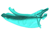
M3#5613D model of the left carpometacarpus of the superb lyrebird, Menura novaehollandia (displayed as a mirror image in the 3DHOP viewer). Type: "3D_surfaces"doi: 10.18563/m3.sf.561 state:published |
Download 3D surface file |

|
M3#5623D model of the mandible of the superb lyrebird, Menura novaehollandiae. Type: "3D_surfaces"doi: 10.18563/m3.sf.562 state:published |
Download 3D surface file |
|
M3#5633D model of the right coracoid of the superb lyrebird, Menura novaehollandiae. Type: "3D_surfaces"doi: 10.18563/m3.sf.563 state:published |
Download 3D surface file |
|
M3#5643D model of the right scapula of the superb lyrebird, Menura novaehollandiae. Type: "3D_surfaces"doi: 10.18563/m3.sf.564 state:published |
Download 3D surface file |
|
M3#5653D model of the right tarsometatarsus of the superb lyrebird, Menura novaehollandiae. Type: "3D_surfaces"doi: 10.18563/m3.sf.565 state:published |
Download 3D surface file |
|
M3#5663D model of the sternum of the superb lyrebird, Menura novaehollandiae. Type: "3D_surfaces"doi: 10.18563/m3.sf.566 state:published |
Download 3D surface file |
|
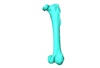
M3#5673D model of the left femur of the superb lyrebird, Menura novaehollandiae (displayed as a mirror image in the 3DHOP viewer). Type: "3D_surfaces"doi: 10.18563/m3.sf.567 state:published |
Download 3D surface file |
|
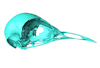
M3#5683D model of the skull of the superb lyrebird, Menura novaehollandiae. Type: "3D_surfaces"doi: 10.18563/m3.sf.568 state:published |
Download 3D surface file |
|
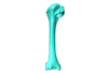
M3#5693D model of the left humerus of the superb lyrebird, Menura novaehollandiae (displayed as a mirror image in the 3DHOP viewer). Type: "3D_surfaces"doi: 10.18563/m3.sf.569 state:published |
Download 3D surface file |

|
M3#5703D model of the synsacrum of the superb lyrebird, Menura novaehollandiae. Type: "3D_surfaces"doi: 10.18563/m3.sf.570 state:published |
Download 3D surface file |
|
M3#5713D model of the left radius of the superb lyrebird, Menura novaehollandiae (displayed as a mirror image in the 3DHOP viewer). Type: "3D_surfaces"doi: 10.18563/m3.sf.571 state:published |
Download 3D surface file |
|
M3#5723D model of the left tibiotarsus of the superb lyrebird, Menura novaehollandiae (displayed as a mirror image in the 3DHOP viewer). Type: "3D_surfaces"doi: 10.18563/m3.sf.572 state:published |
Download 3D surface file |
|
M3#5733D model of the left ulna of the superb lyrebird, Menura novaehollandiae (displayed as a mirror image in the 3DHOP viewer). Type: "3D_surfaces"doi: 10.18563/m3.sf.573 state:published |
Download 3D surface file |
This contribution contains the 3D models described and figured in the following publication: Kassegne K. E., Mourlam M. J., Guinot G., Amoudji Y. Z., Martin J. E., Togbe K. A., Johnson A. K., Hautier L. 2021. First partial cranium of Togocetus from Kpogamé (Togo) and the protocetid diversity in the Togolese phosphate basin. Annales de Paléontologie, Issue 2, April–June 2021, 102488. https://doi.org/10.1016/j.annpal.2021.102488
Togocetus cf. traversei ULDG-KPO1 View specimen
|
M3#768The specimen consists of a partial cranium prepared out of a calcareous phosphate matrix. The partial cranium lacks the anterior part of the rostrum, the cranial roof, and most of the basicranium apart from the left zygomatic process of the squamosal. The maxilla, nasal, palatine, pterygoid, alisphenoid, and squamosal bones are preserved, as well as two incomplete dental rows described hereafter. Type: "3D_surfaces"doi: 10.18563/m3.sf.768 state:published |
Download 3D surface file |
|
M3#770µCT . Resolution: 0.3156mm. This scan can easily be opened with Fiji, MorphoDig, 3DSlicer, or any software that reads .MHD file format. Also, the .RAW file can be opened easily with other software such as Avizo/Amira when providing the correct dimensions (which are enclosed within the file name) Type: "3D_CT"doi: 10.18563/m3.sf.770 state:published |
Download CT data |
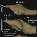
The present 3D Dataset contains 26 3D models analyzed in the study: On the “cartilaginous rider” in the endocasts of turtle brain cavities, published by the authors in the journal Vertebrate Zoology.
Annemys sp. IVPP-V-18106 View specimen
|
M3#7723D surface(s) file to specimen IVPP-V-18106 Type: "3D_surfaces"doi: 10.18563/m3.sf.772 state:published |
Download 3D surface file |
Apalone spinifera FMNH 22178 View specimen

|
M3#7733D surface(s) file to specimen FMNH 22178 Type: "3D_surfaces"doi: 10.18563/m3.sf.773 state:published |
Download 3D surface file |
Caretta caretta NHMUK1940.3.15.1 View specimen
|
M3#7863D surface(s) file to specimen NHMUK1940.3.15.1 Type: "3D_surfaces"doi: 10.18563/m3.sf.786 state:published |
Download 3D surface file |
Chelodina reimanni ZMB 49659 View specimen
|
M3#7743D surface(s) file to specimen ZMB 49659 Type: "3D_surfaces"doi: 10.18563/m3.sf.774 state:published |
Download 3D surface file |
Chelonia mydas ZMB-37416MS View specimen

|
M3#7753D surface(s) file to specimen ZMB-37416MS Type: "3D_surfaces"doi: 10.18563/m3.sf.775 state:published |
Download 3D surface file |
Cuora amboinensis NHMUK69.42.145_4 View specimen
|
M3#7763D surface(s) file to specimen NHMUK69.42.145_4 Type: "3D_surfaces"doi: 10.18563/m3.sf.776 state:published |
Download 3D surface file |
Emydura subglobosa IW92 View specimen
|
M3#7773D surface(s) file to specimen IW92 Type: "3D_surfaces"doi: 10.18563/m3.sf.777 state:published |
Download 3D surface file |
Eubaena cephalica DMNH 96004 View specimen
|
M3#7783D surface(s) file to specimen DMNH 96004 Type: "3D_surfaces"doi: 10.18563/m3.sf.778 state:published |
Download 3D surface file |
Gopherus berlandieri AMNH-73816 View specimen
|
M3#7793D surface(s) file to specimen AMNH-73816 Type: "3D_surfaces"doi: 10.18563/m3.sf.779 state:published |
Download 3D surface file |
Kinixys belliana AMNH-10028 View specimen
|
M3#7803D surface(s) file to specimen AMNH-10028 Type: "3D_surfaces"doi: 10.18563/m3.sf.780 state:published |
Download 3D surface file |
Macrochelys temminckii GPIT-PV-79430 View specimen
|
M3#7813D surface(s) file to specimen GPIT-PV-79430 Type: "3D_surfaces"doi: 10.18563/m3.sf.781 state:published |
Download 3D surface file |
Malacochersus tornieri SMF-58702 View specimen
|
M3#7873D surface(s) file to specimen SMF-58702 Type: "3D_surfaces"doi: 10.18563/m3.sf.787 state:published |
Download 3D surface file |
Naomichelys speciosa FMNH-PR-273 View specimen
|
M3#7823D surface(s) file to specimen FMNH-PR-273 Type: "3D_surfaces"doi: 10.18563/m3.sf.782 state:published |
Download 3D surface file |
Pelodiscus sinensis IW576-2 View specimen
|
M3#7833D surface(s) file to specimen IW576-2 Type: "3D_surfaces"doi: 10.18563/m3.sf.783 state:published |
Download 3D surface file |
Platysternon megacephalum SMF-69684 View specimen
|
M3#7843D surface(s) file to specimen SMF-69684 Type: "3D_surfaces"doi: 10.18563/m3.sf.784 state:published |
Download 3D surface file |
Podocnemis unifilis SMF-55470 View specimen
|
M3#7853D surface(s) file to specimen SMF-55470 Type: "3D_surfaces"doi: 10.18563/m3.sf.785 state:published |
Download 3D surface file |
Proganochelys quenstedtii MB 1910.45.2 View specimen
|
M3#7883D surface(s) file to specimen MB 1910.45.2 Type: "3D_surfaces"doi: 10.18563/m3.sf.788 state:published |
Download 3D surface file |
Proganochelys quenstedtii SMNS 16980 View specimen
|
M3#7893D surface(s) file to specimen SMNS 16980 Type: "3D_surfaces"doi: 10.18563/m3.sf.789 state:published |
Download 3D surface file |
Rhinochelys pulchriceps CAMSM_B55775 View specimen
|
M3#7903D surface(s) file to specimen CAMSM_B55775 Type: "3D_surfaces"doi: 10.18563/m3.sf.790 state:published |
Download 3D surface file |
Rhinoclemmys funereal YPM12174 View specimen
|
M3#7913D surface(s) file to specimen YPM12174 Type: "3D_surfaces"doi: 10.18563/m3.sf.791 state:published |
Download 3D surface file |
Sandownia harrisi MIWG3480 View specimen
|
M3#7923D surface(s) file to specimen MIWG3480 Type: "3D_surfaces"doi: 10.18563/m3.sf.792 state:published |
Download 3D surface file |
Testudo graeca YPM14342 View specimen
|
M3#7933D surface(s) file to specimen YPM14342 Type: "3D_surfaces"doi: 10.18563/m3.sf.793 state:published |
Download 3D surface file |
Testudo hermanni AMNH134518 View specimen
|
M3#7943D surface(s) file to specimen AMNH134518 Type: "3D_surfaces"doi: 10.18563/m3.sf.794 state:published |
Download 3D surface file |
Trachemys scripta NN View specimen
|
M3#7953D surface(s) file to specimen Trachemys scripta Type: "3D_surfaces"doi: 10.18563/m3.sf.795 state:published |
Download 3D surface file |
Xinjiangchelys radiplicatoides IVPP V9539 View specimen
|
M3#7963D surface(s) file to specimen IVPP V9539 Type: "3D_surfaces"doi: 10.18563/m3.sf.796 state:published |
Download 3D surface file |
Chelydra serpentina UFR VP1 View specimen

|
M3#801Brain endocast Type: "3D_surfaces"doi: 10.18563/m3.sf.801 state:published |
Download 3D surface file |
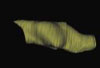
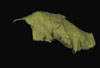
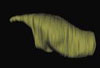
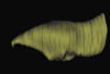
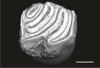
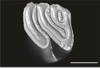
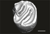
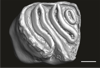
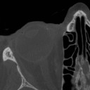
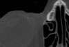
The present 3D Dataset contains the 3D models analyzed in Pochat-Cottilloux Y., Martin J.E., Jouve S., Perrichon G., Adrien J., Salaviale C., de Muizon C., Cespedes R. & Amiot R. (2021). The neuroanatomy of Zulmasuchus querejazus (Crocodylomorpha, Sebecidae) and its implications for the paleoecology of sebecosuchians. The Anatomical Record, https://doi.org/10.1002/ar.24826
Zulmasuchus querejazus MHNC 6672 View specimen
|
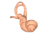
M3#798Left endosseous labyrinth of Z. querejazus (MHNC 6672). Type: "3D_surfaces"doi: 10.18563/m3.sf.798 state:published |
Download 3D surface file |
|
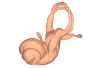
M3#799Reconstruction of the endocranial cavities of Z. querejazus (MHNC 6672). Type: "3D_surfaces"doi: 10.18563/m3.sf.799 state:published |
Download 3D surface file |
|
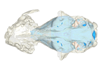
M3#800Three-dimensional reconstruction of the pneumatic cavities within the braincase of Z. querejazus (MHNC 6672) Type: "3D_surfaces"doi: 10.18563/m3.sf.800 state:published |
Download 3D surface file |
This contribution contains the 3D models described and figured in the following publication: Aguirre-Fernández G, Jost J, and Hilfiker S. 2022. First records of extinct kentriodontid and squalodelphinid dolphins from the Upper Marine Molasse (Burdigalian age) of Switzerland and a reappraisal of the Swiss cetacean fauna.
Kentriodon sp. NMBE 5023944 View specimen
|
M3#8583D models of left periotic and bony labyrinth of NMBE 5023944 (Kentriodon sp.) Type: "3D_surfaces"doi: 10.18563/m3.sf.858 state:published |
Download 3D surface file |
Kentriodon sp. NMBE 5023945 View specimen
|
M3#8593D models of right periotic and bony labyrinth of NMBE 5023945 (Kentriodontidae indet.) Type: "3D_surfaces"doi: 10.18563/m3.sf.859 state:published |
Download 3D surface file |
Kentriodon sp. NMBE 5023946 View specimen
|
M3#8603D models of left periotic and bony labyrinth of NMBE 5023946 (Kentriodon sp.) Type: "3D_surfaces"doi: 10.18563/m3.sf.860 state:published |
Download 3D surface file |
Kentriodon sp. NMBE 5036436 View specimen
|
M3#8613D models of right periotic and bony labyrinth of NMBE 5036436 (Kentriodontidae indet.) Type: "3D_surfaces"doi: 10.18563/m3.sf.861 state:published |
Download 3D surface file |
indet. indet. NMBE 5023942 View specimen
|
M3#8623D models of right periotic and bony labyrinth of NMBE 5023942 (Squalodelphinidae indet.) Type: "3D_surfaces"doi: 10.18563/m3.sf.862 state:published |
Download 3D surface file |
indet. indet. NMBE 5023943 View specimen
|
M3#8633D models of left periotic and bony labyrinth of NMBE 5023943 (Squalodelphinidae indet.) Type: "3D_surfaces"doi: 10.18563/m3.sf.863 state:published |
Download 3D surface file |
indet. indet. NMBE 5036437 View specimen
|
M3#8643D models of left periotic and bony labyrinth of NMBE 5036437 (Physeteridae indet.) Type: "3D_surfaces"doi: 10.18563/m3.sf.864 state:published |
Download 3D surface file |
This contribution contains the 3D models of a set of Famennian conodont elements belonging to the species Icriodus alternatus analyzed in the following publication: Girard et al. 2022: Deciphering the morphological variation and its ontogenetic dynamics in the Late Devonian conodont Icriodus alternatus.
Icriodus alternatus UM BUS 031 View specimen
|
M3#887conodont element Type: "3D_surfaces"doi: 10.18563/m3.sf.887 state:published |
Download 3D surface file |
Icriodus alternatus UM BUS 032 View specimen
|
M3#888conodont element Type: "3D_surfaces"doi: 10.18563/m3.sf.888 state:published |
Download 3D surface file |
Icriodus alternatus UM BUS 033 View specimen
|
M3#889conodont element Type: "3D_surfaces"doi: 10.18563/m3.sf.889 state:published |
Download 3D surface file |
Icriodus alternatus UM BUS 034 View specimen
|
M3#890conodont element Type: "3D_surfaces"doi: 10.18563/m3.sf.890 state:published |
Download 3D surface file |
Icriodus alternatus UM BUS 035 View specimen
|
M3#891conodont element Type: "3D_surfaces"doi: 10.18563/m3.sf.891 state:published |
Download 3D surface file |
Icriodus alternatus UM BUS 036 View specimen
|
M3#892conodont element Type: "3D_surfaces"doi: 10.18563/m3.sf.892 state:published |
Download 3D surface file |
Icriodus alternatus UM BUS 037 View specimen
|
M3#893conodont element Type: "3D_surfaces"doi: 10.18563/m3.sf.893 state:published |
Download 3D surface file |
Icriodus alternatus UM BUS 038 View specimen
|
M3#894conodont element Type: "3D_surfaces"doi: 10.18563/m3.sf.894 state:published |
Download 3D surface file |
Icriodus alternatus UM BUS 039 View specimen
|
M3#895conodont element Type: "3D_surfaces"doi: 10.18563/m3.sf.895 state:published |
Download 3D surface file |
Icriodus alternatus UM BUS 040 View specimen
|
M3#896conodont element Type: "3D_surfaces"doi: 10.18563/m3.sf.896 state:published |
Download 3D surface file |
Icriodus alternatus UM BUS 041 View specimen
|
M3#897conodont element Type: "3D_surfaces"doi: 10.18563/m3.sf.897 state:published |
Download 3D surface file |
Icriodus alternatus UM BUS 042 View specimen
|
M3#898conodont element Type: "3D_surfaces"doi: 10.18563/m3.sf.898 state:published |
Download 3D surface file |
Icriodus alternatus UM BUS 043 View specimen
|
M3#899conodont element Type: "3D_surfaces"doi: 10.18563/m3.sf.899 state:published |
Download 3D surface file |
Icriodus alternatus UM BUS 044 View specimen
|
M3#900conodont element Type: "3D_surfaces"doi: 10.18563/m3.sf.900 state:published |
Download 3D surface file |
Icriodus alternatus UM BUS 045 View specimen
|
M3#901conodont element Type: "3D_surfaces"doi: 10.18563/m3.sf.901 state:published |
Download 3D surface file |
This contribution contains the 3D models described and figured in the following publication: Bonis, L. de, Grohé, C., Surault, J., Gardin, A. 2022. Description of the first cranium and endocranial structures of Stenoplesictis minor (Mammalia, Carnivora), an early aeluroid from the Oligocene of the Quercy Phosphorites (southwestern France). Historical Biology. https://doi.org/10.1080/08912963.2022.2045980
Stenoplesictis minor UM-ACQ 6705 View specimen
|
M3#961Endocranium Type: "3D_surfaces"doi: 10.18563/m3.sf.961 state:published |
Download 3D surface file |
|
M3#962Right bony labyrinth Type: "3D_surfaces"doi: 10.18563/m3.sf.962 state:published |
Download 3D surface file |
|
M3#963Left bony labyrinth Type: "3D_surfaces"doi: 10.18563/m3.sf.963 state:published |
Download 3D surface file |
|
M3#964Cranium in transparency with endocranial structures Type: "3D_surfaces"doi: 10.18563/m3.sf.964 state:published |
Download 3D surface file |
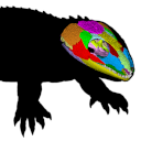
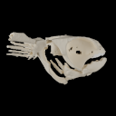
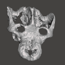
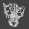
The present 3D Dataset contains the 3D models analyzed in: Abel P., Pommery Y., Ford D. P., Koyabu D., Werneburg I. 2022. Skull sutures and cranial mechanics in the Permian reptile Captorhinus aguti and the evolution of the temporal region in early amniotes. Frontiers in Ecology and Evolution. https://doi.org/10.3389/fevo.2022.841784
Captorhinus aguti OMNH 44816 View specimen
|
M3#965Segmented cranial bone surfaces of OMNH 44816 Type: "3D_surfaces"doi: 10.18563/m3.sf.965 state:published |
Download 3D surface file |
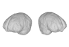
This contribution contains the 3D model of an endocranial cast analyzed in “A 10 ka intentionally deformed human skull from Northeast Asia”. There are many studies on the morphological characteristics of intentional cranial deformation (ICD), but few related 3D models were published. Here, we present the surface model of an intentionally deformed 10 ka human cranium for further research on ICD practice. The 3D model of the endocranial cast of this ICD cranium was discovered near Harbin City, Province Heilongjiang, Northeast China. The fossil preserved only the frontal, parietal, and occipital bones. To complete the endocast model of the specimen, we printed a 3D model and used modeling clay to reconstruct the missing part based on the general form of the modern human endocast morphology.
Homo sapiens IVPP-PA1616 View specimen
|
M3#972The frontal region of the endocast is flattened, probably formed by the constant pressure on the frontal bone during growth. There is a well-developed frontal crest on the endocranial surface. The endocast widens posteriorly from the frontal lobe. The widest point of the endocast is at the lateral border of the parietal lobe. The lower parietal areas display a marked lateral expansion. The overall shape of the endocast is asymmetrical, with the left side of the parietal lobe being more laterally expanded than the right side. Like the frontal lobe, the occipital lobe is also anteroposteriorly flattened. Type: "3D_surfaces"doi: 10.18563/m3.sf.972 state:published |
Download 3D surface file |
|
M3#976The original endocranial cast model (with texture) of IVPP-PA1616. It shows the original structures of the specimen, and was not altered in any way. Type: "3D_surfaces"doi: 10.18563/m3.sf.976 state:published |
Download 3D surface file |
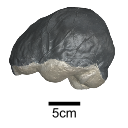
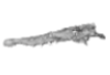
The present 3D dataset contains the 3D models of the holotype of Proterochampsa nodosa that were built and analysed in “Redescription, taxonomic revaluation, and phylogenetic affinities of Proterochampsa nodosa (Archosauriformes: Proterochampsidae), early Late Triassic of Candelaria Sequence (Santa Maria Supersequence)”.
Proterochampsa nodosa MCP 1694-PV View specimen
|
M3#9743D models of Proterochampsa nodosa. Model 1: skull. Model 2: mandible. Model 3: left mandibular ramus. Type: "3D_surfaces"doi: 10.18563/m3.sf.974 state:published |
Download 3D surface file |
The present 3D Dataset contains 3D models of the cranial, visceral, and pectoral endoskeleton of Iniopera, an iniopterygian stem-group holocephalan from the Pennsylvanian of the USA. These data formed the basis for the analyses carried out in Dearden et al. (2023) “Evidence for high-performance suction feeding in the Pennsylvanian stem-group holocephalan Iniopera” PNAS.
Iniopera sp. KUNHM 22060, 158289 View specimen
|
M3#1034plys of the head endoskeleton of Iniopera sp. Type: "3D_surfaces"doi: 10.18563/m3.sf.1034 state:published |
Download 3D surface file |
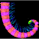
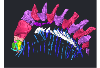
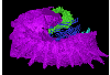
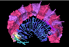
The present 3D Dataset contains the 3D models analyzed in the publication Fossils from the Montceau-les-Mines Lagerstätte (305 Ma) shed light on the anatomy, ecology and phylogeny of Carboniferous millipedes. Authors: Lheritier Mickael, Perroux Maëva, Vannier Jean, Escarguel Gilles, Wesener Thomas, Moritz Leif, Chabard Dominique, Adrien Jerome and Perrier Vincent. Journal of Systematics Palaeontology. https://doi.org/10.1080/14772019.2023.2169891
Amynilyspes fatimae MNHN.F.SOT.2134 View specimen
|
M3#1073Nearly complete specimen. Type: "3D_surfaces"doi: 10.18563/m3.sf.1073 state:published |
Download 3D surface file |
Amynilyspes fatimae MNHN.F.SOT.14983 View specimen
|
M3#1074Nearly complete specimen. Type: "3D_surfaces"doi: 10.18563/m3.sf.1074 state:published |
Download 3D surface file |
Amynilyspes fatimae MNHN.F.SOT.2129 View specimen
|
M3#1075Nearly complete specimen. Type: "3D_surfaces"doi: 10.18563/m3.sf.1075 state:published |
Download 3D surface file |
Blanzilius parriati MNHN.F.SOT.2114A View specimen
|
M3#1076Front part. Type: "3D_surfaces"doi: 10.18563/m3.sf.1076 state:published |
Download 3D surface file |
Blanzilius parriati MNHN.F.SOT.5148 View specimen
|
M3#1077Front part. Type: "3D_surfaces"doi: 10.18563/m3.sf.1077 state:published |
Download 3D surface file |
Blanzilius parriati MNHN.F.SOT.2113 View specimen
|
M3#1078Fragment with legs, sternites and possible tracheal openings. Type: "3D_surfaces"doi: 10.18563/m3.sf.1078 state:published |
Download 3D surface file |
Blanzilius parriati MNHN.F.SOT.81522 View specimen
|
M3#1079Nealry complete specimen. Type: "3D_surfaces"doi: 10.18563/m3.sf.1079 state:published |
Download 3D surface file |
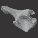
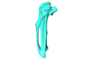
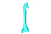
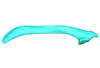
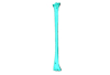
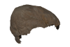
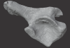
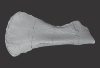
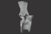
The present 3D Dataset contains the 3D models of an ilium, a vertebra, and a partial scapula of Prestosuchus sp. that were analyzed in “New Loricata remains from the Pinheiros-Chiniquá Sequence (Middle-Upper Triassic), southern Brazil”.
Prestosuchus sp. UFSM11603 View specimen
|
M3#1080Surface scan of a right ilium of Prestosuchus sp. with a 0.4 mm resolution. Type: "3D_surfaces"doi: 10.18563/m3.sf.1080 state:published |
Download 3D surface file |
Prestosuchus sp. UFSM11233 View specimen
|
M3#1081Surface scan of a partial right scapula of Prestosuchus sp. with a 0.4mm resolution. Type: "3D_surfaces"doi: 10.18563/m3.sf.1081 state:published |
Download 3D surface file |
Prestosuchus sp. UFSM11602a View specimen
|
M3#1082Surface scan of a anterior dorsal vertebra of Prestosuchus sp. with a 0.2 mm resolution. Type: "3D_surfaces"doi: 10.18563/m3.sf.1082 state:published |
Download 3D surface file |
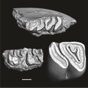
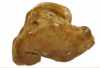
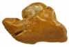
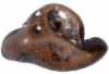
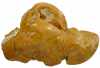
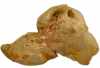
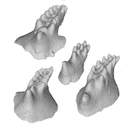
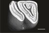
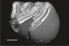
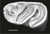
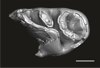
This contribution contains the three-dimensional digital models of a part of the dental fossil material (the large specimens) of caviomorph rodents, discovered in late middle Miocene detrital deposits of the TAR-31 locality in Peruvian Amazonia (San Martín, Peru). These fossils were described, figured and discussed in the following publication: Boivin, Marivaux et al. (2021), Late middle Miocene caviomorph rodents from Tarapoto, Peruvian Amazonia. PLoS ONE 16(11): e0258455. https://doi.org/10.1371/journal.pone.0258455
Microscleromys paradoxalis MUSM 4643 View specimen
|
M3#1115Fragment of left mandibule preserving dp4, m1 and a portion of incisor Type: "3D_surfaces"doi: 10.18563/m3.sf.1115 state:published |
Download 3D surface file |
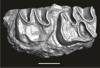
Ricardomys longidens MUSM 4375 View specimen
|
M3#1116Fragment of left maxillary preserving DP4 and M1 (or M1 and M2) Type: "3D_surfaces"doi: 10.18563/m3.sf.1116 state:published |
Download 3D surface file |
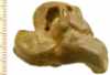
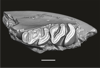
"Scleromys" sp. MUSM 4272 View specimen
|
M3#1117Isolated left upper molar Type: "3D_surfaces"doi: 10.18563/m3.sf.1117 state:published |
Download 3D surface file |
"Scleromys" sp. MUSM 4275 View specimen
|
M3#1118Isolated right upper molar Type: "3D_surfaces"doi: 10.18563/m3.sf.1118 state:published |
Download 3D surface file |
"Scleromys" sp. MUSM 4273 View specimen
|
M3#1119Isolated left upper molar Type: "3D_surfaces"doi: 10.18563/m3.sf.1119 state:published |
Download 3D surface file |
"Scleromys" sp. MUSM 4276 View specimen
|
M3#1120Isolated right upper molar Type: "3D_surfaces"doi: 10.18563/m3.sf.1120 state:published |
Download 3D surface file |
"Scleromys" sp. MUSM 4282 View specimen
|
M3#1121Isolated right lower molar Type: "3D_surfaces"doi: 10.18563/m3.sf.1121 state:published |
Download 3D surface file |
"Scleromys" sp. MUSM 4281 View specimen
|
M3#1122Isolated right lower molar Type: "3D_surfaces"doi: 10.18563/m3.sf.1122 state:published |
Download 3D surface file |
"Scleromys" sp. MUSM 4280 View specimen
|
M3#1123Isolated left p4 Type: "3D_surfaces"doi: 10.18563/m3.sf.1123 state:published |
Download 3D surface file |
"Scleromys" sp. MUSM 4277 View specimen
|
M3#1124Isolated left lower dp4 Type: "3D_surfaces"doi: 10.18563/m3.sf.1124 state:published |
Download 3D surface file |
"Scleromys" sp. MUSM 4279 View specimen
|
M3#1125Isolated right lower dp4 (mesial fragment) Type: "3D_surfaces"doi: 10.18563/m3.sf.1125 state:published |
Download 3D surface file |
gen.indet sp. indet MUSM 4283 View specimen
|
M3#1126Isolated right lower p4 Type: "3D_surfaces"doi: 10.18563/m3.sf.1126 state:published |
Download 3D surface file |
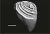
Microscleromys sp. MUSM 4658 View specimen
|
M3#1127Isolated left tarsal bone (astragalus) Type: "3D_surfaces"doi: 10.18563/m3.sf.1127 state:published |
Download 3D surface file |
The present 3D Dataset contains the 3D model of the endocranial cast of Palaeolama sp. from the mid-Pleistocene (~1.2 Mya) of South America, analyzed in Balcarcel et al. 2023.
Palaeolama sp. PIMUZ A/V 4091 View specimen
|
M3#11283D model of a natural endocast Type: "3D_surfaces"doi: 10.18563/m3.sf.1128 state:published |
Download 3D surface file |
The present Dataset contains the micro-CT scan of the head of an anonymous 54 year old female donor, at a voxel resolution of 145µm. The skin of the face has been masked in order to avoid the donor to be recognized.
Homo sapiens UM_HS_2018_09_13 View specimen
|
M3#1152Micro-ct data set Type: "3D_CT"doi: 10.18563/m3.sf.1152 state:published |
Download CT data |
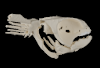
The present 3D Dataset contains the 3D models of a skull and lower jaw of the holotype of Santagnathus mariensis, described in “Old fossil findings in the Upper Triassic rocks of southern Brazil improve diversity of traversodontid cynodonts (Therapsida, Cynodontia)”
Santagnathus mariensis UFRGS-PV-1419-T View specimen
|
M3#1157Skull Type: "3D_surfaces"doi: 10.18563/m3.sf.1157 state:published |
Download 3D surface file |
|
M3#1158Lower jaw Type: "3D_surfaces"doi: 10.18563/m3.sf.1158 state:published |
Download 3D surface file |
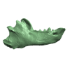
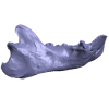
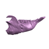
The present 3D Dataset contains the 3D models analyzed in 3D Finite Element Analysis and Geometric Morphometrics of Sloths (Xenarthra, Folivora) Mandibles Show Insights on the Dietary Specializations of Fossil Taxa. Journal of South American Earth Sciences. https://doi.org/10.1016/j.jsames.2023.104445
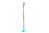
Mylodon darwinii CAV 379 View specimen
|
M3#1159Right hemimandible Type: "3D_surfaces"doi: 10.18563/m3.sf.1159 state:published |
Download 3D surface file |
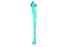
Scelidotherium leptocephalum MNHN-M 137,722 View specimen
|
M3#1160Mandible Type: "3D_surfaces"doi: 10.18563/m3.sf.1160 state:published |
Download 3D surface file |
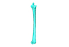
Glossotherium robustum MNHN-M 914 View specimen
|
M3#1161Mandible Type: "3D_surfaces"doi: 10.18563/m3.sf.1161 state:published |
Download 3D surface file |
Lestodon armatus MPAC 899 View specimen
|
M3#1162Mandible Type: "3D_surfaces"doi: 10.18563/m3.sf.1162 state:published |
Download 3D surface file |
Valgipes bucklandi NHMD.Z.M.K. 1/1845:3540 View specimen
|
M3#1163Mandible Type: "3D_surfaces"doi: 10.18563/m3.sf.1163 state:published |
Download 3D surface file |


