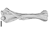
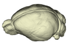
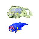
Holotype of Hamadasuchus rebouli
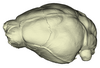
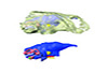
3D model of the holotype specimen of Pebanista yacuruna
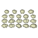
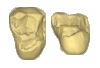
3D models of Eocene–Miocene anuran fossils from Peruvian Amazonia
3D GM dataset of bird skeletal variation
Skeletal embryonic development in the catshark
Bony connexions of the petrosal bone of extant hippos
bony labyrinth (11) , inner ear (10) , South America (8) , Eocene (8) , skull (7) , brain (6) , Oligocene (6)
Maëva Judith Orliac (17) , Lionel Hautier (17) , Bastien Mennecart (12) , Laurent Marivaux (11) , Pierre-Olivier Antoine (11) , Leonardo Kerber (10) , Renaud Lebrun (9)
|
3D model related to the publication: Occurrence of the ground sloth Nothrotheriops (Xenarthra, Folivora) in the Late Pleistocene of Uruguay: New information on its dietary and habitat preferences based on stable isotope analysisLuciano Varela
Published online: 18/05/2023 |
|
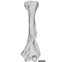
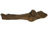
M3#1129Left humerus Type: "3D_surfaces"doi: 10.18563/m3.sf.1129 state:published |
Download 3D surface file |
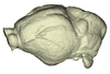
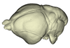
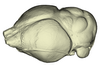
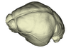
The present 3D Dataset contains the 3D models of extant Chiropteran endocranial casts, documenting 16 of the 19 extant bat families. They are used by Maugoust & Orliac (2023) to assess the correspondences between the brain and brain-surrounding tissues (i.e., neural tissues, blood vessels, meninges) and their imprint on the braincase, allowing for eventually proposing a Chiroptera-scale nomenclature of the endocast.
Balantiopteryx plicata UMMZ 102659 View specimen
|
M3#1132Endocranial cast of the corresponding cranium of Balantiopteryx plicata Type: "3D_surfaces"doi: 10.18563/m3.sf.1132 state:published |
Download 3D surface file |
Nycteris macrotis AMNH M-187705 View specimen
|
M3#1133Endocranial cast of the corresponding cranium of Nycteris macrotis Type: "3D_surfaces"doi: 10.18563/m3.sf.1133 state:published |
Download 3D surface file |
Thyroptera tricolor UMMZ 53240 View specimen
|
M3#1134Endocranial cast of the corresponding cranium of Thyroptera tricolor Type: "3D_surfaces"doi: 10.18563/m3.sf.1134 state:published |
Download 3D surface file |
Noctilio albiventris UMMZ 105827 View specimen
|
M3#1135Endocranial cast of the corresponding cranium of Noctilio albiventris Type: "3D_surfaces"doi: 10.18563/m3.sf.1135 state:published |
Download 3D surface file |
Mormoops blainvillii AMNH M-271513 View specimen
|
M3#1136Endocranial cast of the corresponding cranium of Mormoops blainvillii Type: "3D_surfaces"doi: 10.18563/m3.sf.1136 state:published |
Download 3D surface file |
Macrotus waterhousii UMMZ 95718 View specimen
|
M3#1137Endocranial cast of the corresponding cranium of Macrotus waterhousii Type: "3D_surfaces"doi: 10.18563/m3.sf.1137 state:published |
Download 3D surface file |
Nyctiellus lepidus UMMZ 105767 View specimen
|
M3#1138Endocranial cast of the corresponding cranium of Nyctiellus lepidus Type: "3D_surfaces"doi: 10.18563/m3.sf.1138 state:published |
Download 3D surface file |
Cheiromeles torquatus AMNH M-247585 View specimen
|
M3#1139Endocranial cast of the corresponding cranium of Cheiromeles torquatus Type: "3D_surfaces"doi: 10.18563/m3.sf.1139 state:published |
Download 3D surface file |
Miniopterus schreibersii UMMZ 156998 View specimen
|
M3#1140Endocranial cast of the corresponding cranium of Miniopterus schreibersii Type: "3D_surfaces"doi: 10.18563/m3.sf.1140 state:published |
Download 3D surface file |
Kerivoula pellucida UMMZ 161396 View specimen
|
M3#1141Endocranial cast of the corresponding cranium of Kerivoula pellucida Type: "3D_surfaces"doi: 10.18563/m3.sf.1141 state:published |
Download 3D surface file |
Scotophilus kuhlii UMMZ 157013 View specimen
|
M3#1142Endocranial cast of the corresponding cranium of Scotophilus kuhlii Type: "3D_surfaces"doi: 10.18563/m3.sf.1142 state:published |
Download 3D surface file |
Rhinolophus luctus MNHN CG-2006-87 View specimen
|
M3#1143Endocranial cast of the corresponding cranium of Rhinolophus luctus Type: "3D_surfaces"doi: 10.18563/m3.sf.1143 state:published |
Download 3D surface file |
Triaenops persicus AM RG-38552 View specimen
|
M3#1144Endocranial cast of the corresponding cranium of Triaenops persicus Type: "3D_surfaces"doi: 10.18563/m3.sf.1144 state:published |
Download 3D surface file |
Hipposideros armiger UM ZOOL-762-V View specimen
|
M3#1145Endocranial cast of the corresponding cranium of Hipposideros armiger Type: "3D_surfaces"doi: 10.18563/m3.sf.1145 state:published |
Download 3D surface file |
Lavia frons AM RG-12268 View specimen
|
M3#1146Endocranial cast of the corresponding cranium of Lavia frons Type: "3D_surfaces"doi: 10.18563/m3.sf.1146 state:published |
Download 3D surface file |
Rhinopoma hardwickii AM RG-M31166 View specimen
|
M3#1147Endocranial cast of the corresponding cranium of Rhinopoma hardwickii Type: "3D_surfaces"doi: 10.18563/m3.sf.1147 state:published |
Download 3D surface file |
Sphaerias blanfordi AMNH M-274330 View specimen
|
M3#1148Endocranial cast of the corresponding cranium of Sphaerias blanfordi Type: "3D_surfaces"doi: 10.18563/m3.sf.1148 state:published |
Download 3D surface file |
Rousettus aegyptiacus UMMZ 161026 View specimen
|
M3#1149Endocranial cast of the corresponding cranium of Rousettus aegyptiacus Type: "3D_surfaces"doi: 10.18563/m3.sf.1149 state:published |
Download 3D surface file |
Pteropus pumilus UMMZ 162253 View specimen
|
M3#1150Endocranial cast of the corresponding cranium of Pteropus pumilus Type: "3D_surfaces"doi: 10.18563/m3.sf.1150 state:published |
Download 3D surface file |
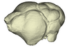
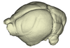
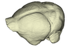
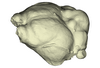
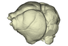
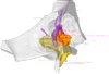
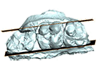
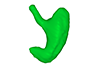
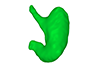
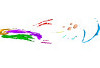
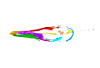
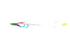
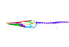
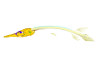
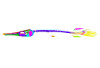
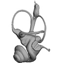
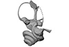
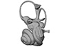
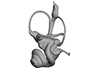
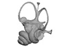
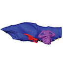
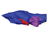
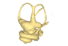
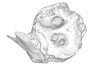
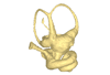
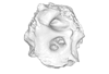
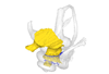
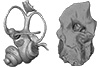
This project presents the osteological connexions of the petrosal bone of the extant Hippopotamidae Hippopotamus amphibius and Choeropsis liberiensis by a virtual osteological dissection of the ear region. The petrosal, the bulla, the sinuses and the major morphological features surrounding the petrosal bone are labelled, both in situ and in an exploded model presenting disassembly views. The directional underwater hearing mode of Hippopotamidae is discussed based on the new observations.
Choeropsis liberiensis UPPal-M09-5-005a View specimen
|
M3#1Labelled compact model of the right ear region of Choeropsis liberiensis (UPPal-M09-5-005a) Type: "3D_surfaces"doi: 10.18563/m3.sf1 state:published |
Download 3D surface file |

|
M3#2Labelled exploded model of the right ear region of Choeropsis liberiensis (UPPal-M09-5-005a) Type: "3D_surfaces"doi: 10.18563/m3.sf2 state:published |
Download 3D surface file |
Hippopotamus amphibius UM N179 View specimen
|
M3#3Labelled compact model of the right ear region of Hippopotamus amphibius (UM N 179) Type: "3D_surfaces"doi: 10.18563/m3.sf3 state:published |
Download 3D surface file |

|
M3#4Labelled exploded model of the right ear region of Hippopotamus amphibius (UM N 179) Type: "3D_surfaces"doi: 10.18563/m3.sf4 state:published |
Download 3D surface file |
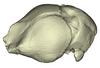
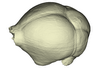
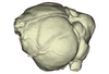
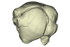
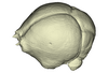
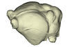
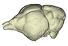
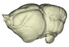
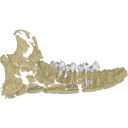
This contribution contains the 3D reconstruction of Canariomys bravoi, described and figured in the following publication: Michaux J., Hautier L., Hutterer R., Lebrun R., Guy F., García-Talavera F., 2012 : Body shape and life style of the extinct rodent Canariomys bravoi (Mammalia, Murinae) from Tenerife, Canary Islands (Spain). Comptes Rendus Palevol 11 (7), 485-494. DOI: 10.1016/j.crpv.2012.06.004
Canariomys bravoi TFMCV872-873 View specimen

|
M3#6This file contains the 3D reconstruction of Canariomys bravoi, described and figured in the following publication: Michaux J., Hautier L., Hutterer R., Lebrun R., Guy F., García-Talavera F., 2012 : Body shape and life style of the extinct rodent Canariomys bravoi (Mammalia, Murinae) from Tenerife, Canary Islands (Spain). Comptes Rendus Palevol 11 (7), 485-494. Type: "3D_surfaces"doi: 10.18563/m3.sf6 state:published |
Download 3D surface file |
This project presents the 3D models of two isolated petrosals from the Oligocene locality of Pech de Fraysse (Quercy, France) here attributed to the genus Prodremotherium Filhol, 1877. Our aim is to describe the petrosal morphology of this Oligocene “early ruminant” as only few data are available in the literature for Oligocene taxa.
Prodremotherium sp. UM PFY 4053 View specimen

|
M3#7Labelled 3D model of right isolated petrosal of Prodremotherium sp. from Pech de Fraysse (Quercy, MP 28) Type: "3D_surfaces"doi: 10.18563/m3.sf7 state:published |
Download 3D surface file |
Prodremotherium sp. UM PFY 4054 View specimen

|
M3#8Labelled 3D model of right isolated petrosal of Prodremotherium sp. from Pech de Fraysse (Quercy, MP 28) Type: "3D_surfaces"doi: 10.18563/m3.sf8 state:published |
Download 3D surface file |
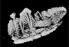
This contribution contains the 3D models described and figured in the following publication: Orliac M.J., Karadenizli L., Antoine P.-O., Sen S. 2015. Small suids (Mammalia, Artiodactyla) from the late Early Miocene of Turkey and a short overview of Early Miocene small suoids in the Old World. Paleontologia electronica 18(2): 18.2.30A: 1-48. https://doi.org/10.26879/547
?Nguruwe galaticum SMT-1 View specimen
|
M3#16fragment of palate with left broken M1-M3 Type: "3D_surfaces"doi: 10.18563/m3.sf16 state:published |
Download 3D surface file |
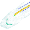
This project presents a µCT dataset and an associated 3D surface model of the holotype of Donrussellia magna (UM PAT 17; Primates, Adapiformes). UM PAT17 is the only known specimen for the species and consists of a well-preserved left lower jaw with p4-m3. It documents one of the oldest European primates, eventually dated near the Paleocene Eocene Thermal Maximum.
Donrussellia magna UM PAT 17 View specimen

|
M3#173D surface file model of UM PAT 17 (type specimen of Donrussellia magna), which is a well preserved left lower jaw with p4-m3. The teeth (and roots) were manually segmented. Type: "3D_surfaces"doi: 10.18563/m3.sf17 state:published |
Download 3D surface file |
|
M3#18CT Scan Data of Donrussellia magna UM PAT 17. Voxel size (in µm): 36µm (isotropic voxels). Dimensions in x,y,z : 594 pixels, 294 pixels, 1038 pixels. Image type : 8-bit voxels. Image format : raw data format (no header). Type: "3D_CT"doi: 10.18563/m3.sf18 state:published |
Download CT data |
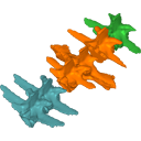
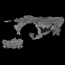
This contribution contains the 3D models described and figured in the following publication: Molnar, JL, Pierce, SE, Bhullar, B-A, Turner, AH, Hutchinson, JR (accepted). Morphological and functional changes in the crocodylomorph vertebral column with increasing aquatic adaptation. Royal Society Open Science.
Protosuchus richardsoni AMNH-VP 3024 View specimen

|
M3#448th and 9th dorsal vertebrae, 1st and 2nd lumbar vertebrae, and 5th lumbar and sacral vertebrae. Type: "3D_surfaces"doi: 10.18563/m3.sf44 state:published |
Download 3D surface file |
Terrestrisuchus gracilis NHM-PV R 7562 View specimen

|
M3#451st and 2nd lumbar vertebrae, and 5th lumbar and sacral vertebrae Type: "3D_surfaces"doi: 10.18563/m3.sf45 state:published |
Download 3D surface file |
Pelagosaurus typus NHM-PV OR 32598 View specimen

|
M3#467th and 8th dorsal vertebrae, 11th and 12th dorsal vertebrae, 15th dorsal vertebra and sacral vertebra. Type: "3D_surfaces"doi: 10.18563/m3.sf46 state:published |
Download 3D surface file |
Metriorhynchus superciliosus NHM-PV R 2054 View specimen

|
M3#476th and 7th dorsal vertebrae, 10th and 11th dorsal vertebrae, 17th dorsal vertebra and sacral vertebra Type: "3D_surfaces"doi: 10.18563/m3.sf47 state:published |
Download 3D surface file |
Crocodylus niloticus FNC0 View specimen

|
M3#487th and 8th dorsal vertebrae, 1st and 2nd lumbar vertebrae, 5th lumbar vertebra and sacral vertebra. Type: "3D_surfaces"doi: 10.18563/m3.sf48 state:published |
Download 3D surface file |
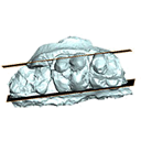
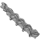
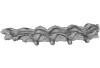
Current knowledge on the skeletogenesis of Chondrichthyes is scarce compared with their extant sister group, the bony fishes. Most of the previously described developmental tables in Chondrichthyes have focused on embryonic external morphology only. Due to its small body size and relative simplicity to raise eggs in laboratory conditions, the small-spotted catshark Scyliorhinus canicula has emerged as a reference species to describe developmental mechanisms in the Chondrichthyes lineage. Here we investigate the dynamic of mineralization in a set of six embryonic specimens using X-ray microtomography and describe the developing units of both the dermal skeleton (teeth and dermal scales) and endoskeleton (vertebral axis). This preliminary data on skeletogenesis in the catshark sets the first bases to a more complete investigation of the skeletal developmental in Chondrichthyes. It should provide comparison points with data known in osteichthyans and could thus be used in the broader context of gnathostome skeletal evolution.
Scyliorhinus canicula SC6_2_2015_03_20 View specimen

|
M3#50Mineralized skeleton of a 6,2 cm long embryo of Scyliorhinus canicula Type: "3D_surfaces"doi: 10.18563/m3.sf.50 state:published |
Download 3D surface file |
Scyliorhinus canicula SC6_7_2015_03_20 View specimen

|
M3#51Mineralized skeleton of a 6,7 cm long embryo of Scyliorhinus canicula Type: "3D_surfaces"doi: 10.18563/m3.sf.51 state:published |
Download 3D surface file |
Scyliorhinus canicula SC7_1_2015_04_03 View specimen

|
M3#52Mineralized skeleton of a 7,1 cm long embryo of Scyliorhinus canicula Type: "3D_surfaces"doi: 10.18563/m3.sf.52 state:published |
Download 3D surface file |
Scyliorhinus canicula SC7_5_2015_03_13 View specimen

|
M3#53Mineralized skeleton of a 7,5 cm long embryo of Scyliorhinus canicula Type: "3D_surfaces"doi: 10.18563/m3.sf.53 state:published |
Download 3D surface file |
Scyliorhinus canicula SC8_2015_03_20 View specimen

|
M3#54Mineralized skeleton of a 8 cm long embryo of Scyliorhinus canicula Type: "3D_surfaces"doi: 10.18563/m3.sf.54 state:published |
Download 3D surface file |
Scyliorhinus canicula SC10_2015_02_27 View specimen

|
M3#55Mineralized skeleton of a 10 cm long embryo of Scyliorhinus canicula Type: "3D_surfaces"doi: 10.18563/m3.sf.55 state:published |
Download 3D surface file |
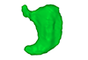
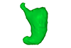
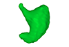
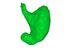
The present 3D Dataset contains the 3D models analyzed in: Kaigai N et al. Morphogenesis and three-dimensional movement of the stomach during the human embryonic period, Anat Rec (Hoboken). 2014 May;297(5):791-797. doi: 10.1002/ar.22833.
Homo sapiens KC-CS16STM27159 View specimen
|
M3#56computationally reconstructed stomach of the human embryo (M3#56_KC-CS16STM27159) at Carnegie Stage 16 (Crown Rump Length= 9.9mm). Type: "3D_surfaces"doi: 10.18563/m3.sf56 state:published |
Download 3D surface file |
Homo sapiens KC-CS17STM20383 View specimen
|
M3#57computationally reconstructed stomach of the human embryo (M3#57_KC-CS17STM20383) at Carnegie Stage 17 (Crown Rump Length= 12.3mm). Type: "3D_surfaces"doi: 10.18563/m3.sf57 state:published |
Download 3D surface file |
Homo sapiens KC-CS18STM21807 View specimen
|
M3#58computationally reconstructed stomach of the human embryo (M3#58_KC-CS18STM21807) at Carnegie Stage 18 (Crown Rump Length= 14.7mm). Type: "3D_surfaces"doi: 10.18563/m3.sf58 state:published |
Download 3D surface file |
Homo sapiens KC-CS19STM17998 View specimen
|
M3#59computationally reconstructed stomach of the human embryo (M3#59_KC-CS19STM17998) at Carnegie Stage 19 (Crown Rump Length was unmeasured ). Type: "3D_surfaces"doi: 10.18563/m3.sf59 state:published |
Download 3D surface file |
Homo sapiens KC-CS20STM20785 View specimen
|
M3#60computationally reconstructed stomach of the human embryo (M3#60_KC-CS20STM20785) at Carnegie Stage 20 (Crown Rump Length= 18.7 mm). Type: "3D_surfaces"doi: 10.18563/m3.sf60 state:published |
Download 3D surface file |
Homo sapiens KC-CS21STM24728 View specimen
|
M3#61computationally reconstructed stomach of the human embryo (M3#61_KC-CS21STM24728) at Carnegie Stage 21 (Crown Rump Length= 20.9 mm). Type: "3D_surfaces"doi: 10.18563/m3.sf61 state:published |
Download 3D surface file |
Homo sapiens KC-CS22STM26438 View specimen
|
M3#62computationally reconstructed stomach of the human embryo (M3#62_KC-CS22STM26438) at Carnegie Stage 22 (Crown Rump Length= 21.5 mm). Type: "3D_surfaces"doi: 10.18563/m3.sf62 state:published |
Download 3D surface file |
Homo sapiens KC-CS23STM20018 View specimen
|
M3#63computationally reconstructed stomach of the human embryo (M3#63_KC-CS23STM20018) at Carnegie Stage 23 (Crown Rump Length= 23.1 mm). Type: "3D_surfaces"doi: 10.18563/m3.sf63 state:published |
Download 3D surface file |
Using X-ray microtomography, we describe the ossification events during the larval development of a non-teleost actinopterygian species: the Cuban gar Atractosteus tristoechus from the order Lepisosteiformes. We provide a detailed developmental series for each anatomical structure, covering a large sequence of mineralization events going from an early stage (13 days post-hatching, 21mm total length) to an almost fully ossified larval stage (118dph or 87mm in standard length). With this work, we expect to bring new developmental data to be used in further comparative studies with other lineages of bony vertebrates. We also hope that the on-line publication of these twelve successive 3D reconstructions, fully labelled and flagged, will be an educational tool for all students in comparative anatomy.
Atractosteus tristoechus At1-13dph View specimen
|
M3#94At1-13dph : 13 dph larvae, 21 mm TL Type: "3D_surfaces"doi: 10.18563/m3.sf.94 state:published |
Download 3D surface file |
Atractosteus tristoechus At2-16dph View specimen
|
M3#95Atractosteus tristoechus larva, 16 dph, 26mm SL. Type: "3D_surfaces"doi: 10.18563/m3.sf.95 state:published |
Download 3D surface file |
Atractosteus tristoechus At3-19dph View specimen

|
M3#96Atractosteus tristoechus larva, 19 dph, 27mm SL. Type: "3D_surfaces"doi: 10.18563/m3.sf.96 state:published |
Download 3D surface file |
Atractosteus tristoechus At4-22dph View specimen
|
M3#97Atractosteus tristoechus larva, 22dph, 30mm SL. Type: "3D_surfaces"doi: 10.18563/m3.sf.97 state:published |
Download 3D surface file |
Atractosteus tristoechus At5-26dph View specimen
|
M3#98Atractosteus tristoechus larva, 26 dph, 32mm SL. Type: "3D_surfaces"doi: 10.18563/m3.sf.98 state:published |
Download 3D surface file |
Atractosteus tristoechus At6-31dph View specimen
|
M3#99Atractosteus tristoechus larva, 31 dph, 39mm SL. Type: "3D_surfaces"doi: 10.18563/m3.sf.99 state:published |
Download 3D surface file |
Atractosteus tristoechus At7-37dph View specimen
|
M3#100Atractosteus tristoechus larva, 37 dph, 43mm SL. Type: "3D_surfaces"doi: 10.18563/m3.sf.100 state:published |
Download 3D surface file |
Atractosteus tristoechus At8-52dph View specimen
|
M3#101Atractosteus tristoechus larva, 52 dph, 46mm SL. Type: "3D_surfaces"doi: 10.18563/m3.sf.101 state:published |
Download 3D surface file |
Atractosteus tristoechus At9-74dph View specimen
|
M3#102Atractosteus tristoechus larva, 74 dph, 61mm SL. Not all structures are colored, only newly ossified ones. Type: "3D_surfaces"doi: 10.18563/m3.sf.102 state:published |
Download 3D surface file |
Atractosteus tristoechus At10-89dph View specimen
|
M3#103Atractosteus tristoechus larva, 89 dph, 63mm SL. Not all structures are colored, only newly ossified ones. You may find the tag file in the At1-13dph reconstruction data. Type: "3D_surfaces"doi: 10.18563/m3.sf.103 state:published |
Download 3D surface file |
Atractosteus tristoechus At11-104dph View specimen
|
M3#104Atractosteus tristoechus larva, 104 dph, 70mm SL. Not all structures are colored, only newly ossified ones. Type: "3D_surfaces"doi: 10.18563/m3.sf.104 state:published |
Download 3D surface file |
Atractosteus tristoechus At12-118dph View specimen
|
M3#105Atractosteus tristoechus larva, 118 dph, 87mm SL. Type: "3D_surfaces"doi: 10.18563/m3.sf.105 state:published |
Download 3D surface file |
The present 3D Dataset contains the 3D models analyzed in Neogene sloth assemblages (Mammalia, Pilosa) of the Cocinetas Basin (La Guajira, Colombia): implications for the Great American Biotic Interchange. Palaeontology. doi: 10.1111/pala.12244
cf. Nothrotherium indet. MUN STRI 12924 View specimen
|
M3#106Fragmentary basicranium with posterior portion of the skull roof. Type: "3D_surfaces"doi: 10.18563/m3.sf.106 state:published |
Download 3D surface file |
indet. indet. MUN STRI 16535 View specimen
|
M3#107Complete left ulna of a Scelidotheriinae gen. et sp. indet. Type: "3D_surfaces"doi: 10.18563/m3.sf.107 state:published |
Download 3D surface file |
This contribution contains the 3D model described and figured in the following publication: Albino, A., Carrillo-Briceño, J. D. & Neenan, J. M. 2016. An enigmatic aquatic snake from the Cenomanian of northern South America. PeerJ 4:e2027 http://dx.doi.org/10.7717/peerj.2027
Lunaophis aquaticus MCNC-1827-F View specimen
|
M3#116Articulated precloacal vertebrae of Lunaophis aquaticus Type: "3D_surfaces"doi: 10.18563/m3.sf.116 state:published |
Download 3D surface file |
The present 3D Dataset contains the 3D models analyzed in the article Mennecart, B., and L. Costeur. 2016. A Dorcatherium (Mammalia, Ruminantia, Middle Miocene) petrosal bone and the tragulid ear region. Journal of Vertebrate Paleontology 36(6), 1211665(1)-1211665(7). DOI: 10.1080/02724634.2016.1211665.
Tragulus javanicus 10028 View specimen
|
M3#1193D surface of the left bony labyrinth of Tragulus javanicus NMB 10028 Type: "3D_surfaces"doi: 10.18563/m3.sf.119 state:published |
Download 3D surface file |
Moschiola meminna C.2453 View specimen
|
M3#1203D surface of the left bony labyrinth of Moschiola meminna NMB C.2453 Type: "3D_surfaces"doi: 10.18563/m3.sf.120 state:published |
Download 3D surface file |
Hyemoschus aquaticus C.1930 View specimen
|
M3#1223D surface of the right bony labyrinth of Hyemoschus aquaticus NMB C.1930 Type: "3D_surfaces"doi: 10.18563/m3.sf.122 state:published |
Download 3D surface file |
Dorcatherium crassum San.15053 View specimen
|
M3#1233D surface of the right bony labyrinth of Dorcatherium crassum NMB San.15053 Type: "3D_surfaces"doi: 10.18563/m3.sf.123 state:published |
Download 3D surface file |
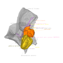
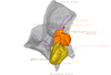
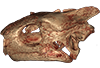
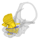
The present publication contains the µCT dataset and the 3D models analyzed in the following publication: Mautner, A.-K., A. E. Latimer, U. Fritz, and T. M. Scheyer. An updated description of the osteology of the pancake tortoise Malacochersus tornieri (Testudines: Testudinidae) with special focus on intraspecific variation. Journal of Morphology. https://doi.org/10.1002/jmor.20640
Malacochersus tornieri ZM 100.102 View specimen
|
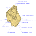
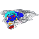
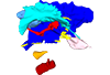
M3#129Virtual brain and inner ear endocast of Malacochersus tornieri (ZM 100.102; Zoological Museum of The University of Zurich). This virtual model is accompanied by the 3D dataset. Blue, endocranium; red, blood vessels; purple, semicircular canals; yellow, cranial nerves. Type: "3D_surfaces"doi: 10.18563/m3.sf.129 state:published |
Download 3D surface file |
|
M3#1303D dataset of skull of Malacochersus tornieri (ZM 100.102) Type: "3D_CT"doi: 10.18563/m3.sf.130 state:published |
Download CT data |
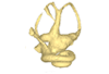
This contribution contains the 3D models described and figured in the publication entitled "The petrosal and bony labyrinth of Diplobune minor, an enigmatic Artiodactyla from the Oligocene of Western Europe" by Orliac, Araújo, and Lihoreau published in Journal of Morphology (Orliac et al. 2017) https://doi.org/10.1002/jmor.20702.
Diplobune minor UM ITD 1079 View specimen
|
M3#138right bony labyrinth of Diplobune minor from Itardies, France Type: "3D_surfaces"doi: 10.18563/m3.sf.138 state:published |
Download 3D surface file |
|
M3#139right isolated petrosal of Diplobune minor from Itardies, France Type: "3D_surfaces"doi: 10.18563/m3.sf.139 state:published |
Download 3D surface file |
Diplobune minor UM ITD 1080 View specimen
|
M3#140left bony labyrinth of Diplobune minor from Itardies, France Type: "3D_surfaces"doi: 10.18563/m3.sf.140 state:published |
Download 3D surface file |
|
M3#141left isolated petrosal of Diplobune minor from Itardies, France Type: "3D_surfaces"doi: 10.18563/m3.sf.141 state:published |
Download 3D surface file |
Diplobune minor UM ITD 1081 View specimen
|
M3#142right bony labyrinth and associated nerves and veins of Diplobune minor from Itardies, France Type: "3D_surfaces"doi: 10.18563/m3.sf.142 state:published |
Download 3D surface file |
|
M3#143right isolated petrosal of Diplobune minor from Itardies, France Type: "3D_surfaces"doi: 10.18563/m3.sf.143 state:published |
Download 3D surface file |
Diplobune minor UM ITD 1083 View specimen
|
M3#144left bony labyrinth of Diplobune minor from Itardies, France Type: "3D_surfaces"doi: 10.18563/m3.sf.144 state:published |
Download 3D surface file |
|
M3#145left petrosal of Diplobune minor from Itardies, France Type: "3D_surfaces"doi: 10.18563/m3.sf.145 state:published |
Download 3D surface file |
The present 3D Dataset contains the 3D models analyzed in: "a giant dapediid from the Late Triassic of Switzerland and insights into neopterygian phylogeny", Royal Society Open Science, https://doi.org/10.1098/rsos.180497
Scopulipiscis saxciput PIMUZ A/I 3026 View specimen
|
M3#1773D surfaces of the skull and endocranial spaces inside neurocranium, including the aortic canal, braincase, fossa bridgei, lateral cranial canal, nerves and other passageways, notochord, posterior myodome, and right semicircular canals. Type: "3D_surfaces"doi: 10.18563/m3.sf.177 state:published |
Download 3D surface file |

|
M3#178Scan of the neurocranium of PIMUZ A/I 3026 Type: "3D_CT"doi: 10.18563/m3.sf.178 state:published |
Download CT data |
This contribution contains the 3D models described and figured in the following publication: Mennecart B., de Perthuis Ad., Rössner G.E., Guzmán J.A., de Perthuis Au., Costeur L. The first French tragulid skull (Mammalia, Ruminantia, Tragulidae) and associated tragulid remains from the Middle Miocene of Contres (Loir-et-Cher, France). Comptes Rendus Palévol. https://doi.org/10.1016/j.crpv.2017.08.004
Dorcatherium crassum NMB Fa.213.abg View specimen
|
M3#181The 3D surface files of the specimen NMB Fa.213 are the reconstructions of the main skull fragments, the right petrosal bone, and the left bony labyrinth. Type: "3D_surfaces"doi: 10.18563/m3.sf.181 state:published |
Download 3D surface file |
This contribution contains the 3D model described and figured in the following publication: New turtles from the Late Cretaceous of Monte Alto-SP, Brazil, including cranial osteology, neuroanatomy and phylogenetic position of a new taxon. PalZ. https://doi.org/10.1007/s12542-017-0397-x
Yuraramirim montealtensis 04-0008/89 View specimen
|
M3#2783D surfaces related to specimen MPMA 04-0008/89. Type: "3D_surfaces"doi: 10.18563/m3.sf.278 state:published |
Download 3D surface file |
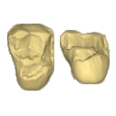
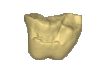
This contribution contains the 3D models of the isolated teeth of Canaanimico amazonensis, a new stem platyrrhine primate, described and figured in the following publication: Marivaux et al. (2016), Neotropics provide insights into the emergence of New World monkeys: new dental evidence from the late Oligocene of Peruvian Amazonia. Journal of Human Evolution. http://dx.doi.org/10.1016/j.jhevol.2016.05.011
Canaanimico amazonensis MUSM-2499 View specimen
|
M3#2893D model of left upper M2 Type: "3D_surfaces"doi: 10.18563/m3.sf.289 state:published |
Download 3D surface file |
Canaanimico amazonensis MUSM-2500 View specimen
|
M3#2903D model of left upper M1 (lingual part) Type: "3D_surfaces"doi: 10.18563/m3.sf.290 state:published |
Download 3D surface file |


