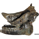
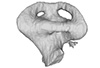
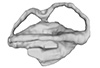
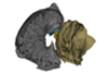
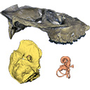
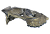
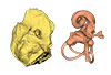
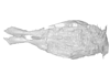
Holotype of Hamadasuchus rebouli
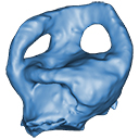
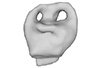
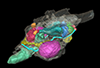
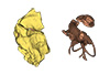
3D model of the holotype specimen of Pebanista yacuruna
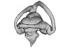
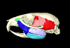
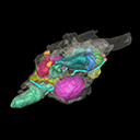
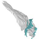
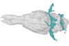
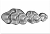
3D models of the endocranial anatomy of Voay robustus and comparative specimens
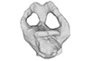
3D GM dataset of bird skeletal variation
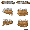
Skeletal embryonic development in the catshark
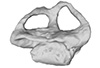
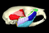
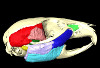
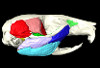
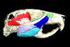
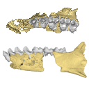
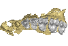
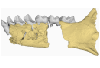
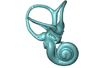
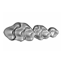
Bony connexions of the petrosal bone of extant hippos
bony labyrinth (11) , inner ear (10) , Eocene (8) , South America (8) , skull (7) , Oligocene (6) , phylogeny (6)
Maëva Judith Orliac (17) , Lionel Hautier (17) , Bastien Mennecart (12) , Laurent Marivaux (11) , Pierre-Olivier Antoine (11) , Leonardo Kerber (10) , Renaud Lebrun (9)
|
3D models related to the publication: New data on Amynodontidae (Mammalia, Perissodactyla) from the Eocene of Eastern Europe: phylogenetic and palaeobiogeographic implicationsJérémy Tissier
Published online: 12/03/2018 |
|
M3#297Incomplete skull with left M3. Type: "3D_surfaces"doi: 10.18563/m3.sf.297 state:published |
Download 3D surface file |
Sellamynodon zimborensis UBB MPS 15795 View specimen

|
M3#298Mandible with complete molar and premolar rows, lacking symphysis. Type: "3D_surfaces"doi: 10.18563/m3.sf.298 state:published |
Download 3D surface file |
Amynodontopsis aff. bodei UBB MPS V545 View specimen
|
M3#299Maxillary fragment with M1-3. Type: "3D_surfaces"doi: 10.18563/m3.sf.299 state:published |
Download 3D surface file |
Amynodontopsis aff. bodei UBB MPS V546 View specimen
|
M3#300Unworn m1/2 on mandible fragment. Type: "3D_surfaces"doi: 10.18563/m3.sf.300 state:published |
Download 3D surface file |
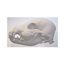
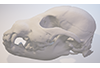
This contribution comprises the 3D models of three wolf pup skulls, which were used for the publication by Geiger et al. 2017 on Neomorphosis and heterochrony of skull shape in dog domestication.
Canis lupus CLL2 View specimen
|
M3#3123d model of a wolf pup skull Type: "3D_surfaces"doi: 10.18563/m3.sf.312 state:published |
Download 3D surface file |
Canis lupus CLL4 View specimen
|
M3#3133d model of a wolf pup skull Type: "3D_surfaces"doi: 10.18563/m3.sf.313 state:published |
Download 3D surface file |
Canis lupus CLL5 View specimen
|
M3#3143d model of a wolf pup skull Type: "3D_surfaces"doi: 10.18563/m3.sf.314 state:published |
Download 3D surface file |
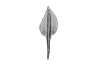
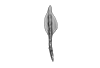
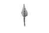
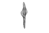
The present 3D Dataset contains the 3D models analyzed in "Neenan, J. M., Reich, T., Evers, S., Druckenmiller, P. S., Voeten, D. F. A. E., Choiniere, J. N., Barrett, P. M., Pierce, S. E. and Benson, R. B. J. Evolution of the sauropterygian labyrinth with increasingly pelagic lifestyles. Current Biology, 27." https://doi.org/10.1016/j.cub.2017.10.069
Amblyrhynchus cristatus OUMNH 11616 View specimen
|
M3#322Right labyrinth of Amblyrhynchus cristatus (OUMNH 11616). Type: "3D_surfaces"doi: 10.18563/m3.sf.322 state:published |
Download 3D surface file |
Augustasaurus hagdorni FMNH PR 1974 View specimen
|
M3#333Right labyrinth model of Augustasaurus FMNH PR 1974 Type: "3D_surfaces"doi: 10.18563/m3.sf.333 state:published |
Download 3D surface file |
Callawayasaurus colombiensis UCMP V-38349 / UCMP V-125328 View specimen
|
M3#331Composite left labyrinth of Callawayasaurus. The majority of the model is from the holotype (UCMP V-38349), but the anterior portion is formed from the right labyrinth (reflected) from the paratype (UCMP V-125328). Type: "3D_surfaces"doi: 10.18563/m3.sf.331 state:published |
Download 3D surface file |
Lepidochelys olivacea SMNS 11070 View specimen
|
M3#330Left labyrinth model of Lepidochelys SMNS 11070 Type: "3D_surfaces"doi: 10.18563/m3.sf.330 state:published |
Download 3D surface file |
Macrochelys temminckii FMNH 22111 View specimen
|
M3#334Left labyrinth model of Macrochelys FMNH 22111 Type: "3D_surfaces"doi: 10.18563/m3.sf.334 state:published |
Download 3D surface file |
Macroplata tenuiceps NHMUK R 5488 View specimen
|
M3#328Left labyrinth of Macroplata NHMUK R 5488 Type: "3D_surfaces"doi: 10.18563/m3.sf.328 state:published |
Download 3D surface file |
Microcleidus homalospondylus NHMUK 36184 View specimen
|
M3#327Right labyrinth model of Microcleidus NHMUK 36184 Type: "3D_surfaces"doi: 10.18563/m3.sf.327 state:published |
Download 3D surface file |
Nothosaurus sp. NME 16/4 View specimen
|
M3#326Right labyrinth model of Nothosaurus sp. NME 16/4 Type: "3D_surfaces"doi: 10.18563/m3.sf.326 state:published |
Download 3D surface file |
Peloneustes philarchus NHMUK R 3803 View specimen
|
M3#325Left labyrinth model of Peloneustes philarchus NHMUK R 3803 Type: "3D_surfaces"doi: 10.18563/m3.sf.325 state:published |
Download 3D surface file |
Placodus gigas UMO BT 13 View specimen
|
M3#324Right labyrinth model of Placodus gigas UMO BT 13 Type: "3D_surfaces"doi: 10.18563/m3.sf.324 state:published |
Download 3D surface file |
Puppigerus camperi NHMUK R 38955 View specimen
|
M3#323Left labyrinth model of Puppigerus NHMUK R 38955 Type: "3D_surfaces"doi: 10.18563/m3.sf.323 state:published |
Download 3D surface file |
Simosaurus gaillardoti GPIT RE/09313 View specimen
|
M3#332Right labyrinth model of Simosaurus GPIT RE/09313 Type: "3D_surfaces"doi: 10.18563/m3.sf.332 state:published |
Download 3D surface file |
Libonectes morgani SMUSMP 69120 View specimen
|
M3#335Right labyrinth model of Libonected morgani (SMUSMP 69120) Type: "3D_surfaces"doi: 10.18563/m3.sf.335 state:published |
Download 3D surface file |
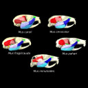
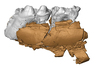
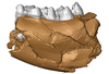
The present 3D Dataset contains the 3D models analyzed in the article entitled "One skull to rule them all? Descriptive and comparative anatomy of the masticatory apparatus in five mice species based on traditional and digital dissections" (Ginot et al. 2018, Journal of Morphology, https://doi.org/10.1002/jmor.20845).
Mus cervicolor R7314 View specimen
|
M3#343.ply surfaces of the skull and masticatory muscles of Mus cervicolor. Created with MorphoDig, .pos and .ntw files also included. Scans were obtained thanks to the Institut des Sciences de l'Evolution de Montpellier MRI platform. Type: "3D_surfaces"doi: 10.18563/m3.sf.343 state:published |
Download 3D surface file |
Mus caroli R7264 View specimen
|
M3#344.ply surfaces of the skull and masticatory muscles of Mus caroli. Created with MorphoDig, .pos and .ntw files also included. Scans were obtained thanks to the Institut des Sciences de l'Evolution de Montpellier MRI platform. Type: "3D_surfaces"doi: 10.18563/m3.sf.344 state:published |
Download 3D surface file |
Mus fragilicauda R7260 View specimen
|
M3#345.ply surfaces of the skull and masticatory muscles of Mus fragilicauda. Created with MorphoDig, .pos and .ntw files also included. Scans were obtained thanks to the Institut des Sciences de l'Evolution de Montpellier MRI platform. Type: "3D_surfaces"doi: 10.18563/m3.sf.345 state:published |
Download 3D surface file |
Mus pahari R7226 View specimen
|
M3#346.ply surfaces of the skull and masticatory muscles of Mus pahari. Created with MorphoDig, .pos and .ntw files also included. Scans were obtained thanks to the Institut des Sciences de l'Evolution de Montpellier MRI platform. Type: "3D_surfaces"doi: 10.18563/m3.sf.346 state:published |
Download 3D surface file |
Mus minutoides minutoides-1 View specimen
|
M3#347.ply surfaces of the skull and masticatory muscles of Mus minutoides. Created with MorphoDig, .pos and .ntw files also included. Scans were obtained thanks to the Institut des Sciences de l'Evolution de Montpellier MRI platform. Type: "3D_surfaces"doi: 10.18563/m3.sf.347 state:published |
Download 3D surface file |
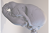
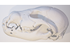
The present 3D Dataset contains the 3D models analyzed in the publication “Systematic and locomotor diversification of the Adapis group (Primates, Adapiformes) in the late Eocene of the Quercy (Southwest France), revealed by humeral remains”. In this paper, twenty humeral specimens from the old and new Quercy collections attributed to the fossil primates Adapis and Palaeolemur are described and analysed together. In this dataset only the scans of the fossils belonging to the collections of Université de Montpellier are provided.
In our paper (Marigó et al., 2019) we provide a qualitative and quantitative analysis of the different humeri, revealing that high variability is present within the “Adapis group” sample. Six different morphotypes are identified, confirming that what has often been called “Adapis parisiensis” is a mix of different species that present different locomotor adaptations.
Adapis sp. UM ROS 2-95 View specimen
|
M3#356Complete right humerus ROS 2-95 attributed to the Adapis group Type: "3D_surfaces"doi: 10.18563/m3.sf.356 state:published |
Download 3D surface file |
Adapis sp. UM ROS 2-536 View specimen
|
M3#357Proximal end of the right humerus ROS 2-536 attributed to the Adapis group Type: "3D_surfaces"doi: 10.18563/m3.sf.357 state:published |
Download 3D surface file |
Adapis sp. UM ROS 2-534 View specimen
|
M3#358Distal end of the left humerus ROS 2-534 attributed to the Adapis group Type: "3D_surfaces"doi: 10.18563/m3.sf.358 state:published |
Download 3D surface file |
Adapis sp. UM ROS 2-535 View specimen
|
M3#359Distal end of the left humerus ROS 2-535 attributed to the Adapis group Type: "3D_surfaces"doi: 10.18563/m3.sf.359 state:published |
Download 3D surface file |
Adapis sp. UM ROS 2-80 View specimen
|
M3#360Proximal end of the right humerus ROS 2-80 attributed to the Adapis group Type: "3D_surfaces"doi: 10.18563/m3.sf.360 state:published |
Download 3D surface file |
Adapis sp. UM ROS 2-79 View specimen
|
M3#361Distal end of the right humerus ROS 2-79 attributed to the Adapis group Type: "3D_surfaces"doi: 10.18563/m3.sf.361 state:published |
Download 3D surface file |
Adapis sp. UM ECA 1364 View specimen
|
M3#362Distal end of the left humerus ECA 1364 attributed to the Adapis group Type: "3D_surfaces"doi: 10.18563/m3.sf.362 state:published |
Download 3D surface file |
Adapis sp. UM ACQ-262 View specimen

|
M3#3733D model of ACQ 262. Humerus Type: "3D_surfaces"doi: 10.18563/m3.sf373 state:published |
Download 3D surface file |
This contribution contains the 3D models of the fossil remains (maxilla, dentary, and talus) attributed to Djebelemur martinezi, a ca. 50 Ma primate from Tunisia (Djebel Chambi), described and figured in the following publication: Marivaux et al. (2013), Djebelemur, a tiny pre-tooth-combed primate from the Eocene of Tunisia: a glimpse into the origin of crown strepsirhines. PLoS ONE. https://doi.org/10.1371/journal.pone.0080778
Djebelemur martinezi CBI-1-544 View specimen
|
M3#365CBI-1-544, left maxilla preserving P3-M3 and alveoli for P2 and C1 Type: "3D_surfaces"doi: 10.18563/m3.sf.365 state:published |
Download 3D surface file |
Djebelemur martinezi CBI-1-567 View specimen
|
M3#363Isolated left upper P4 Type: "3D_surfaces"doi: 10.18563/m3.sf.363 state:published |
Download 3D surface file |
Djebelemur martinezi CBI-1-565-577-587-580 View specimen
|
M3#366- CBI-1-565, a damaged right mandible, which consists of three isolated pieces found together and reassembled here: the anterior part of the dentary bears the p3 and m1, and alveoli for p4, p2 and c, while the posterior part preserves m3 and a portion of the ascending ramus; the m2 was found isolated but in the same small calcareous block treated by acid processing. - CBI-1-577, isolated right lower p4. - CBI-1-587, isolated left lower p2 (reversed). - CBI-1-580, isolated left lower canine (reversed). Type: "3D_surfaces"doi: 10.18563/m3.sf.366 state:published |
Download 3D surface file |
Djebelemur martinezi CBI-1-545 View specimen
|
M3#364Right Talus Type: "3D_surfaces"doi: 10.18563/m3.sf.364 state:published |
Download 3D surface file |
This contribution contains the 3D model of the fossil talus of a small-bodied anthropoid primate (Platyrrhini, Cebidae, Cebinae) discovered from lower Miocene deposits of Peruvian Amazonia (MD-61 locality, Upper Madre de Dios Basin). This fossil was described and figured in the following publication: Marivaux et al. (2012), A platyrrhine talus from the early Miocene of Peru (Amazonian Madre de Dios Sub-Andean Zone). Journal of Human Evolution. http://dx.doi.org/10.1016/j.jhevol.2012.07.005
Cebinae indet. sp. MUSM-2024 View specimen
|
M3#380Right talus 3D surface of a Miocene Cebinae indet. primate Type: "3D_surfaces"doi: 10.18563/m3.sf.380 state:published |
Download 3D surface file |
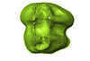
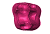
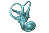
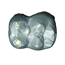
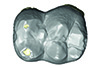
The present 3D Dataset contains the 3D models of the enamel-dentine junctions of upper third molars and of the bony labyrinths of the extant cercopithecoid specimens analyzed in the following publication: Beaudet, A., Dumoncel, J., Thackeray, J.F., Bruxelles, L., Duployer, B., Tenailleau, C., Bam, L., Hoffman, J., de Beer, F., Braga, J.: Upper third molar internal structural organization and semicircular canal morphology in Plio-Pleistocene South African cercopithecoids. Journal of Human Evolution 95, 104-120. https://doi.org/10.1016/j.jhevol.2016.04.004
Cercocebus atys 81.007-M-0041 View specimen
|
M3#4453D model of the enamel-dentine junction of the right upper third molar. Type: "3D_surfaces"doi: 10.18563/m3.sf.445 state:published |
Download 3D surface file |
Cercocebus torquatus 73.018-M-0359 View specimen
|
M3#4463D model of the enamel-dentine junction of the right upper third molar. Type: "3D_surfaces"doi: 10.18563/m3.sf.446 state:published |
Download 3D surface file |

|
M3#4963D model of the left bony labyrinth. Type: "3D_surfaces"doi: 10.18563/m3.sf.496 state:published |
Download 3D surface file |
Mandrillus leucophaeus 73.029-M-0106 View specimen
|
M3#4473D model of the enamel-dentine junction of the right upper third molar. Type: "3D_surfaces"doi: 10.18563/m3.sf.447 state:published |
Download 3D surface file |

|
M3#4703D model of the right bony labyrinth. Type: "3D_surfaces"doi: 10.18563/m3.sf.470 state:published |
Download 3D surface file |
Lophocebus albigena 73.029-M-0109 View specimen
|
M3#4483D model of the enamel-dentine junction of the right upper third molar. Type: "3D_surfaces"doi: 10.18563/m3.sf.448 state:published |
Download 3D surface file |
|
M3#4713D model of the right bony labyrinth. Type: "3D_surfaces"doi: 10.18563/m3.sf.471 state:published |
Download 3D surface file |
Piliocolobus foai 91.060-M-0071 View specimen
|
M3#4493D model of the enamel-dentine junction of the right upper third molar. Type: "3D_surfaces"doi: 10.18563/m3.sf.449 state:published |
Download 3D surface file |

|
M3#4723D model of the right bony labyrinth. Type: "3D_surfaces"doi: 10.18563/m3.sf.472 state:published |
Download 3D surface file |
Colobus guereza 1215 View specimen
|
M3#4503D model of the enamel-dentine junction of the right upper third molar. Type: "3D_surfaces"doi: 10.18563/m3.sf.450 state:published |
Download 3D surface file |

|
M3#4733D model of the right bony labyrinth. Type: "3D_surfaces"doi: 10.18563/m3.sf.473 state:published |
Download 3D surface file |
Colobus guereza 2800 View specimen
|
M3#4513D model of the enamel-dentine junction of the right upper third molar. Type: "3D_surfaces"doi: 10.18563/m3.sf.451 state:published |
Download 3D surface file |
|
M3#4743D model of the right bony labyrinth. Type: "3D_surfaces"doi: 10.18563/m3.sf.474 state:published |
Download 3D surface file |
Papio cynocephalus kindae 3503 View specimen
|
M3#4523D model of the enamel-dentine junction of the right upper third molar. Type: "3D_surfaces"doi: 10.18563/m3.sf.452 state:published |
Download 3D surface file |

|
M3#4753D model of the right bony labyrinth. Type: "3D_surfaces"doi: 10.18563/m3.sf.475 state:published |
Download 3D surface file |
Erythrocebus patas 8452 View specimen
|
M3#4533D model of the enamel-dentine junction of the right upper third molar. Type: "3D_surfaces"doi: 10.18563/m3.sf.453 state:published |
Download 3D surface file |

|
M3#4763D model of the right bony labyrinth. Type: "3D_surfaces"doi: 10.18563/m3.sf.476 state:published |
Download 3D surface file |
Papio cynocephalus kindae 17979 View specimen
|
M3#4543D model of the enamel-dentine junction of the right upper third molar. Type: "3D_surfaces"doi: 10.18563/m3.sf.454 state:published |
Download 3D surface file |

|
M3#4773D model of the right bony labyrinth. Type: "3D_surfaces"doi: 10.18563/m3.sf.477 state:published |
Download 3D surface file |
Colobus angolensis 25456 View specimen
|
M3#4553D model of the enamel-dentine junction of the right upper third molar. Type: "3D_surfaces"doi: 10.18563/m3.sf.455 state:published |
Download 3D surface file |

|
M3#4783D model of the right bony labyrinth. Type: "3D_surfaces"doi: 10.18563/m3.sf.478 state:published |
Download 3D surface file |
Chlorocebus pygerythrus 37477 View specimen
|
M3#4563D model of the enamel-dentine junction of the right upper third molar. Type: "3D_surfaces"doi: 10.18563/m3.sf.456 state:published |
Download 3D surface file |
|
M3#4813D model of the right bony labyrinth. Type: "3D_surfaces"doi: 10.18563/m3.sf.481 state:published |
Download 3D surface file |
Chlorocebus pygerythrus 37478 View specimen
|
M3#4573D model of the enamel-dentine junction of the right upper third molar. Type: "3D_surfaces"doi: 10.18563/m3.sf.457 state:published |
Download 3D surface file |

|
M3#4823D model of the right bony labyrinth. Type: "3D_surfaces"doi: 10.18563/m3.sf.482 state:published |
Download 3D surface file |
Lophocebus albigena 37572 View specimen
|
M3#4583D model of the enamel-dentine junction of the right upper third molar. Type: "3D_surfaces"doi: 10.18563/m3.sf.458 state:published |
Download 3D surface file |

|
M3#4833D model of the right bony labyrinth. Type: "3D_surfaces"doi: 10.18563/m3.sf.483 state:published |
Download 3D surface file |
Lophocebus albigena 37579 View specimen
|
M3#4593D model of the enamel-dentine junction of the right upper third molar. Type: "3D_surfaces"doi: 10.18563/m3.sf.459 state:published |
Download 3D surface file |
Erythrocebus patas OST.2002-26 View specimen
|
M3#4603D model of the enamel-dentine junction of the right upper third molar. Type: "3D_surfaces"doi: 10.18563/m3.sf.460 state:published |
Download 3D surface file |
|
M3#4843D model of the right bony labyrinth. Type: "3D_surfaces"doi: 10.18563/m3.sf.484 state:published |
Download 3D surface file |
Mandrillus sphinx OST.AC.488 View specimen
|
M3#4613D model of the enamel-dentine junction of the right upper third molar. Type: "3D_surfaces"doi: 10.18563/m3.sf.461 state:published |
Download 3D surface file |
|
M3#4853D model of the left bony labyrinth. Type: "3D_surfaces"doi: 10.18563/m3.sf.485 state:published |
Download 3D surface file |
Macaca mulatta OST.AC.492 View specimen
|
M3#4623D model of the enamel-dentine junction of the right upper third molar. Type: "3D_surfaces"doi: 10.18563/m3.sf.462 state:published |
Download 3D surface file |
|
M3#4863D model of the right bony labyrinth. Type: "3D_surfaces"doi: 10.18563/m3.sf.486 state:published |
Download 3D surface file |
Chlorocebus aethiops OST.AC.523 View specimen
|
M3#4633D model of the enamel-dentine junction of the right upper third molar. Type: "3D_surfaces"doi: 10.18563/m3.sf.463 state:published |
Download 3D surface file |
|
M3#4913D model of the right bony labyrinth. Type: "3D_surfaces"doi: 10.18563/m3.sf.491 state:published |
Download 3D surface file |
Cercopithecus cephus OST.AC.533 View specimen
|
M3#4643D model of the enamel-dentine junction of the right upper third molar. Type: "3D_surfaces"doi: 10.18563/m3.sf.464 state:published |
Download 3D surface file |
|
M3#4933D model of the right bony labyrinth. Type: "3D_surfaces"doi: 10.18563/m3.sf.493 state:published |
Download 3D surface file |
Chlorocebus aethiops OST.AC.540 View specimen
|
M3#4653D model of the enamel-dentine junction of the right upper third molar. Type: "3D_surfaces"doi: 10.18563/m3.sf.465 state:published |
Download 3D surface file |

|
M3#4943D model of the right bony labyrinth. Type: "3D_surfaces"doi: 10.18563/m3.sf.494 state:published |
Download 3D surface file |
Mandrillus sphinx OST.AC.543 View specimen
|
M3#4663D model of the enamel-dentine junction of the right upper third molar. Type: "3D_surfaces"doi: 10.18563/m3.sf.466 state:published |
Download 3D surface file |
|
M3#4953D model of the right bony labyrinth. Type: "3D_surfaces"doi: 10.18563/m3.sf.495 state:published |
Download 3D surface file |
Cercocebus torquatus 73.018-M-389 View specimen

|
M3#4683D model of the right bony labyrinth. Type: "3D_surfaces"doi: 10.18563/m3.sf.468 state:published |
Download 3D surface file |
Mandrillus leucophaeus 73.029-M-0105 View specimen
|
M3#4693D model of the right bony labyrinth. Type: "3D_surfaces"doi: 10.18563/m3.sf.469 state:published |
Download 3D surface file |
Mandrillus leucophaeus 28425 View specimen

|
M3#4793D model of the right bony labyrinth. Type: "3D_surfaces"doi: 10.18563/m3.sf.479 state:published |
Download 3D surface file |
Cercocebus atys 28998 View specimen
|
M3#4803D model of the right bony labyrinth. Type: "3D_surfaces"doi: 10.18563/m3.sf.480 state:published |
Download 3D surface file |
Macaca sylvanus OST.AC.493 View specimen

|
M3#4873D model of the right bony labyrinth. Type: "3D_surfaces"doi: 10.18563/m3.sf.487 state:published |
Download 3D surface file |
Chlorocebus aethiops OST.AC.508 View specimen

|
M3#4883D model of the left bony labyrinth. Type: "3D_surfaces"doi: 10.18563/m3.sf.488 state:published |
Download 3D surface file |
Cercopithecus cephus OST.AC.515 View specimen

|
M3#4893D model of the right bony labyrinth. Type: "3D_surfaces"doi: 10.18563/m3.sf.489 state:published |
Download 3D surface file |
Colobus guereza OST.AC.519 View specimen

|
M3#4903D model of the right bony labyrinth. Type: "3D_surfaces"doi: 10.18563/m3.sf.490 state:published |
Download 3D surface file |
Macaca sp. OST.AC.532 View specimen

|
M3#4923D model of the left bony labyrinth. Type: "3D_surfaces"doi: 10.18563/m3.sf.492 state:published |
Download 3D surface file |
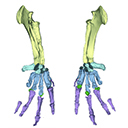
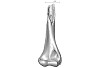
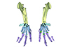
The present 3D Dataset contains the 3D model analyzed in Vautrin et al. (2019), Palaeontology, From limb to fin: an Eocene protocetid forelimb from Senegal sheds new light on the early locomotor evolution of early cetaceans.
?Carolinacetus indet. SNTB 2011-01 View specimen
|
M3#3983D model of an articulated forelimb of a Carolinacetus-like protocetid from Senegal Type: "3D_surfaces"doi: 10.18563/m3.sf.398 state:published |
Download 3D surface file |
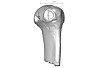
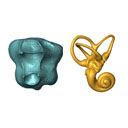
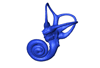
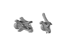
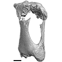
This contribution includes the 3D models of the reconstructed ossicular chain of the cainotheriid Caenomeryx filholi from the late Oligocene locality of Pech Desse (MP28, Quercy, France) described and figured in the publication of Assemat et al. (2020). It represents the oldest ossicular chain reconstruction for a Paleogene terrestrial artiodactyl species.
Caenomeryx filholi UM PDS 3353 View specimen
|
M3#508reconstruction of the middle ear with petrosal, bulla, stapes, incus, malleus Type: "3D_surfaces"doi: 10.18563/m3.sf.508 state:published |
Download 3D surface file |
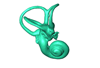
The present Dataset contains the 3D model of the male genital organs of greater horseshoe bat, Rhinolophus ferrumequinum. This is the first detailed 3D structure of the soft-tissue genital organs of bats. The 3D model was generated using microCT and techniques of virtual reconstruction.
Rhinolophus ferrumequinum JP18-006 View specimen
|
M3#521The genital organs of male greater horseshoe bat. Type: "3D_surfaces"doi: 10.18563/m3.sf.521 state:published |
Download 3D surface file |

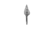
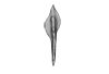
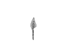
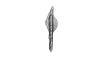
This contribution contains the 3D models of the set of Famennian conodont elements belonging to the species Polygnathus glaber and Polygnathus communis analyzed in the following publication: Renaud et al. 2021: Patterns of bilateral asymmetry and allometry in Late Devonian Polygnathus. Palaeontology. https://doi.org/10.1111/pala.12513
Polygnathus glaber UM BUS 001 View specimen
|
M3#574right P1 element Type: "3D_surfaces"doi: 10.18563/m3.sf.574 state:published |
Download 3D surface file |
Polygnathus glaber UM BUS 002 View specimen
|
M3#575right P1 element Type: "3D_surfaces"doi: 10.18563/m3.sf.575 state:published |
Download 3D surface file |
Polygnathus glaber UM BUS 003 View specimen

|
M3#576right P1 element Type: "3D_surfaces"doi: 10.18563/m3.sf.576 state:published |
Download 3D surface file |
Polygnathus glaber UM BUS 004 View specimen
|
M3#577left P1 element Type: "3D_surfaces"doi: 10.18563/m3.sf.577 state:published |
Download 3D surface file |
Polygnathus glaber UM BUS 005 View specimen

|
M3#578left P1 element Type: "3D_surfaces"doi: 10.18563/m3.sf.578 state:published |
Download 3D surface file |
Polygnathus glaber UM BUS 006 View specimen
|
M3#579right P1 element Type: "3D_surfaces"doi: 10.18563/m3.sf.579 state:published |
Download 3D surface file |
Polygnathus glaber UM BUS 007 View specimen
|
M3#580right P1 element Type: "3D_surfaces"doi: 10.18563/m3.sf.580 state:published |
Download 3D surface file |
Polygnathus glaber UM BUS 008 View specimen
|
M3#581left P1 element Type: "3D_surfaces"doi: 10.18563/m3.sf.581 state:published |
Download 3D surface file |
Polygnathus glaber UM BUS 009 View specimen
|
M3#582left P1 element Type: "3D_surfaces"doi: 10.18563/m3.sf.582 state:published |
Download 3D surface file |
Polygnathus glaber UM BUS 010 View specimen
|
M3#583right P1 element Type: "3D_surfaces"doi: 10.18563/m3.sf.583 state:published |
Download 3D surface file |
Polygnathus glaber UM BUS 011 View specimen

|
M3#584right P1 element Type: "3D_surfaces"doi: 10.18563/m3.sf.584 state:published |
Download 3D surface file |
Polygnathus glaber UM BUS 012 View specimen
|
M3#585right P1 element Type: "3D_surfaces"doi: 10.18563/m3.sf.585 state:published |
Download 3D surface file |
Polygnathus glaber UM BUS 013 View specimen

|
M3#586left P1 element Type: "3D_surfaces"doi: 10.18563/m3.sf.586 state:published |
Download 3D surface file |
Polygnathus glaber UM BUS 014 View specimen
|
M3#587left P1 element Type: "3D_surfaces"doi: 10.18563/m3.sf.587 state:published |
Download 3D surface file |
Polygnathus glaber UM BUS 015 View specimen

|
M3#588left P1 element Type: "3D_surfaces"doi: 10.18563/m3.sf.588 state:published |
Download 3D surface file |
Polygnathus glaber UM BUS 016 View specimen
|
M3#589right P1 element Type: "3D_surfaces"doi: 10.18563/m3.sf.589 state:published |
Download 3D surface file |
Polygnathus glaber UM BUS 017 View specimen

|
M3#590left P1 element Type: "3D_surfaces"doi: 10.18563/m3.sf.590 state:published |
Download 3D surface file |
Polygnathus glaber UM BUS 018 View specimen

|
M3#591left P1 element Type: "3D_surfaces"doi: 10.18563/m3.sf.591 state:published |
Download 3D surface file |
Polygnathus glaber UM BUS 019 View specimen
|
M3#592left P1 element Type: "3D_surfaces"doi: 10.18563/m3.sf.592 state:published |
Download 3D surface file |
Polygnathus glaber UM BUS 020 View specimen

|
M3#593left P1 element Type: "3D_surfaces"doi: 10.18563/m3.sf.593 state:published |
Download 3D surface file |
Polygnathus glaber UM BUS 021 View specimen

|
M3#594right P1 element Type: "3D_surfaces"doi: 10.18563/m3.sf.594 state:published |
Download 3D surface file |
Polygnathus glaber UM BUS 022 View specimen
|
M3#595left P1 element Type: "3D_surfaces"doi: 10.18563/m3.sf.595 state:published |
Download 3D surface file |
Polygnathus glaber UM BUS 023 View specimen

|
M3#596left P1 element Type: "3D_surfaces"doi: 10.18563/m3.sf.596 state:published |
Download 3D surface file |
Polygnathus glaber UM BUS 024 View specimen
|
M3#597left P1 element Type: "3D_surfaces"doi: 10.18563/m3.sf.597 state:published |
Download 3D surface file |
Polygnathus glaber UM BUS 025 View specimen

|
M3#598left P1 element Type: "3D_surfaces"doi: 10.18563/m3.sf.598 state:published |
Download 3D surface file |
Polygnathus glaber UM BUS 026 View specimen

|
M3#599left P1 element Type: "3D_surfaces"doi: 10.18563/m3.sf.599 state:published |
Download 3D surface file |
Polygnathus glaber UM BUS 027 View specimen

|
M3#600right P1 element Type: "3D_surfaces"doi: 10.18563/m3.sf.600 state:published |
Download 3D surface file |
Polygnathus glaber UM BUS 028 View specimen
|
M3#601right P1 element Type: "3D_surfaces"doi: 10.18563/m3.sf.601 state:published |
Download 3D surface file |
Polygnathus glaber UM BUS 029 View specimen

|
M3#602right P1 element Type: "3D_surfaces"doi: 10.18563/m3.sf.602 state:published |
Download 3D surface file |
Polygnathus glaber UM BUS 030 View specimen
|
M3#603right P1 element Type: "3D_surfaces"doi: 10.18563/m3.sf.603 state:published |
Download 3D surface file |
Polygnathus communis UM CTB 001 View specimen
|
M3#604right P1 element Type: "3D_surfaces"doi: 10.18563/m3.sf.604 state:published |
Download 3D surface file |
Polygnathus communis UM CTB 002 View specimen

|
M3#605right P1 element Type: "3D_surfaces"doi: 10.18563/m3.sf.605 state:published |
Download 3D surface file |
Polygnathus communis UM CTB 003 View specimen
|
M3#606right P1 element Type: "3D_surfaces"doi: 10.18563/m3.sf.606 state:published |
Download 3D surface file |
Polygnathus communis UM CTB 004 View specimen

|
M3#607right P1 element Type: "3D_surfaces"doi: 10.18563/m3.sf.607 state:published |
Download 3D surface file |
Polygnathus communis UM CTB 005 View specimen
|
M3#608left P1 element Type: "3D_surfaces"doi: 10.18563/m3.sf.608 state:published |
Download 3D surface file |
Polygnathus communis UM CTB 006 View specimen
|
M3#609left P1 element Type: "3D_surfaces"doi: 10.18563/m3.sf.609 state:published |
Download 3D surface file |
Polygnathus communis UM CTB 007 View specimen

|
M3#610left P1 element Type: "3D_surfaces"doi: 10.18563/m3.sf.610 state:published |
Download 3D surface file |
Polygnathus communis UM CTB 008 View specimen
|
M3#611left P1 element Type: "3D_surfaces"doi: 10.18563/m3.sf.611 state:published |
Download 3D surface file |
Polygnathus communis UM CTB 009 View specimen

|
M3#612right P1 element Type: "3D_surfaces"doi: 10.18563/m3.sf.612 state:published |
Download 3D surface file |
Polygnathus communis UM CTB 010 View specimen

|
M3#613left P1 element Type: "3D_surfaces"doi: 10.18563/m3.sf.613 state:published |
Download 3D surface file |
Polygnathus communis UM CTB 011 View specimen

|
M3#614right P1 element Type: "3D_surfaces"doi: 10.18563/m3.sf.614 state:published |
Download 3D surface file |
Polygnathus communis UM CTB 012 View specimen

|
M3#615right P1 element Type: "3D_surfaces"doi: 10.18563/m3.sf.615 state:published |
Download 3D surface file |
Polygnathus communis UM CTB 013 View specimen

|
M3#616right P1 element Type: "3D_surfaces"doi: 10.18563/m3.sf.616 state:published |
Download 3D surface file |
Polygnathus communis UM CTB 014 View specimen

|
M3#617right P1 element Type: "3D_surfaces"doi: 10.18563/m3.sf.617 state:published |
Download 3D surface file |
Polygnathus communis UM CTB 015 View specimen

|
M3#618right P1 element Type: "3D_surfaces"doi: 10.18563/m3.sf.618 state:published |
Download 3D surface file |
Polygnathus communis UM CTB 016 View specimen

|
M3#619left P1 element Type: "3D_surfaces"doi: 10.18563/m3.sf.619 state:published |
Download 3D surface file |
Polygnathus communis UM CTB 017 View specimen

|
M3#620right P1 element Type: "3D_surfaces"doi: 10.18563/m3.sf.620 state:published |
Download 3D surface file |
Polygnathus communis UM CTB 018 View specimen

|
M3#621right P1 element Type: "3D_surfaces"doi: 10.18563/m3.sf.621 state:published |
Download 3D surface file |
Polygnathus communis UM CTB 019 View specimen

|
M3#622right P1 element Type: "3D_surfaces"doi: 10.18563/m3.sf.622 state:published |
Download 3D surface file |
Polygnathus communis UM CTB 020 View specimen

|
M3#623right P1 element Type: "3D_surfaces"doi: 10.18563/m3.sf.623 state:published |
Download 3D surface file |
Polygnathus communis UM CTB 021 View specimen

|
M3#624left P1 element Type: "3D_surfaces"doi: 10.18563/m3.sf.624 state:published |
Download 3D surface file |
Polygnathus communis UM CTB 022 View specimen

|
M3#625left element Type: "3D_surfaces"doi: 10.18563/m3.sf.625 state:published |
Download 3D surface file |
Polygnathus communis UM CTB 023 View specimen

|
M3#626left P1 element Type: "3D_surfaces"doi: 10.18563/m3.sf.626 state:published |
Download 3D surface file |
Polygnathus communis UM CTB 024 View specimen

|
M3#627left P1 element Type: "3D_surfaces"doi: 10.18563/m3.sf.627 state:published |
Download 3D surface file |
Polygnathus communis UM CTB 025 View specimen

|
M3#628left P1 element Type: "3D_surfaces"doi: 10.18563/m3.sf.628 state:published |
Download 3D surface file |
Polygnathus communis UM CTB 026 View specimen

|
M3#629left P1 element Type: "3D_surfaces"doi: 10.18563/m3.sf.629 state:published |
Download 3D surface file |
Polygnathus communis UM CTB 027 View specimen

|
M3#630left P1 element Type: "3D_surfaces"doi: 10.18563/m3.sf.630 state:published |
Download 3D surface file |
Polygnathus communis UM CTB 028 View specimen

|
M3#631left P1 element Type: "3D_surfaces"doi: 10.18563/m3.sf.631 state:published |
Download 3D surface file |
Polygnathus communis UM CTB 029 View specimen
|
M3#632left P1 element Type: "3D_surfaces"doi: 10.18563/m3.sf.632 state:published |
Download 3D surface file |
Polygnathus communis UM CTB 030 View specimen

|
M3#633left P1 element Type: "3D_surfaces"doi: 10.18563/m3.sf.633 state:published |
Download 3D surface file |
Polygnathus communis UM CTB 031 View specimen
|
M3#634left P1 element Type: "3D_surfaces"doi: 10.18563/m3.sf.634 state:published |
Download 3D surface file |
Polygnathus communis UM CTB 032 View specimen
|
M3#635left P1 element Type: "3D_surfaces"doi: 10.18563/m3.sf.635 state:published |
Download 3D surface file |
Polygnathus communis UM CTB 033 View specimen

|
M3#636left P1 element Type: "3D_surfaces"doi: 10.18563/m3.sf.636 state:published |
Download 3D surface file |
Polygnathus communis UM CTB 034 View specimen

|
M3#637right P1 element Type: "3D_surfaces"doi: 10.18563/m3.sf.637 state:published |
Download 3D surface file |
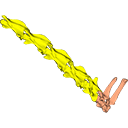
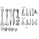
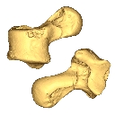
This contribution contains the 3D models analyzed in Müller et al. (2021) “Pushing the boundary? Testing the ‘functional elongation hypothesis’ of the giraffe’s neck”.
Aepyceros melampus ZFMK 2001.278 View specimen

|
M3#643Vertebrae C7, T1 Type: "3D_surfaces"doi: 10.18563/m3.sf.643 state:published |
Download 3D surface file |
Giraffa camelopardalis ZMB 66393 View specimen

|
M3#644Vertebrae Type: "3D_surfaces"doi: 10.18563/m3.sf.644 state:published |
Download 3D surface file |
Giraffa camelopardalis ZSM 1967/17 View specimen

|
M3#645Vertebrae Type: "3D_surfaces"doi: 10.18563/m3.sf.645 state:published |
Download 3D surface file |
Giraffa camelopardalis ZSM 1981/19 View specimen
|
M3#646C3, C4, C5, C6, C7, T1, T2 Type: "3D_surfaces"doi: 10.18563/m3.sf.646 state:published |
Download 3D surface file |
Giraffa camelopardalis KMDA M-10861 View specimen

|
M3#647C3, C4, C5, C6, C7, T1, T2. Acquired via laser scanner. Type: "3D_surfaces"doi: 10.18563/m3.sf.647 state:published |
Download 3D surface file |
Giraffa camelopardalis SMF 84214 View specimen

|
M3#648C7, T1. Warning : photogrammetric models (unit scale is CM, not MM). Type: "3D_surfaces"doi: 10.18563/m3.sf.648 state:published |
Download 3D surface file |
Giraffa camelopardalis SMF 78299 View specimen

|
M3#649C7, T1. Warning : unscaled photogrammetric 3D models (unknown size). Type: "3D_surfaces"doi: 10.18563/m3.sf.649 state:published |
Download 3D surface file |
Giraffa camelopardalis SMF o. N View specimen

|
M3#650C7, T1. Warning : unscaled photogrammetric 3D models (unknown size). Type: "3D_surfaces"doi: 10.18563/m3.sf.650 state:published |
Download 3D surface file |
Giraffa camelopardalis SMNS 19138 View specimen

|
M3#671C7, T1. Warning : unscaled photogrammetric 3D models (unknown size). Type: "3D_surfaces"doi: 10.18563/m3.sf.671 state:published |
Download 3D surface file |
Okapia johnstoni ZMB 62086 View specimen

|
M3#651C3, C4, C5, C6, C7, T1, T2 Type: "3D_surfaces"doi: 10.18563/m3.sf.651 state:published |
Download 3D surface file |
Okapia johnstoni ZMB 70325 View specimen

|
M3#652C3, C4, C5, C6, C7, T1, T2 Type: "3D_surfaces"doi: 10.18563/m3.sf.652 state:published |
Download 3D surface file |
Sivatherium giganteum NHMUK 15707 View specimen
|
M3#653C7. Warning : unscaled photogrammetric 3D model (unknown size). Type: "3D_surfaces"doi: 10.18563/m3.sf.653 state:published |
Download 3D surface file |
Sivatherium giganteum NHMUK 15297 View specimen

|
M3#654T1. Warning : unscaled photogrammetric 3D model (unknown size). Type: "3D_surfaces"doi: 10.18563/m3.sf.654 state:published |
Download 3D surface file |
Cervus elaphus ZMB 47502 View specimen

|
M3#655C3, C4, C5, C6, C7, T1, T2 Type: "3D_surfaces"doi: 10.18563/m3.sf.655 state:published |
Download 3D surface file |
Axis axis SMF 1450 View specimen

|
M3#656C7, T1 Type: "3D_surfaces"doi: 10.18563/m3.sf.656 state:published |
Download 3D surface file |
Cervus nippon SMF 4368 View specimen

|
M3#657C7, T1 Type: "3D_surfaces"doi: 10.18563/m3.sf.657 state:published |
Download 3D surface file |
Capreolus capreolus SMF 79852 View specimen

|
M3#658C7, T1 Type: "3D_surfaces"doi: 10.18563/m3.sf.658 state:published |
Download 3D surface file |
Capreolus capreolus ZFMK 67.237 View specimen

|
M3#659C7, T1 Type: "3D_surfaces"doi: 10.18563/m3.sf.659 state:published |
Download 3D surface file |
Muntiacus reevesi SMF 92954 View specimen

|
M3#660C7, T1 Type: "3D_surfaces"doi: 10.18563/m3.sf.660 state:published |
Download 3D surface file |
Muntiacus reevesi SMF 92332 View specimen

|
M3#661C7, T1 Type: "3D_surfaces"doi: 10.18563/m3.sf.661 state:published |
Download 3D surface file |
Alces alces SMF 35549 View specimen

|
M3#662C7, T1 Type: "3D_surfaces"doi: 10.18563/m3.sf.662 state:published |
Download 3D surface file |
Dama dama ZFMK 86.125 View specimen

|
M3#663C7, T1 Type: "3D_surfaces"doi: 10.18563/m3.sf.663 state:published |
Download 3D surface file |
Antilope cervicapra ZMB 78829 View specimen

|
M3#664C3, C4, C5, C6, C7, T1, T2 Type: "3D_surfaces"doi: 10.18563/m3.sf.664 state:published |
Download 3D surface file |
Bison bonasus SMNS 2998 View specimen

|
M3#665C7, T1. Warning : unscaled photogrammetric 3D models (unknown size). Type: "3D_surfaces"doi: 10.18563/m3.sf.665 state:published |
Download 3D surface file |
Nanger dama SMF 74435 View specimen

|
M3#666C7, T1 Type: "3D_surfaces"doi: 10.18563/m3.sf.666 state:published |
Download 3D surface file |
Litocranius walleri SMF 23747 View specimen

|
M3#667C7, T1 Type: "3D_surfaces"doi: 10.18563/m3.sf.667 state:published |
Download 3D surface file |
Litocranius walleri SMF 23749 View specimen

|
M3#669C7, T1 Type: "3D_surfaces"doi: 10.18563/m3.sf.669 state:published |
Download 3D surface file |
Tragelaphus eurycerus SMF 95875 View specimen

|
M3#670C7, T1 Type: "3D_surfaces"doi: 10.18563/m3.sf.670 state:published |
Download 3D surface file |
Bos javanicus SMF 64934 View specimen

|
M3#672C7, T1 Type: "3D_surfaces"doi: 10.18563/m3.sf.672 state:published |
Download 3D surface file |
Ovis aries musimon ZFMK 1982.338 View specimen

|
M3#673C7, T1 Type: "3D_surfaces"doi: 10.18563/m3.sf.673 state:published |
Download 3D surface file |
Rupicapra rupicapra ZFMK 72.367 View specimen

|
M3#674C7, T1 Type: "3D_surfaces"doi: 10.18563/m3.sf.674 state:published |
Download 3D surface file |
Kobus ellipsiprymnus SMNS 4443 View specimen

|
M3#675C7, T1 Type: "3D_surfaces"doi: 10.18563/m3.sf.675 state:published |
Download 3D surface file |
Sylvicapra grimmia SMNS 15292 View specimen

|
M3#676C7, T1 Type: "3D_surfaces"doi: 10.18563/m3.sf.676 state:published |
Download 3D surface file |
Syncerus caffer SMNS 7347 View specimen

|
M3#677C7, T1. Warning : unscaled photogrammetric 3D models (unknown size). Type: "3D_surfaces"doi: 10.18563/m3.sf.677 state:published |
Download 3D surface file |
Procapra gutturosa SMNS 5796 View specimen

|
M3#678C7, T1 Type: "3D_surfaces"doi: 10.18563/m3.sf.678 state:published |
Download 3D surface file |
Damaliscus pygargus SMNS 21617 View specimen

|
M3#679C7, T1 Type: "3D_surfaces"doi: 10.18563/m3.sf.679 state:published |
Download 3D surface file |
Madoqua kirkii SMNS 4432 View specimen

|
M3#680C7, T1 Type: "3D_surfaces"doi: 10.18563/m3.sf.680 state:published |
Download 3D surface file |
Bubalus mindorensis SMNS 2054 View specimen

|
M3#681C7, T1. Warning : unscaled photogrammetric 3D models (unknown size). Type: "3D_surfaces"doi: 10.18563/m3.sf.681 state:published |
Download 3D surface file |
Capra hircus dom. SMNS 51328 View specimen

|
M3#682C7, T1 Type: "3D_surfaces"doi: 10.18563/m3.sf.682 state:published |
Download 3D surface file |
Connochaetes taurinus SMNS 4442 View specimen

|
M3#683C7, T1. Warning : unscaled photogrammetric 3D models (unknown size). Type: "3D_surfaces"doi: 10.18563/m3.sf.683 state:published |
Download 3D surface file |
Antilocapra americana ZSM 1964/218 View specimen

|
M3#684C3, C4, C5, C6, C7, T1, T2 Type: "3D_surfaces"doi: 10.18563/m3.sf.684 state:published |
Download 3D surface file |
Antilocapra americana ZMB 77281 View specimen

|
M3#685C7, T1 Type: "3D_surfaces"doi: 10.18563/m3.sf.685 state:published |
Download 3D surface file |
Moschus moschiferus ZMB 62080 View specimen

|
M3#686C3, C4, C5, C6, C7, T1, T2 Type: "3D_surfaces"doi: 10.18563/m3.sf.686 state:published |
Download 3D surface file |
Moschus moschiferus ZMB 60367 View specimen

|
M3#687C7, T1 Type: "3D_surfaces"doi: 10.18563/m3.sf.687 state:published |
Download 3D surface file |
Moschus moschiferus ZMB 51830 View specimen

|
M3#688C7, T1 Type: "3D_surfaces"doi: 10.18563/m3.sf.688 state:published |
Download 3D surface file |
Tragulus javanicus SMF 82179 View specimen

|
M3#689C7, T1 Type: "3D_surfaces"doi: 10.18563/m3.sf.689 state:published |
Download 3D surface file |
Tragulus javanicus ZMB 86222 View specimen

|
M3#690C7, T1 Type: "3D_surfaces"doi: 10.18563/m3.sf.690 state:published |
Download 3D surface file |
Tragulus sp. ZMB o. N. View specimen

|
M3#691C7, T1 Type: "3D_surfaces"doi: 10.18563/m3.sf.691 state:published |
Download 3D surface file |
Hyemoschus aquaticus ZMB 71071 View specimen

|
M3#692C7, T1 Type: "3D_surfaces"doi: 10.18563/m3.sf.692 state:published |
Download 3D surface file |
Hyemoschus aquaticus ZMB 103235 View specimen

|
M3#693C7, T1 Type: "3D_surfaces"doi: 10.18563/m3.sf.693 state:published |
Download 3D surface file |
Vicugna vicugna SMF 94752 View specimen

|
M3#694C7, T1 Type: "3D_surfaces"doi: 10.18563/m3.sf.694 state:published |
Download 3D surface file |
Camelus dromedarius SMF 70473 View specimen

|
M3#695C7, T1. Warning : unscaled photogrammetric 3D models (unknown size). Type: "3D_surfaces"doi: 10.18563/m3.sf.695 state:published |
Download 3D surface file |
Camelus bactrianus SMF 25542 View specimen

|
M3#696C7, T1. Warning : unscaled photogrammetric 3D models (unknown size). Type: "3D_surfaces"doi: 10.18563/m3.sf.696 state:published |
Download 3D surface file |
Lama glama SMNS 31175 View specimen

|
M3#697C7, T1 Type: "3D_surfaces"doi: 10.18563/m3.sf.697 state:published |
Download 3D surface file |
Vicugna pacos SMNS 46255 View specimen

|
M3#698C7, T1 Type: "3D_surfaces"doi: 10.18563/m3.sf.698 state:published |
Download 3D surface file |
Vicugna pacos SMNS 7349 View specimen

|
M3#699C7, T1 Type: "3D_surfaces"doi: 10.18563/m3.sf.699 state:published |
Download 3D surface file |
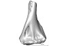
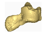
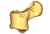
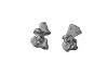
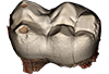
The present 3D Dataset contains the 3D models analyzed in Mennecart B., Métais G., Costeur L., Ginsburg L, and Rössner G. 2021, Reassessment of the enigmatic ruminant Miocene genus Amphimoschus Bourgeois, 1873 (Mammalia, Artiodactyla, Pecora). PlosOne. https://doi.org/10.1371/journal.pone.0244661
Amphimoschus ponteleviensis MNHN.F.AR3266 View specimen
|
M3#701Surface scan of the cast of the skull of Amphimoschus ponteleviensis MNHN.F.AR3266 from Artenay (France) Type: "3D_surfaces"doi: 10.18563/m3.sf.701 state:published |
Download 3D surface file |
|
M3#702Right petrosal bone and bony labyrinth of the skull MNHN.F.AR3266 from Artenay (France) Type: "3D_surfaces"doi: 10.18563/m3.sf.702 state:published |
Download 3D surface file |
Amphimoschus ponteleviensis SMNS40693 View specimen
|
M3#704Left petrosal bone and bony labyrinth of the skull SMNS40693 from Langenau 1 (Germany) Type: "3D_surfaces"doi: 10.18563/m3.sf.704 state:published |
Download 3D surface file |
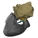
The present 3D Dataset contains the 3D models of the endocranial cast of two specimens of Indohyus indirae described in the article entitled “The endocranial cast of Indohyus (Artiodactyla, Raoellidae): the origin of the cetacean brain” (Orliac and Thewissen, 2021). They represent the cast of the main cavity of the braincase as well as associated intraosseous sinuses.
Indohyus indirae RR 207 View specimen
|
M3#710cast of the main endocranial cavity and associated intraosseous sinuses Type: "3D_surfaces"doi: 10.18563/m3.sf.710 state:published |
Download 3D surface file |
Indohyus indirae RR 601 View specimen
|
M3#711casts of the main endocranial cavity Type: "3D_surfaces"doi: 10.18563/m3.sf.711 state:published |
Download 3D surface file |
This contribution contains 3D models of upper molar rows of house mice (Mus musculus domesticus). The erupted part of the right row is presented for specimens belonging to four groups: wild-trapped mice, wild-derived lab offspring, a typical laboratory strain (Swiss) and hybrids between wild-derived and Swiss mice. These models are analyzed in the following publication: Savriama et al 2021: Wild versus lab house mice: Effects of age, diet, and genetics on molar geometry and topography. https://doi.org/10.1111/joa.13529
Mus musculus BW_03 View specimen

|
M3#736BW_03 Type: "3D_surfaces"doi: 10.18563/m3.sf.736 state:published |
Download 3D surface file |
Mus musculus BW_04 View specimen

|
M3#752BW_04 Type: "3D_surfaces"doi: 10.18563/m3.sf.752 state:published |
Download 3D surface file |
Mus musculus BW_06 View specimen

|
M3#753BW_06 Type: "3D_surfaces"doi: 10.18563/m3.sf.753 state:published |
Download 3D surface file |
Mus musculus BW_07 View specimen

|
M3#754BW_07 Type: "3D_surfaces"doi: 10.18563/m3.sf.754 state:published |
Download 3D surface file |
Mus musculus BW_08 View specimen

|
M3#755BW_08 Type: "3D_surfaces"doi: 10.18563/m3.sf.755 state:published |
Download 3D surface file |
Mus musculus BW_11 View specimen

|
M3#756BW_11 Type: "3D_surfaces"doi: 10.18563/m3.sf.756 state:published |
Download 3D surface file |
Mus musculus BW_12 View specimen

|
M3#757BW_12 Type: "3D_surfaces"doi: 10.18563/m3.sf.757 state:published |
Download 3D surface file |
Mus musculus Blab_035 View specimen

|
M3#758Blab_035 Type: "3D_surfaces"doi: 10.18563/m3.sf.758 state:published |
Download 3D surface file |
Mus musculus Blab_046 View specimen

|
M3#759Blab_046 Type: "3D_surfaces"doi: 10.18563/m3.sf.759 state:published |
Download 3D surface file |
Mus musculus Blab_054 View specimen

|
M3#760Blab_054 Type: "3D_surfaces"doi: 10.18563/m3.sf.760 state:published |
Download 3D surface file |
Mus musculus Blab_056 View specimen

|
M3#761Blab_056 Type: "3D_surfaces"doi: 10.18563/m3.sf.761 state:published |
Download 3D surface file |
Mus musculus Blab_082 View specimen

|
M3#762Blab_082 Type: "3D_surfaces"doi: 10.18563/m3.sf.762 state:published |
Download 3D surface file |
Mus musculus Blab_086 View specimen

|
M3#763Blab_086 Type: "3D_surfaces"doi: 10.18563/m3.sf.763 state:published |
Download 3D surface file |
Mus musculus Blab_092 View specimen

|
M3#764Blab_092 Type: "3D_surfaces"doi: 10.18563/m3.sf.764 state:published |
Download 3D surface file |
Mus musculus Blab_319 View specimen

|
M3#751Blab_319 Type: "3D_surfaces"doi: 10.18563/m3.sf.751 state:published |
Download 3D surface file |
Mus musculus Blab_325 View specimen

|
M3#750Blab_325 Type: "3D_surfaces"doi: 10.18563/m3.sf.750 state:published |
Download 3D surface file |
Mus musculus Blab_329 View specimen

|
M3#737Blab_329 Type: "3D_surfaces"doi: 10.18563/m3.sf.737 state:published |
Download 3D surface file |
Mus musculus Blab_330 View specimen

|
M3#738Blab_330 Type: "3D_surfaces"doi: 10.18563/m3.sf.738 state:published |
Download 3D surface file |
Mus musculus Blab_F2a View specimen

|
M3#739Blab_F2a Type: "3D_surfaces"doi: 10.18563/m3.sf.739 state:published |
Download 3D surface file |
Mus musculus Blab_F2b View specimen

|
M3#740Blab_F2b Type: "3D_surfaces"doi: 10.18563/m3.sf.740 state:published |
Download 3D surface file |
Mus musculus Blab_BB3w View specimen

|
M3#741Blab_BB3w Type: "3D_surfaces"doi: 10.18563/m3.sf.741 state:published |
Download 3D surface file |
Mus musculus hyb_BS01 View specimen

|
M3#742hyb_BS01 Type: "3D_surfaces"doi: 10.18563/m3.sf.742 state:published |
Download 3D surface file |
Mus musculus hyb_BS02 View specimen

|
M3#743hyb_BS02 Type: "3D_surfaces"doi: 10.18563/m3.sf.743 state:published |
Download 3D surface file |
Mus musculus hyb_SB01 View specimen

|
M3#744hyb_SB01 Type: "3D_surfaces"doi: 10.18563/m3.sf.744 state:published |
Download 3D surface file |
Mus musculus hyb_SB02 View specimen

|
M3#745hyb_SB02 Type: "3D_surfaces"doi: 10.18563/m3.sf.745 state:published |
Download 3D surface file |
Mus musculus SW_001 View specimen

|
M3#746SW_001 Type: "3D_surfaces"doi: 10.18563/m3.sf.746 state:published |
Download 3D surface file |
Mus musculus SW_002 View specimen
|
M3#747SW_002 Type: "3D_surfaces"doi: 10.18563/m3.sf.747 state:published |
Download 3D surface file |
Mus musculus SW_005 View specimen

|
M3#748SW_005 Type: "3D_surfaces"doi: 10.18563/m3.sf.748 state:published |
Download 3D surface file |
Mus musculus SW_0ter View specimen

|
M3#749SW_0ter Type: "3D_surfaces"doi: 10.18563/m3.sf.749 state:published |
Download 3D surface file |
Mus musculus SW_343 View specimen

|
M3#765SW_343 Type: "3D_surfaces"doi: 10.18563/m3.sf.765 state:published |
Download 3D surface file |
The present 3D Dataset contains the 3D models analyzed in Mennecart B., Wazir W.A., Sehgal R.K., Patnaik R., Singh N.P., Kumar N, and Nanda A.C. 2021. New remains of Nalamaeryx (Tragulidae, Mammalia) from the Ladakh Himalaya and their phylogenetical and palaeoenvironmental implications. Historical Biology. https://doi.org/10.1080/08912963.2021.2014479
Nalameryx savagei WIMF/A4801 View specimen
|
M3#766Nalameryx savagei, Partial lower right jaw preserving m2 and m3. Type: "3D_surfaces"doi: 10.18563/m3.sf.766 state:published |
Download 3D surface file |
Nalameryx savagei WIMF/A4802 View specimen
|
M3#767Nalameryx savagei, partial lower right jaw preserving m2 and m3 Type: "3D_surfaces"doi: 10.18563/m3.sf.767 state:published |
Download 3D surface file |
This contribution contains the 3D models described and figured in the following publication: Hautier L, Tabuce R, Kassegne KE, Amoudji YZ, Mourlam M, Orliac M, Quillévéré F, Charruault A-L, Johnson AKC, Guinot G. 2021. New middle Eocene proboscidean from Togo illuminates the early evolution of the elephantiform-like dental pattern.
Dagbatitherium tassyi ULDG-DAG1 View specimen
|
M3#7693D model of a molar of Dagbatitherium tassyi. Type: "3D_surfaces"doi: 10.18563/m3.sf.769 state:published |
Download 3D surface file |
|
M3#771µCT scan of a molar of Dagbatitherium tassyi. Type: "3D_CT"doi: 10.18563/m3.sf.771 state:published |
Download CT data |
The present 3D Dataset contains the 3D model analyzed in Presence of the ground sloth Valgipes bucklandi (Xenarthra, Folivora, Scelidotheriinae) in southern Uruguay during the Late Pleistocene: Ecological and biogeographical implications. Quaternary International. https://doi.org/10.1016/j.quaint.2021.06.011
Valgipes bucklandi CAV 1573 View specimen
|
M3#797Left tibia-fibula Type: "3D_surfaces"doi: 10.18563/m3.sf.797 state:published |
Download 3D surface file |
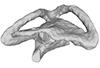
The present 3D Dataset contains the 3D models of Carboniferous-Permian chondrichthyan neurocrania analyzed in “Phylogenetic implications of the systematic reassessment of Xenacanthiformes and ‘Ctenacanthiformes’ (Chondrichthyes) neurocrania from the Carboniferous-Permian Autun Basin (France)”.
cf. Triodus sp MNHN.F.AUT811 View specimen
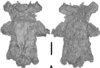
|
M3#834MHNH.F.AUT811 (isolated neurocranium) in dorsal view. Type: "3D_surfaces"doi: 10.18563/m3.sf.834 state:published |
Download 3D surface file |
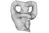
indet indet MNHN.F.AUT812 View specimen
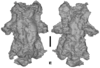
|
M3#835MHNH.F.AUT812 (isolated neurocranium) in dorsal view. Type: "3D_surfaces"doi: 10.18563/m3.sf.835 state:published |
Download 3D surface file |
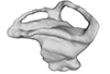
indet indet MNHN.F.AUT813 View specimen
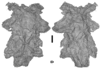
|
M3#836MHNH.F.AUT813 (isolated neurocranium) in dorsal view. Type: "3D_surfaces"doi: 10.18563/m3.sf.836 state:published |
Download 3D surface file |
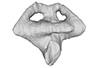
cf. Triodus sp MNHN.F.AUT814 View specimen
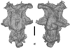
|
M3#837MHNH.F.AUT814 (isolated neurocranium) in dorsal view. Type: "3D_surfaces"doi: 10.18563/m3.sf.837 state:published |
Download 3D surface file |
cf. Triodus sp MHNE.2021.9.1 View specimen
|
M3#838MHNE.2021.9.1 (isolated neurocranium) in dorsal view. Type: "3D_surfaces"doi: 10.18563/m3.sf.838 state:published |
Download 3D surface file |


