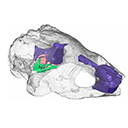
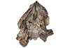
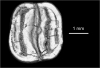
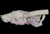
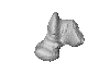
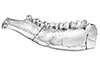
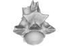
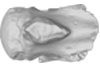
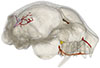
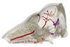
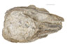
Holotype of Hamadasuchus rebouli
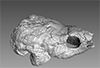
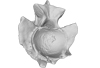
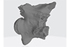
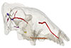
3D model of the holotype specimen of Pebanista yacuruna
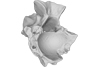
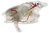
3D models of Eocene–Miocene anuran fossils from Peruvian Amazonia
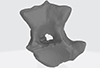
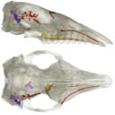
3D GM dataset of bird skeletal variation
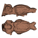
Skeletal embryonic development in the catshark
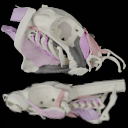
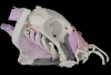
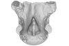
Bony connexions of the petrosal bone of extant hippos
bony labyrinth (11) , inner ear (10) , South America (8) , Eocene (8) , skull (7) , brain (6) , Oligocene (6)
Maëva Judith Orliac (17) , Lionel Hautier (17) , Bastien Mennecart (12) , Laurent Marivaux (11) , Pierre-Olivier Antoine (11) , Leonardo Kerber (10) , Renaud Lebrun (9)
|
3D model related to the publication: The endocranial anatomy of the stem turtle Naomichelys speciosa from the Early Cretaceous of North AmericaAriana Paulina-Carabajal
Published online: 10/09/2019 |
|
M3#428FMNH_PR273_1 - Naomichlys speciosa - skull Type: "3D_surfaces"doi: 10.18563/m3.sf.428 state:published |
Download 3D surface file |
The present 3D Dataset contains the 3D model of a left dentary with m1-m3 analyzed in “A new fossil of Tayassuidae (Mammalia: Certartiodactyla) from the Pleistocene of northern Brazil”. The 3D model was generated using a laser scanning.
cf. Pecari tajacu UFSM 11606 View specimen
|
M3#498Left dentary with m1-m3 Type: "3D_surfaces"doi: 10.18563/m3.sf.498 state:published |
Download 3D surface file |
This contribution contains the 3D models described and figured in the following publication: Paulina-Carabajal, A. and Nieto, M. N. In press. Brief comment on the brain and inner ear of Giganotosaurus carolinii (Dinosauria: Theropoda) based on CT scans. Ameghiniana. https://doi.org/10.5710/AMGH.25.10.2019.3237
Giganotosaurus carolinii MUCPv-CH-1 View specimen
|
M3#504The current file contents 3D models of the braincase, brain, left and right inner ears Type: "3D_surfaces"doi: 10.18563/m3.sf.504 state:published |
Download 3D surface file |
The present 3D Dataset contains the 3D models described and figured in the following publication: Grohé C., Bonis L. de, Chaimanee Y., Chavasseau O., Rugbumrung M., Yamee C., Suraprasit K., Gibert C., Surault J., Blondel C., Jaeger J.-J. 2020. The late middle Miocene Mae Moh Basin of northern Thailand: the richest Neogene assemblage of Carnivora from Southeast Asia and a paleobiogeographic analysis of Miocene Asian carnivorans. American Museum Novitates. http://digitallibrary.amnh.org/handle/2246/7223
Siamogale bounosa MM-54 View specimen
|
M3#5053D model of the skull of Siamogale bounosa The zip file contains: - the 3D surface in PLY - the orientation files in .pos and .ori - the project in .ntw Type: "3D_surfaces"doi: 10.18563/m3.sf.505 state:published |
Download 3D surface file |
Vishnuonyx maemohensis MM-78 View specimen
|
M3#5063D model of the skull of Vishnuonyx maemohensis The zip file contains: - the 3D surface in PLY - the orientation files in .pos and .ori - the project in .ntw Type: "3D_surfaces"doi: 10.18563/m3.sf.506 state:published |
Download 3D surface file |
|
M3#5073D model of the reconstructed upper teeth of Vishnuonyx maemohensis The zip file contains: - the 3D surface in PLY - the orientation files in .pos and .ori - the project in .ntw Type: "3D_surfaces"doi: 10.18563/m3.sf.507 state:published |
Download 3D surface file |
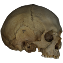
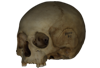
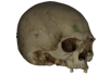
This contribution contains the 3D models described and figured in the following publications:
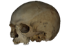
- Marini E., Lussu P., 2020. A virtual physical anthropology lab. Teaching in the time of coronavirus, in prep.;
- Lussu P., Bratzu D., Marini E., 2020. Cloud-based ultra close-range digital photogrammetry: validation of an approach for the effective virtual reconstruction of skeletal remains, in prep.
Homo sapiens MSAE 59 View specimen

|
M3#509MSAE 59 Type: "3D_surfaces"doi: 10.18563/m3.sf.509 state:published |
Download 3D surface file |
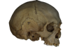
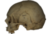
Homo sapiens MSAE 62 View specimen
|
M3#510MSAE 62 Type: "3D_surfaces"doi: 10.18563/m3.sf.510 state:published |
Download 3D surface file |
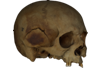
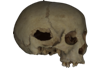
Homo sapiens MSAE 63 View specimen
|
M3#512MSAE 63 Type: "3D_surfaces"doi: 10.18563/m3.sf.512 state:published |
Download 3D surface file |
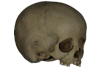
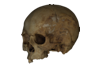
Homo sapiens MSAE 78 View specimen
|
M3#514MSAE 78 Type: "3D_surfaces"doi: 10.18563/m3.sf.514 state:published |
Download 3D surface file |
Homo sapiens MSAE 95 View specimen
|
M3#515MSAE 95 Type: "3D_surfaces"doi: 10.18563/m3.sf.515 state:published |
Download 3D surface file |
Homo sapiens MSAE 1852 View specimen
|
M3#516MSAE 1852 Type: "3D_surfaces"doi: 10.18563/m3.sf.516 state:published |
Download 3D surface file |
Homo sapiens MSAE 6426 View specimen
|
M3#517MSAE 6426 Type: "3D_surfaces"doi: 10.18563/m3.sf.517 state:published |
Download 3D surface file |
Homo sapiens MSAE 6428 View specimen
|
M3#518MSAE 6428 Type: "3D_surfaces"doi: 10.18563/m3.sf.518 state:published |
Download 3D surface file |
Homo sapiens MSAE 6992 View specimen
|
M3#519MSAE 6992 Type: "3D_surfaces"doi: 10.18563/m3.sf.519 state:published |
Download 3D surface file |
Homo sapiens MSAE 7688 View specimen
|
M3#520MSAE 7688 Type: "3D_surfaces"doi: 10.18563/m3.sf.520 state:published |
Download 3D surface file |
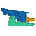
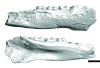
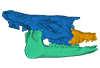
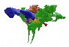
The present 3D Dataset contains two 3D models described in Tissier et al. (https://doi.org/10.1098/rsos.200633): the only known complete mandible of the early-branching rhinocerotoid Epiaceratherium magnum Uhlig, 1999, and a hypothetical reconstruction of the complete archetypic skull of Epiaceratherium Heissig, 1969, created by merging three cranial parts from three distinct Epiaceratherium species.
Epiaceratherium magnum NMB.O.B.928 View specimen
|
M3#5343D surface model of the mandible NMB.O.B.928 of Epiaceratherium magnum, with texture file. Type: "3D_surfaces"doi: 10.18563/m3.sf.534 state:published |
Download 3D surface file |
Epiaceratherium magnum NMB.O.B.928 + MJSN POI007–245 + NMB.I.O.43 View specimen
|
M3#535Archetypal reconstruction of the skull of Epiaceratherium, generated by 3D virtual association of the cranium of E. delemontense (MJSN POI007–245, in blue), mandible of E. magnum (NMB.O.B.928, green) and snout of E. bolcense (NMB.I.O.43, in orange). Type: "3D_surfaces"doi: 10.18563/m3.sf.535 state:published |
Download 3D surface file |
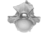
This contribution contains the 3D surface model of the holotype cranium of the Late Jurassic thalassochelydian turtle Solnhofia brachyrhyncha described and figured in the publication of Anquetin and Püntener (2020).
Solnhofia brachyrhyncha MJSN BAN001-2.1 View specimen
|
M3#536Textured 3D surface model of the holotype cranium of the Late Jurassic turtle Solnhofia brachyrhyncha Type: "3D_surfaces"doi: 10.18563/m3.sf.536 state:published |
Download 3D surface file |
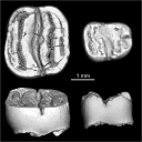
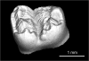
This contribution provides the raw files for the μCT-scan data and renderings of the three-dimensional digital models of two fossil teeth of a geomyin geomorph rodent (Caribeomys merzeraudi), discovered from lower Oligocene deposits of Puerto Rico, San Sebastian Formation (locality LACM Loc. 8060). These fossils were described, figured and discussed in the following publication: Marivaux et al. (2021), An unpredicted ancient colonization of the West Indies by North American rodents: dental evidence of a geomorph from the early Oligocene of Puerto Rico. Papers in Palaeontology. https://doi.org/10.1002/spp2.1388
Caribeomys merzeraudi LACM 162478 View specimen
|
M3#712Right lower dp4: isolated deciduous premolar. The specimen was scanned with a resolution of 5 µm using a μ-CT-scanning station EasyTom 150 / Rx Solutions (Montpellier RIO Imaging, ISE-M, Montpellier, France). AVIZO 7.1 (Visualization Sciences Group) software was used for visualization, segmentation, and 3D rendering. This isolated tooth was prepared within a “labelfield” module of AVIZO, using the segmentation threshold selection tool. Type: "3D_surfaces"doi: 10.18563/m3.sf.712 state:published |
Download 3D surface file |
|
M3#7145µm µCT data set . Right lower dp4: isolated deciduous premolar. The specimen was scanned with a resolution of 5 µm using a μ-CT-scanning station EasyTom 150 / Rx Solutions (Montpellier RIO Imaging, ISE-M, Montpellier, France). Type: "3D_CT"doi: 10.18563/m3.sf.714 state:published |
Download CT data |
Caribeomys merzeraudi LACM 162449 View specimen
|
M3#713Right lower molar (m1 or m2). The specimen was scanned with a resolution of 4.5 µm using a μ-CT-scanning station EasyTom 150 / Rx Solutions (Montpellier RIO Imaging, ISE-M, Montpellier, France). AVIZO 7.1 (Visualization Sciences Group) software was used for visualization, segmentation, and 3D rendering. This isolated tooth was prepared within a “labelfield” module of AVIZO, using the segmentation threshold selection tool. Type: "3D_surfaces"doi: 10.18563/m3.sf.713 state:published |
Download 3D surface file |
|
M3#715µCT data at 4.5µm Type: "3D_CT"doi: 10.18563/m3.sf.715 state:published |
Download CT data |
This contribution contains 3D models of the cranial skeleton and muscles in an elephantfish (Callorhinchus milii) and a catshark (Scyliorhinus canicula), based on synchrotron tomographic scans. These datasets were analyzed and described in Dearden et al. (2021) “The morphology and evolution of chondrichthyan cranial muscles: a digital dissection of the elephantfish Callorhinchus milii and the catshark Scyliorhinus canicula.” Journal of Anatomy.
Callorhinchus milii 001 View specimen
|
M3#7083D models of the cranial skeleton and muscles of Callorhinchus milii, created using Mimics. Type: "3D_surfaces"doi: 10.18563/m3.sf.708 state:published |
Download 3D surface file |
Scyliorhinus canicula 002 View specimen
|
M3#7093D models of the cranial skeleton and muscles of Scyliorhinus canicula, created using Mimics. Type: "3D_surfaces"doi: 10.18563/m3.sf.709 state:published |
Download 3D surface file |
The present 3D Dataset contains the 3D model of a specimen of Metamynodon planifrons (UNISTRA.2015.0.1106) described and figured in: Veine-Tonizzo, L., Tissier, J., Bukhsianidze, M., Vasilyan, D., Becker, D., 2023, Cranial morphology and phylogenetic relationships of Amynodontidae Scott & Osborn, 1883 (Perissodactyla, Rhinocerotoidea).
Metamynodon planifrons UNISTRA.2015.0.1106 View specimen
|
M3#716Textured 3D surface model of the skull of the specimen UNISTRA.2015.0.1106 with right C1 and both rows of P2-M3. Type: "3D_surfaces"doi: 10.18563/m3.sf.716 state:published |
Download 3D surface file |
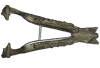
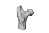
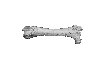
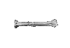
This contribution contains the 3D models of postcranial bones (humerus, ulna, innominate, femur, tibia, astragalus, navicular, and metatarsal III) described and figured in the following publication: “Postcranial morphology of the extinct rodent Neoepiblema (Rodentia: Chinchilloidea): insights into the paleobiology of neoepiblemids”.
Neoepiblema acreensis UFAC 3549 View specimen
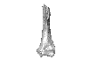
|
M3#719UFAC 3549, left humerus missing the proximal region. Type: "3D_surfaces"doi: 10.18563/m3.sf.719 state:published |
Download 3D surface file |
Neoepiblema acreensis UFAC 5076 View specimen
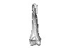
|
M3#720UFAC 5076, right humerus missing the proximal region. Type: "3D_surfaces"doi: 10.18563/m3.sf.720 state:published |
Download 3D surface file |
Neoepiblema acreensis UFAC 1939 View specimen
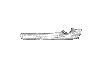
|
M3#721UFAC 1939, right ulna missing the olecranon epiphysis and the distal region. Type: "3D_surfaces"doi: 10.18563/m3.sf.721 state:published |
Download 3D surface file |
Neoepiblema acreensis UFAC 3697 View specimen
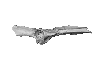
|
M3#722UFAC 3697, right innominate bone. Type: "3D_surfaces"doi: 10.18563/m3.sf.722 state:published |
Download 3D surface file |
Neoepiblema acreensis UFAC 2574 View specimen
|
M3#723UFAC 2574, proximal region of a left femur. Type: "3D_surfaces"doi: 10.18563/m3.sf.723 state:published |
Download 3D surface file |
Neoepiblema acreensis UFAC 2937 View specimen
|
M3#724UFAC 2937, right femur with damaged proximal region. Type: "3D_surfaces"doi: 10.18563/m3.sf.724 state:published |
Download 3D surface file |
Neoepiblema acreensis UFAC 2210 View specimen
|
M3#725UFAC 2210, distal region of a right femur. Type: "3D_surfaces"doi: 10.18563/m3.sf.725 state:published |
Download 3D surface file |
Neoepiblema acreensis UFAC 1887 View specimen
|
M3#726UFAC 1887, right tibia Type: "3D_surfaces"doi: 10.18563/m3.sf.726 state:published |
Download 3D surface file |
Neoepiblema acreensis UFAC 1840 View specimen
|
M3#727UFAC 1840, left astragalus. Type: "3D_surfaces"doi: 10.18563/m3.sf.727 state:published |
Download 3D surface file |
Neoepiblema acreensis UFAC 2549 View specimen
|
M3#728UFAC 2549, right astragalus. Type: "3D_surfaces"doi: 10.18563/m3.sf.728 state:published |
Download 3D surface file |
Neoepiblema acreensis UFAC 3672 View specimen
|
M3#729UFAC 3672, right navicular. Type: "3D_surfaces"doi: 10.18563/m3.sf.729 state:published |
Download 3D surface file |
Neoepiblema acreensis UFAC 2116 View specimen
|
M3#730UFAC 2116, left metatarsal III. Type: "3D_surfaces"doi: 10.18563/m3.sf.730 state:published |
Download 3D surface file |
Neoepiblema horridula UFAC 3260 View specimen
|
M3#731UFAC 3260, fragmented left innominate. Type: "3D_surfaces"doi: 10.18563/m3.sf.731 state:published |
Download 3D surface file |
Neoepiblema horridula UFAC 2620 View specimen
|
M3#732UFAC 2620, distal region of a right femur. Type: "3D_surfaces"doi: 10.18563/m3.sf.732 state:published |
Download 3D surface file |
Neoepiblema horridula UFAC 2737 View specimen
|
M3#733UFAC 2737, proximal region of right femur. Type: "3D_surfaces"doi: 10.18563/m3.sf.733 state:published |
Download 3D surface file |
Neoepiblema horridula UFAC 3202 View specimen
|
M3#734UFAC 3202, right tibia, missing the proximalmost and distal portions. Type: "3D_surfaces"doi: 10.18563/m3.sf.734 state:published |
Download 3D surface file |
Neoepiblema horridula UFAC 3212 View specimen
|
M3#735UFAC 3212, left astragalus. Type: "3D_surfaces"doi: 10.18563/m3.sf.735 state:published |
Download 3D surface file |
The present 3D Dataset contains the 3D model analyzed in Hendrickx, C. and Bell, P. R. 2021. The scaly skin of the abelisaurid Carnotaurus sastrei (Theropoda: Ceratosauria) from the Upper Cretaceous of Patagonia. Cretaceous Research. https://doi.org/10.1016/j.cretres.2021.104994
Carnotaurus sastrei MACN 894 View specimen
|
M3#8023D reconstruction of the biggest patch of skin (~1200 cm2) from the anterior tail region of the holotype of Carnotaurus, which is the largest single patch of squamous integument available for any saurischian. The skin consists of medium to large (up to 65 mm in diameter) conical feature scales surrounded by a network of low and small (< 14 mm) irregular basement scales separated by narrow interstitial tissue. Type: "3D_surfaces"doi: 10.18563/m3.sf.802 state:published |
Download 3D surface file |
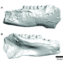
The present 3D Dataset contains the 3D models of the holotype mandible and referred fragmented skull of the new species Amphimoschus xishuiensis analyzed in the article Li, Y.-K., Mennecart, B., Aiglstorfer, M., Ni, X.-J., Li, Q., Deng, T. 2021. The early evolution of cranial appendages in Bovoidea revealed by new species of Amphimoschus (Mammalia: Ruminantia) from China. Zoological Journal of the Linnean Society https://doi.org/10.1093/zoolinnean/zlab053
Amphimoschus xishuiensis IVPP V 25521.1 View specimen
|
M3#803the holotype, a right hemimandible with tooth row p2 to m3 Type: "3D_surfaces"doi: 10.18563/m3.sf.803 state:published |
Download 3D surface file |
Amphimoschus xishuiensis IVPP V 25521.2 View specimen
|
M3#804referred material, anterior part of a skull with right P4-M3 and left P3-M2 Type: "3D_surfaces"doi: 10.18563/m3.sf.804 state:published |
Download 3D surface file |
This contribution contains the 3D models described and figured in the following publication: Georgalis, G.L., G. Guinot, K.E. Kassegne, Y.Z. Amoudji, A.K.C. Johnson, H. Cappetta and L. Hautier. 2021. An assemblage of giant aquatic snakes (Serpentes, Palaeophiidae) from the Eocene of Togo. Swiss Journal of Palaeontology 140, https://doi.org/10.1186/s13358-021-00236-w
Palaeophis africanus UM KPO 21 View specimen
|
M3#821Trunk vertebra UM KPO 21 of Palaeophis africanus Type: "3D_surfaces"doi: 10.18563/m3.sf.821 state:published |
Download 3D surface file |
Palaeophis africanus UM KPO 22 View specimen
|
M3#822Trunk vertebra UM KPO 22 of Palaeophis africanus from the Eocene of Togo Type: "3D_surfaces"doi: 10.18563/m3.sf.822 state:published |
Download 3D surface file |
Palaeophis africanus UM KPO 23 View specimen
|
M3#823Trunk vertebra UM KPO 23 of Palaeophis africanus Type: "3D_surfaces"doi: 10.18563/m3.sf.823 state:published |
Download 3D surface file |
Palaeophis africanus UM KPO 24 View specimen
|
M3#824Trunk vertebra UM KPO 24 of Palaeophis africanus Type: "3D_surfaces"doi: 10.18563/m3.sf.824 state:published |
Download 3D surface file |
Palaeophis africanus UM KPO 25 View specimen
|
M3#825Trunk vertebra UM KPO 25 of Palaeophis africanus Type: "3D_surfaces"doi: 10.18563/m3.sf.825 state:published |
Download 3D surface file |
Palaeophis africanus UM KPO 26 View specimen
|
M3#826Trunk vertebra UM KPO 26 of Palaeophis africanus Type: "3D_surfaces"doi: 10.18563/m3.sf.826 state:published |
Download 3D surface file |
Palaeophis africanus UM KPO 27 View specimen
|
M3#827Trunk vertebra UM KPO 27 of Palaeophis africanus Type: "3D_surfaces"doi: 10.18563/m3.sf.827 state:published |
Download 3D surface file |
Palaeophis africanus UM KPO 28 View specimen
|
M3#828Trunk vertebra UM KPO 28 of Palaeophis africanus Type: "3D_surfaces"doi: 10.18563/m3.sf.828 state:published |
Download 3D surface file |
Palaeophis africanus UM KPO 29 View specimen
|
M3#829Trunk vertebra UM KPO 29 of Palaeophis africanus Type: "3D_surfaces"doi: 10.18563/m3.sf.829 state:published |
Download 3D surface file |
Palaeophis africanus UM KPO 30 View specimen
|
M3#830Trunk vertebra UM KPO 30 of Palaeophis africanus Type: "3D_surfaces"doi: 10.18563/m3.sf.830 state:published |
Download 3D surface file |
Palaeophis africanus UM KPO 31 View specimen
|
M3#831Trunk vertebra UM KPO 28 of Palaeophis africanus Type: "3D_surfaces"doi: 10.18563/m3.sf.831 state:published |
Download 3D surface file |
Palaeophis africanus UM KPO 32 View specimen
|
M3#832Trunk vertebra UM KPO 32 of Palaeophis africanus Type: "3D_surfaces"doi: 10.18563/m3.sf.832 state:published |
Download 3D surface file |
Palaeophis africanus UM KPO 33 View specimen
|
M3#833Trunk vertebra UM KPO 33 of Palaeophis africanus Type: "3D_surfaces"doi: 10.18563/m3.sf.833 state:published |
Download 3D surface file |
Palaeophis africanus UM KPO 34 View specimen

|
M3#839Trunk vertebra UM KPO 34 of Palaeophis africanus Type: "3D_surfaces"doi: 10.18563/m3.sf.839 state:published |
Download 3D surface file |
Palaeophis africanus UM KPO 35 View specimen
|
M3#840Trunk vertebra UM KPO 35 of Palaeophis africanus Type: "3D_surfaces"doi: 10.18563/m3.sf.840 state:published |
Download 3D surface file |
Palaeophis africanus UM KPO 36 View specimen
|
M3#841Trunk vertebra UM KPO 36 of Palaeophis africanus Type: "3D_surfaces"doi: 10.18563/m3.sf.841 state:published |
Download 3D surface file |
Palaeophis africanus UM KPO 37 View specimen
|
M3#842Trunk vertebra UM KPO 37 of Palaeophis africanus Type: "3D_surfaces"doi: 10.18563/m3.sf.842 state:published |
Download 3D surface file |
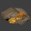
The present 3D Dataset contains the 3D models analyzed in the following publication: Le Verger K., González Ruiz L.R., Billet G. 2021. Comparative anatomy and phylogenetic contribution of intracranial osseous canals and cavities in armadillos and glyptodonts (Xenarthra, Cingulata). Journal of Anatomy 00: 1-30 p. https://doi.org/10.1111/joa.13512
Bradypus tridactylus MNHN ZM-MO-1999-1065 View specimen
|
M3#844Bradypus tridactylus MNHN ZM-MO-1999-1065: cranium, cranial canals & alveolar cavities. Type: "3D_surfaces"doi: 10.18563/m3.sf.844 state:published |
Download 3D surface file |
Tamandua tetradactyla NHMUK ZD-1903.7.7.135 View specimen
|
M3#845Tamandua tetradactyla NHMUK ZD-1903.7.7.135: cranium, cranial canals & alveolar cavities. Type: "3D_surfaces"doi: 10.18563/m3.sf.845 state:published |
Download 3D surface file |
Dasypus novemcinctus AMNH 33150 View specimen
|
M3#846Dasypus novemcinctus AMNH 33150: cranium, cranial canals & alveolar cavities. Type: "3D_surfaces"doi: 10.18563/m3.sf.846 state:published |
Download 3D surface file |
Dasypus novemcinctus AMNH 133261 View specimen
|
M3#847Dasypus novemcinctus AMNH 133261: cranium, cranial canals & alveolar cavities. Type: "3D_surfaces"doi: 10.18563/m3.sf.847 state:published |
Download 3D surface file |
Dasypus novemcinctus AMNH 133328 View specimen
|
M3#848Dasypus novemcinctus AMNH 133328: cranium, cranial canals & alveolar cavities. Type: "3D_surfaces"doi: 10.18563/m3.sf.848 state:published |
Download 3D surface file |
Zaedyus pichiy ZMB-MAM-49039 View specimen
|
M3#849Zaedyus pichiy ZMB-MAM-49039: cranium, cranial canals & alveolar cavities. Type: "3D_surfaces"doi: 10.18563/m3.sf.849 state:published |
Download 3D surface file |
Zaedyus pichiy MHNG 1627.053 View specimen

|
M3#850Zaedyus pichiy MHNG 1627.053: cranium, cranial canals & alveolar cavities. Type: "3D_surfaces"doi: 10.18563/m3.sf.850 state:published |
Download 3D surface file |
Zaedyus pichiy MHNG 1276.076 View specimen
|
M3#851Zaedyus pichiy MHNG 1276.076: cranium, cranial canals & alveolar cavities. Type: "3D_surfaces"doi: 10.18563/m3.sf.851 state:published |
Download 3D surface file |
Cabassous unicinctus NBC_ZMA.MAM.26326.a View specimen
|
M3#852Cabassous unicinctus NBC ZMA.MAM.26326.a: cranium, cranial canals & alveolar cavities. Type: "3D_surfaces"doi: 10.18563/m3.sf.852 state:published |
Download 3D surface file |
Cabassous unicinctus MNHN-CG-1999-1044 View specimen
|
M3#853Cabassous unicinctus MNHN-CG-1999-1044: cranium, cranial canals & alveolar cavities. Type: "3D_surfaces"doi: 10.18563/m3.sf.853 state:published |
Download 3D surface file |
Vassallia maxima FMNH P14424 View specimen
|
M3#854Vassallia maxima FMNH P14424: cranium, cranial canals & alveolar cavities. Type: "3D_surfaces"doi: 10.18563/m3.sf.854 state:published |
Download 3D surface file |
Glyptodon sp. MNHN-F-PAM-759 View specimen
|
M3#855Glyptodon sp. MNHN-F-PAM-759: cranium, cranial canals & alveolar cavities. Type: "3D_surfaces"doi: 10.18563/m3.sf.855 state:published |
Download 3D surface file |
Glyptodon sp. MNHN-F-PAM-760 View specimen

|
M3#856Glyptodon sp. MNHN-F-PAM-760: cranium, cranial canals & alveolar cavities. Type: "3D_surfaces"doi: 10.18563/m3.sf.856 state:published |
Download 3D surface file |
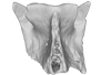
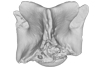
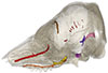
Our knowledge of the external brain morphology of the late Eocene artiodactyl ungulate Mixtotherium, relies on a plaster model realized on a specimen from the Victor Brun Museum in Montauban (France) and described by Dechaseaux (1973). Here, based on micro CT-scan data, we virtually reconstruct the 3D cast of the empty cavity of the partial cranium MA PHQ 716 from the Victor Brun Museum and compare it to the plaster model illustrated and described by Dechaseaux (1973). Indeed, the specimen from which the original plaster endocast originates was not identified by Dechaseaux by a specimen number. We confirm here that the studied specimen was indeed the one described and illustrated by Dechaseaux (1973). We also reconstruct a second, more detailed, model providing additional morphological and quantitative observations made available by micro CT scan investigation such as precisions on the neopallium folding and endocranial volumes.
Mixtotherium cuspidatum MA PHQ 716 View specimen
|
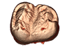
M3#857endocast of the brain cavity Type: "3D_surfaces"doi: 10.18563/m3.sf.857 state:published |
Download 3D surface file |
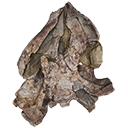
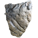
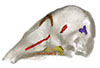
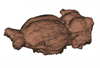
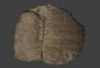
Turtles are one of the most impressive vertebrates. Much of the body is either hidden in a shell or can be drawn into it. Turtles impress with their individual longevity and their often peaceful disposition. Also, with their resilience, they have survived all extinction events since their emergence in the Late Triassic. Today's diversity of shapes is impressive and ranges from the large and high domed Galapagos turtles to the hamster-sized flat pancake turtles. The holotype of one of the oldest fossil turtles, Proganochelys quenstedtii, is housed in the paleontological collection in Tübingen/Germany. Since its discovery some years before 1873, P. quenstedtii has represented the 'prototype' of the turtle and has had an eventful scientific history. It was found in Neuenhaus (Häfner-Neuhausen in Schönbuch forest), Baden-Württemberg, Germany, and stems from Löwenstein-Formation (Weißer Keupersandstein), Late Triassic. The current catalogue number is GPIT-PV-30000. The specimen is listed in the historical inventory “Tübinger Petrefaktenverzeichnis 1841 bis 1896, [folio 326v.]“, as “[catalogue number: PV]16549, Schildkröte Weiser Keupersandstein Hafnerhausen” [turtle from White Keuper Sandstone]. Another, more recent synonym is “GPIT/RE/9396”. The same specimen was presented as uncatalogued by Gaffney (1990). Here we provide a surface scan of the steinkern for easier access of this famous specimen to the scientific community.
Proganochelys quenstedtii GPIT-PV-30000 View specimen
|
M3#967This the surface model of the steinkern of the shell of Proganochelys quenstedtii. Type: "3D_surfaces"doi: 10.18563/m3.sf.967 state:published |
Download 3D surface file |
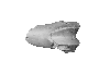
The present 3D Dataset contains the 3D models analyzed in Benites-Palomino A., Velez-Juarbe J., Altamirano-Sierra A., Collareta A., Carrillo-Briceño J., and Urbina M. 2022. Sperm whales (Physeteroidea) from the Pisco Formation, Peru, and their Trophic role as fat-sources for Late Miocene sharks.
Scaphokogia cochlearis MUSM 978 View specimen
|
M3#977juvenile Scaphokogia cochlearis Type: "3D_surfaces"doi: 10.18563/m3.sf.977 state:published |
Download 3D surface file |
This contribution contains the 3D model(s) described and figured in the following publication: Carolina A. Hoffmann, P. G. Rodrigues, M. B. Soares & M. B. Andrade. 2021. Brain endocast of two non-mammaliaform cynodonts from southern Brazil: an ontogenetic and evolutionary approach, Historical Biology, 33:8, 1196-1207, https://doi.org/10.1080/08912963.2019.1685512
Probelesodon kitchingi MCP 1600 PV View specimen
|
M3#9783D model of the brain endocast of Probelesodon kitchingi. Type: "3D_surfaces"doi: 10.18563/m3.sf.978 state:published |
Download 3D surface file |
Massetognathus ochagaviae MCP 3871 PV View specimen
|
M3#9793D model of the brain endocast of Massetognathus ochagaviae. Type: "3D_surfaces"doi: 10.18563/m3.sf.979 state:published |
Download 3D surface file |
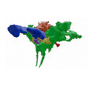
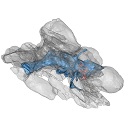
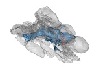
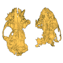
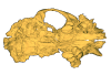
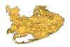
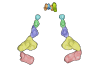
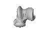
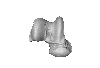
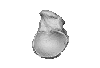
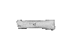
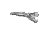
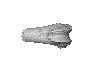
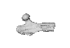
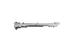
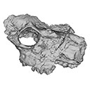
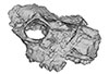
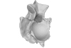
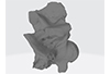
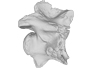
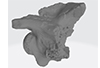
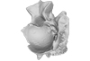
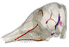
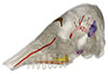
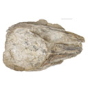
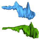
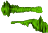
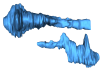
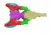
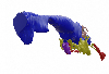
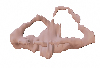
The present 3D Dataset contains the 3D models analyzed in Pochat-Cottilloux Y., Rinder N., Perrichon G., Adrien J., Amiot R., Hua S. & Martin J. E. (2023). The neuroanatomy and pneumaticity of Hamadasuchus from the Cretaceous of Morocco and its significance for the paleoecology of Peirosauridae and other altirostral crocodylomorphs. Journal of Anatomy, https://doi.org/10.1111/joa.13887
Hamadasuchus sp. UCBL-FSL 532408 View specimen
|
M3#10943D volume reconstruction of the braincase osteology Type: "3D_surfaces"doi: 10.18563/m3.sf.1094 state:published |
Download 3D surface file |
|
M3#10963D volume reconstruction of the endocast Type: "3D_surfaces"doi: 10.18563/m3.sf.1096 state:published |
Download 3D surface file |
|
M3#10973D volume reconstruction of the labyrinths Type: "3D_surfaces"doi: 10.18563/m3.sf.1097 state:published |
Download 3D surface file |
|
M3#10983D volume reconstruction of the pneumatic cavities Type: "3D_surfaces"doi: 10.18563/m3.sf.1098 state:published |
Download 3D surface file |


