3D models of early strepsirrhine primate teeth from North Africa
3D models of Pontognathus ignotus and Massetognathus pascuali
3D models of Protosilvestria sculpta and Coloboderes roqueprunetherion
3D GM dataset of bird skeletal variation
Skeletal embryonic development in the catshark
Bony connexions of the petrosal bone of extant hippos
bony labyrinth (11) , inner ear (10) , Eocene (8) , South America (8) , Paleobiogeography (7) , skull (7) , phylogeny (6)
Lionel Hautier (22) , Maëva Judith Orliac (21) , Laurent Marivaux (16) , Rodolphe Tabuce (14) , Bastien Mennecart (13) , Pierre-Olivier Antoine (12) , Renaud Lebrun (11)

|
3D model related to the publication: New remains of Chambius kasserinensis from the Eocene of Tunisia and evaluation of proposed affinities for Macroscelidea (Mammalia, Afrotheria)Rodolphe Tabuce
Published online: 23/03/2017 Keywords: Herodotiinae; Macroscelidea; Maxilla https://doi.org/10.18563/m3.3.2.e1 Cite this article: Rodolphe Tabuce, 2017. 3D model related to the publication: New remains of Chambius kasserinensis from the Eocene of Tunisia and evaluation of proposed affinities for Macroscelidea (Mammalia, Afrotheria). MorphoMuseuM 3 (2)-e1. doi: 10.18563/m3.3.2.e1 Export citationAbstract This contribution contains the 3D model of the holotype of Chambius kasserinensis, the basalmost ‘elephant-shrew’ figured in the following publication: New remains of Chambius kasserinensis from the Eocene of Tunisia and evaluation of proposed affinities for Macroscelidea (Mammalia, Afrotheria). https://doi.org/10.1080/08912963.2017.1297433 See original publication M3 article infos Published in Volume 03, Issue 02 (2017) |
|

|
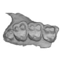
M3#1463D model of the holotype maxilla of Chambius kasserinensis. The 3D surface was extracted manually from the limestone matrix within AVIZO 9.2 Type: "3D_surfaces"doi: 10.18563/m3.sf.146 Data citation: Rodolphe Tabuce, 2017. M3#146. doi: 10.18563/m3.sf.146 state:published |
Download 3D surface file |