3D models of Cainotheriids Ossicular chain
3D models of Kalakocetus, the earliest Cetacea
The specimens of Speothos pacivorus
3D GM dataset of bird skeletal variation
Skeletal embryonic development in the catshark
Bony connexions of the petrosal bone of extant hippos
bony labyrinth (14) , inner ear (11) , Eocene (11) , geometric morphometrics (10) , CT-scan (10) , Oligocene (9) , Micro-CT (9)
Lionel Hautier (25) , Maëva Judith Orliac (24) , Laurent Marivaux (18) , Renaud Lebrun (15) , Rodolphe Tabuce (15) , Pierre-Olivier Antoine (13) , Bastien Mennecart (13)
MorphoMuseuM Volume 03, Issue 04
<< prev. article next article >>
|
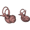
3D dataset3D models related to the publication: Comparative anatomy of the bony labyrinth of the bats Platalina genovensium (Phyllostomidae, Lonchophyllinae) and Tomopeas ravus (Molossidae, Tomopeatinae)Paul M. Velazco
Published online: 09/04/2018 |

|
M3#276Right bony labyrinth surface positioned (.PLY) Labels associated (.FLG) Type: "3D_surfaces"doi: 10.18563/m3.sf.276 state:published |
Download 3D surface file |
Tomopeas ravus 278525 View specimen

|
M3#277Right bony labyrinth surface (.PLY) Labels associated (.FLG) Type: "3D_surfaces"doi: 10.18563/m3.sf.277 state:published |
Download 3D surface file |
Ekdale, E.G. 2013. Comparative anatomy of the bony labyrinth (inner ear) of placental mammals. PLoS ONE 10(8), e0137149. https://doi.org/10.1371/journal.pone.0066624
Lebrun, R. 2014. ISE-MeshTools, a 3D interactive fossil reconstruction freeware. 12th Annual Meeting of EAVP, Torino, Italy.
Velazco, P. M., Grohé, C. 2017. Comparative anatomy of the bony labyrinth of the bats Tomopeas ravus (Molossidae, Tomopeatinae) and Platalina genovensium (Phyllostomidae, Lonchophyllinae). Biotempo 14(2). https://doi.org/10.31381/biotempo.v14i2.1404
Velazco, P. M., Cadenillas, R., Centty, O., Huamani, L., Zamora, H. 2013. New records of Platalina genovensium Thomas, 1928 (Chiroptera, Phyllostomidae, Lonchophyllinae) and Tomopeas ravus Miller, 1900 (Chiroptera, Molossidae, Tomopeatinae). Mastozoología Neotropical 20(2), 425-434.
Paúl M Velazco, Kerry A Kline, Sergio Solari and Meredith J Hamilton (2019).
Tomopeas ravus (Chiroptera: Molossidae). Mammalian Species. https://doi.org/10.1093/mspecies/sez011
Gonzalo Ossa, Hugo T Zamora, Paúl M Velazco, Sergio Solari and Meredith J Hamilton (2020). Platalina genovensium(Chiroptera: Phyllostomidae). Mammalian Species. https://doi.org/10.1093/mspecies/seaa008