3D models of Cainotheriids Ossicular chain
Explodable 3D Dog Skull for Veterinary Education
3D models of Kalakocetus, the earliest Cetacea
3D GM dataset of bird skeletal variation
Skeletal embryonic development in the catshark
Bony connexions of the petrosal bone of extant hippos
bony labyrinth (14) , inner ear (11) , Eocene (11) , geometric morphometrics (10) , CT-scan (10) , Oligocene (9) , Micro-CT (9)
Lionel Hautier (25) , Maëva Judith Orliac (24) , Laurent Marivaux (18) , Renaud Lebrun (15) , Rodolphe Tabuce (15) , Pierre-Olivier Antoine (13) , Bastien Mennecart (13)
MorphoMuseuM Volume 05, issue 04
<< prev. article next article >>
|
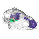
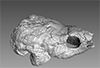
3D dataset3D model related to the publication: The endocranial anatomy of the stem turtle Naomichelys speciosa from the Early Cretaceous of North AmericaAriana Paulina-Carabajal
Published online: 10/09/2019 |
|
M3#428FMNH_PR273_1 - Naomichlys speciosa - skull Type: "3D_surfaces"doi: 10.18563/m3.sf.428 state:published |
Download 3D surface file |
Hay, O.P., 1908. The Fossil Turtles of North America. Carnegie Institution of Washington, No 75, 810 p.
Joyce, W.G., Chapman, S.D., Moody, R.T. and Walker, C.A., 2011. The skull of the solemydid turtle Helochelydra nopcsai from the Early Cretaceous of the Isle of Wight (UK) and a review of Solemydidae. Special Papers in Palaeontology 86, 75–97.
Joyce, W.G., Sterli, J. and Chapman, S.D., 2014. The skeletal morphology of the solemydid turtle Naomichelys speciosa from the Early Cretaceous of Texas. Journal of Paleontology 88, 1257–1287. https://doi.org/10.1666/14-002
Lautenschlager, S., Ferreira, G.S., and Werneburg, I., 2018. Sensory evolution and ecology of early turtles revealed by digital endocranial reconstructions. Frontiers in Ecology and Evolution 6, https://doi.org/10.3389/fevo.2018.00007
Paulina-Carabajal, A., Sterli, J., Werneburg, I., 2019. The endocranial morphology of the stem turtle Naomichelys speciosa from the Early Cretaceous of North America. Acta Palaeontologica Polonica, https://doi.org/10.4202/app.00606.2019
Ingmar Werneburg, Serjoscha W. Evers and Gabriel Ferreira (2021). On the “cartilaginous rider” in the endocasts of turtle brain cavities. Vertebrate Zoology. https://doi.org/10.3897/vz.71.e66756
Ingmar Werneburg, Serjoscha W. Evers and Gabriel S. Ferreira (2021). 3D models related to the publication: On the “cartilaginous rider” in the endocasts of turtle brain cavities. MorphoMuseuM. https://doi.org/10.18563/journal.m3.146