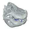
Explodable 3D Dog Skull for Veterinary Education
3D Topography and food processing in Palmatolepis
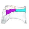
3D models of the paratympanic sinus system, the endocast and the neurovascular bony canal of the maxilla, premaxilla and the jugal of Leidyosuchus canadensis and Stangerochampsa mccabei
3D GM dataset of bird skeletal variation
Skeletal embryonic development in the catshark
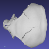
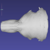
Bony connexions of the petrosal bone of extant hippos
bony labyrinth (14) , inner ear (11) , geometric morphometrics (10) , Eocene (10) , CT-scan (9) , Micro-CT (9) , Miocene (8)
Lionel Hautier (24) , Maëva Judith Orliac (21) , Laurent Marivaux (17) , Rodolphe Tabuce (14) , Pierre-Olivier Antoine (13) , Bastien Mennecart (13) , Renaud Lebrun (12)