|
The endocranial cast of a 10 ka intentionally deformed human cranium from China
Published online: 27/07/2022
Keywords:
endocranial cast; intentional cranial deformation; Northeast China
https://doi.org/10.18563/journal.m3.169
Cite this article:
Yin Qiyu, Li Qiang, Ma Ming, Zhang Wei and Ni Xijun, 2022. The endocranial cast of a 10 ka intentionally deformed human cranium from China. MorphoMuseuM 8:169. doi: 10.18563/journal.m3.169
Export citation
Abstract
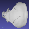
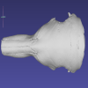
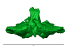
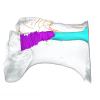
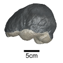
This contribution contains the 3D model of an endocranial cast analyzed in “A 10 ka intentionally deformed human skull from Northeast Asia”. There are many studies on the morphological characteristics of intentional cranial deformation (ICD), but few related 3D models were published. Here, we present the surface model of an intentionally deformed 10 ka human cranium for further research on ICD practice. The 3D model of the endocranial cast of this ICD cranium was discovered near Harbin City, Province Heilongjiang, Northeast China. The fossil preserved only the frontal, parietal, and occipital bones. To complete the endocast model of the specimen, we printed a 3D model and used modeling clay to reconstruct the missing part based on the general form of the modern human endocast morphology.
M3 article infos
Published in Volume 08, issue 03 (2022)
|
PDF
|