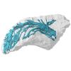
3D models of Cainotheriids Ossicular chain
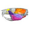
Explodable 3D Dog Skull for Veterinary Education
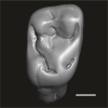
3D models of teeth and neurovascular system in the rostrum of Spinosauridae (theropod dinosaurs)
3D GM dataset of bird skeletal variation
Skeletal embryonic development in the catshark
Bony connexions of the petrosal bone of extant hippos
bony labyrinth (14) , inner ear (11) , Eocene (11) , geometric morphometrics (10) , CT-scan (10) , Oligocene (9) , Micro-CT (9)
Lionel Hautier (25) , Maëva Judith Orliac (24) , Laurent Marivaux (19) , Bastien Mennecart (16) , Renaud Lebrun (15) , Rodolphe Tabuce (15) , Pierre-Olivier Antoine (13)
Page 1 of 1, showing 2 record(s) out of 2 total
|
3D models related to the publication: Triassic turtles of France: the first European Rhaetian stem turtle taxon (clades Testudinata, Proterochersidae)Tomasz Szczygielski
Published online: 20/08/2026 |
|
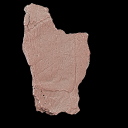
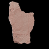
M3#1907Left axillary region and base of the anterior plastral lobe (left hyoplastron, small medial fragment of the right hyoplastron, part of the posterior process of the entoplastron) Type: "3D_surfaces"doi: 10.18563/m3.sf.1907 state:in_press |
Download 3D surface file |
Bartassochersis lopezi UM-PDE2-02 View specimen
|
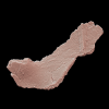
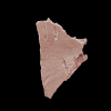
M3#1908Posterior plastral lobe Type: "3D_surfaces"doi: 10.18563/m3.sf.1908 state:in_press |
Download 3D surface file |
Bartassochersis lopezi UM-PDE2-03 View specimen
|
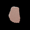
M3#1909First vertebral scute region, right side Type: "3D_surfaces"doi: 10.18563/m3.sf.1909 state:in_press |
Download 3D surface file |
Bartassochersis lopezi UM-PDE2-04 View specimen
|
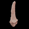
M3#1910?Carapace fragment Type: "3D_surfaces"doi: 10.18563/m3.sf.1910 state:in_press |
Download 3D surface file |
Bartassochersis lopezi UM-PDE2-05 View specimen
|
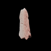
M3#1911Left scapula Type: "3D_surfaces"doi: 10.18563/m3.sf.1911 state:in_press |
Download 3D surface file |
Bartassochersis lopezi UM-PDE2-06 View specimen
|
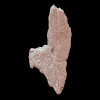
M3#1912Ungual Type: "3D_surfaces"doi: 10.18563/m3.sf.1912 state:in_press |
Download 3D surface file |
Bartassochersis lopezi UM-PDE2-08 View specimen
|
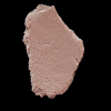
M3#1913Right hypoplastron Type: "3D_surfaces"doi: 10.18563/m3.sf.1913 state:in_press |
Download 3D surface file |
Bartassochersis lopezi UM-PDE2-09 View specimen
|
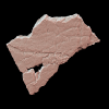
M3#1914Indeterminate Type: "3D_surfaces"doi: 10.18563/m3.sf.1914 state:in_press |
Download 3D surface file |
Bartassochersis lopezi UM-PDE2-10 View specimen
|
M3#1915?Mesoplastron/hypoplastron Type: "3D_surfaces"doi: 10.18563/m3.sf.1915 state:in_press |
Download 3D surface file |
Bartassochersis lopezi UM-PDE2-11 View specimen
|
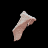
M3#1916Left inguinal periphery (peripheral, hyoplastron) Type: "3D_surfaces"doi: 10.18563/m3.sf.1916 state:in_press |
Download 3D surface file |
Bartassochersis lopezi UM-PDE2-12 View specimen
|
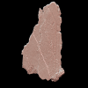
M3#1917?Left side of a vertebral scute area with two partial pleural scute areas Type: "3D_surfaces"doi: 10.18563/m3.sf.1917 state:in_press |
Download 3D surface file |
Bartassochersis lopezi UM-PDE2-13 View specimen
|
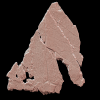
M3#1918?Posterior pleural and supramarginal of marginal areas Type: "3D_surfaces"doi: 10.18563/m3.sf.1918 state:in_press |
Download 3D surface file |
Bartassochersis lopezi UM-PDE2-14 View specimen
|
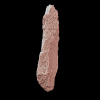
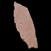
M3#1919Posterior peripherals Type: "3D_surfaces"doi: 10.18563/m3.sf.1919 state:in_press |
Download 3D surface file |
Bartassochersis lopezi UM-PDE2-15A View specimen
|
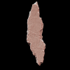
M3#1920?Left edge of the anterior plastral lobe Type: "3D_surfaces"doi: 10.18563/m3.sf.1920 state:in_press |
Download 3D surface file |
Bartassochersis lopezi UM-PDE2-15B View specimen
|
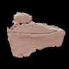
M3#1921Indeterminate Type: "3D_surfaces"doi: 10.18563/m3.sf.1921 state:in_press |
Download 3D surface file |
Bartassochersis lopezi UM-PDE2-16 View specimen
|
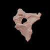
M3#1922Indeterminate Type: "3D_surfaces"doi: 10.18563/m3.sf.1922 state:in_press |
Download 3D surface file |
Bartassochersis lopezi UM-PDE2-17 View specimen
|
M3#1923?Bridge fragment Type: "3D_surfaces"doi: 10.18563/m3.sf.1923 state:in_press |
Download 3D surface file |
Bartassochersis lopezi UM-PDE2-19 View specimen
|
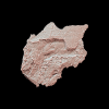
M3#1924Right bridge region Type: "3D_surfaces"doi: 10.18563/m3.sf.1924 state:in_press |
Download 3D surface file |
Bartassochersis lopezi UM-PDE2-20 View specimen
|
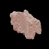
M3#1925Left femoral bulge Type: "3D_surfaces"doi: 10.18563/m3.sf.1925 state:in_press |
Download 3D surface file |
Bartassochersis lopezi UM-PDE2-21 View specimen
|
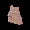
M3#1926Indeterminate Type: "3D_surfaces"doi: 10.18563/m3.sf.1926 state:in_press |
Download 3D surface file |
 , Christina Kyriakouli and Tomasz Szczygielski
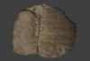
 Turtles are one of the most impressive vertebrates. Much of the body is either hidden in a shell or can be drawn into it. Turtles impress with their individual longevity and their often peaceful disposition. Also, with their resilience, they have survived all extinction events since their emergence in the Late Triassic. Today's diversity of shapes is impressive and ranges from the large and high domed Galapagos turtles to the hamster-sized flat pancake turtles. The holotype of one of the oldest fossil turtles, Proganochelys quenstedtii, is housed in the paleontological collection in Tübingen/Germany. Since its discovery some years before 1873, P. quenstedtii has represented the 'prototype' of the turtle and has had an eventful scientific history. It was found in Neuenhaus (Häfner-Neuhausen in Schönbuch forest), Baden-Württemberg, Germany, and stems from Löwenstein-Formation (Weißer Keupersandstein), Late Triassic. The current catalogue number is GPIT-PV-30000. The specimen is listed in the historical inventory “Tübinger Petrefaktenverzeichnis 1841 bis 1896, [folio 326v.]“, as “[catalogue number: PV]16549, Schildkröte Weiser Keupersandstein Hafnerhausen” [turtle from White Keuper Sandstone]. Another, more recent synonym is “GPIT/RE/9396”. The same specimen was presented as uncatalogued by Gaffney (1990). Here we provide a surface scan of the steinkern for easier access of this famous specimen to the scientific community.
Proganochelys quenstedtii GPIT-PV-30000 View specimen
|
M3#967This the surface model of the steinkern of the shell of Proganochelys quenstedtii. Type: "3D_surfaces"doi: 10.18563/m3.sf.967 state:published |
Download 3D surface file |
Page 1 of 1, showing 2 record(s) out of 2 total