Explodable 3D Dog Skull for Veterinary Education
3D models of teeth and neurovascular system in the rostrum of Spinosauridae (theropod dinosaurs)
3D models of Ocnotherium skull
3D GM dataset of bird skeletal variation
Skeletal embryonic development in the catshark
Bony connexions of the petrosal bone of extant hippos
bony labyrinth (14) , inner ear (11) , Eocene (11) , geometric morphometrics (10) , CT-scan (10) , Oligocene (9) , Micro-CT (9)
Lionel Hautier (25) , Maëva Judith Orliac (24) , Laurent Marivaux (19) , Bastien Mennecart (16) , Renaud Lebrun (15) , Rodolphe Tabuce (15) , Pierre-Olivier Antoine (13)
Page 1 of 1, showing 4 record(s) out of 4 total
|
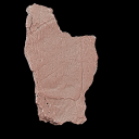
3D models related to the publication: Triassic turtles of France: the first European Rhaetian stem turtle taxon (clades Testudinata, Proterochersidae)Tomasz Szczygielski
Published online: 20/08/2026 |
|
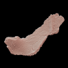
M3#1907Left axillary region and base of the anterior plastral lobe (left hyoplastron, small medial fragment of the right hyoplastron, part of the posterior process of the entoplastron) Type: "3D_surfaces"doi: 10.18563/m3.sf.1907 state:in_press |
Download 3D surface file |
Bartassochersis lopezi UM-PDE2-02 View specimen
|
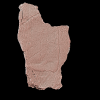
M3#1908Posterior plastral lobe Type: "3D_surfaces"doi: 10.18563/m3.sf.1908 state:in_press |
Download 3D surface file |
Bartassochersis lopezi UM-PDE2-03 View specimen
|
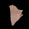
M3#1909First vertebral scute region, right side Type: "3D_surfaces"doi: 10.18563/m3.sf.1909 state:in_press |
Download 3D surface file |
Bartassochersis lopezi UM-PDE2-04 View specimen
|
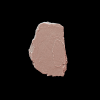
M3#1910?Carapace fragment Type: "3D_surfaces"doi: 10.18563/m3.sf.1910 state:in_press |
Download 3D surface file |
Bartassochersis lopezi UM-PDE2-05 View specimen
|
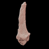
M3#1911Left scapula Type: "3D_surfaces"doi: 10.18563/m3.sf.1911 state:in_press |
Download 3D surface file |
Bartassochersis lopezi UM-PDE2-06 View specimen
|
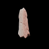
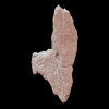
M3#1912Ungual Type: "3D_surfaces"doi: 10.18563/m3.sf.1912 state:in_press |
Download 3D surface file |
Bartassochersis lopezi UM-PDE2-08 View specimen
|
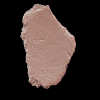
M3#1913Right hypoplastron Type: "3D_surfaces"doi: 10.18563/m3.sf.1913 state:in_press |
Download 3D surface file |
Bartassochersis lopezi UM-PDE2-09 View specimen
|
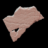
M3#1914Indeterminate Type: "3D_surfaces"doi: 10.18563/m3.sf.1914 state:in_press |
Download 3D surface file |
Bartassochersis lopezi UM-PDE2-10 View specimen
|
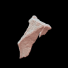
M3#1915?Mesoplastron/hypoplastron Type: "3D_surfaces"doi: 10.18563/m3.sf.1915 state:in_press |
Download 3D surface file |
Bartassochersis lopezi UM-PDE2-11 View specimen
|
M3#1916Left inguinal periphery (peripheral, hyoplastron) Type: "3D_surfaces"doi: 10.18563/m3.sf.1916 state:in_press |
Download 3D surface file |
Bartassochersis lopezi UM-PDE2-12 View specimen
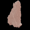
|
M3#1917?Left side of a vertebral scute area with two partial pleural scute areas Type: "3D_surfaces"doi: 10.18563/m3.sf.1917 state:in_press |
Download 3D surface file |
Bartassochersis lopezi UM-PDE2-13 View specimen
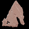
|
M3#1918?Posterior pleural and supramarginal of marginal areas Type: "3D_surfaces"doi: 10.18563/m3.sf.1918 state:in_press |
Download 3D surface file |
Bartassochersis lopezi UM-PDE2-14 View specimen
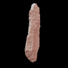
|
M3#1919Posterior peripherals Type: "3D_surfaces"doi: 10.18563/m3.sf.1919 state:in_press |
Download 3D surface file |
Bartassochersis lopezi UM-PDE2-15A View specimen
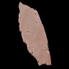
|
M3#1920?Left edge of the anterior plastral lobe Type: "3D_surfaces"doi: 10.18563/m3.sf.1920 state:in_press |
Download 3D surface file |
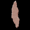
Bartassochersis lopezi UM-PDE2-15B View specimen
|
M3#1921Indeterminate Type: "3D_surfaces"doi: 10.18563/m3.sf.1921 state:in_press |
Download 3D surface file |
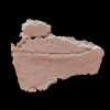
Bartassochersis lopezi UM-PDE2-16 View specimen
|
M3#1922Indeterminate Type: "3D_surfaces"doi: 10.18563/m3.sf.1922 state:in_press |
Download 3D surface file |
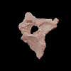
Bartassochersis lopezi UM-PDE2-17 View specimen
|
M3#1923?Bridge fragment Type: "3D_surfaces"doi: 10.18563/m3.sf.1923 state:in_press |
Download 3D surface file |
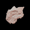
Bartassochersis lopezi UM-PDE2-19 View specimen
|
M3#1924Right bridge region Type: "3D_surfaces"doi: 10.18563/m3.sf.1924 state:in_press |
Download 3D surface file |
Bartassochersis lopezi UM-PDE2-20 View specimen
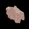
|
M3#1925Left femoral bulge Type: "3D_surfaces"doi: 10.18563/m3.sf.1925 state:in_press |
Download 3D surface file |
Bartassochersis lopezi UM-PDE2-21 View specimen
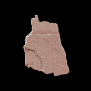
|
M3#1926Indeterminate Type: "3D_surfaces"doi: 10.18563/m3.sf.1926 state:in_press |
Download 3D surface file |
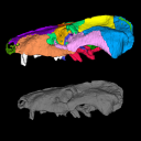
 , Agustín Martinelli and Marco Brandalise de Andrade
 The present 3D Dataset contains the 3D model analyzed in the following publication: Carolina A. Hoffmann, A. G. Martinelli & M. B. Andrade. 2023. Anatomy of the holotype of “Probelesodon” kitchingi revisited, a chiniquodontid cynodont (Synapsida, Probainognathia) from the early Late Triassic of southern Brazil, Journal of Paleontology
Probelesodon kitchingi MCP 1600 PV View specimen
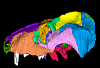
|
M3#11513D models of the skull with segmented bones and without the segmentation. colormap and orientation files also added. Type: "3D_surfaces"doi: 10.18563/m3.sf.1151 state:published |
Download 3D surface file |
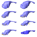 , Marc Jones , José D. Ferreira and Annie Hsiou
 The present 3D Dataset contains the 3D models analyzed in the following manuscript: L. Roese-Miron, M.E.H. Jones, J.D. Ferreira and A.S. Hsiou., 2023. Virtual endocasts of Clevosaurus brasiliensis and the tuatara: Rhynchocephalian neuroanatomy and the oldest endocranial record for Lepidosauria.
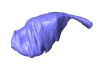
Sphenodon punctatus CM 30660 View specimen
|
M3#10993D surface model of the cranial endocast of specimen CM 30660 (Sphenodon punctatus). Type: "3D_surfaces"doi: 10.18563/m3.sf.1099 state:published |
Download 3D surface file |
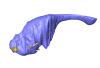
Sphenodon punctatus KCLZJ 001 View specimen
|
M3#11003D surface models of the cranial endocast and the initial trunks of the cranial nerves of specimen KCLZJ 001 (Sphenodon punctatus). Type: "3D_surfaces"doi: 10.18563/m3.sf.1100 state:published |
Download 3D surface file |
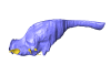
Sphenodon punctatus LDUCZ x0036 View specimen
|
M3#11013D surface models of the cranial endocast and the initial trunks of the cranial nerves of specimen LDUCZ x0036 (Sphenodon punctatus). Type: "3D_surfaces"doi: 10.18563/m3.sf.1101 state:published |
Download 3D surface file |
Sphenodon punctatus LDUCZ x1126 View specimen
|
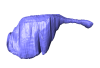
M3#11023D surface model of the cranial endocast of specimen LDUCZ x1126 (Sphenodon punctatus). Type: "3D_surfaces"doi: 10.18563/m3.sf.1102 state:published |
Download 3D surface file |
Clevosaurus brasiliensis MCN PV 2852 View specimen
|
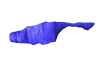
M3#11033D surface model of the cranial endocast of specimen MCN PV 2852 (Clevosaurus brasiliensis). Type: "3D_surfaces"doi: 10.18563/m3.sf.1103 state:published |
Download 3D surface file |
Sphenodon punctatus SAMA 70524 View specimen
|
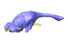
M3#11043D surface models of the cranial endocast, brain, endosseous labyrinth and initial trunks of the cranial nerves of specimen SAMA 70524 (Sphenodon punctatus). Type: "3D_surfaces"doi: 10.18563/m3.sf.1104 state:published |
Download 3D surface file |
Sphenodon punctatus SU1 View specimen
|
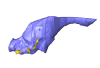
M3#11053D surface models of the cranial endocast and the initial trunks of the cranial nerves of specimen SU1 (Sphenodon punctatus). Type: "3D_surfaces"doi: 10.18563/m3.sf.1105 state:published |
Download 3D surface file |
Sphenodon punctatus YPM HERR 009194 View specimen
|
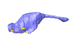
M3#11063D surface models of the cranial endocast and the initial trunks of the cranial nerves of specimen YPM HERR 009194 (Sphenodon punctatus). Type: "3D_surfaces"doi: 10.18563/m3.sf.1106 state:published |
Download 3D surface file |
 and Sam Giles
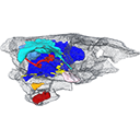
 The present 3D Dataset contains the 3D models analyzed in: "a giant dapediid from the Late Triassic of Switzerland and insights into neopterygian phylogeny", Royal Society Open Science, https://doi.org/10.1098/rsos.180497
Scopulipiscis saxciput PIMUZ A/I 3026 View specimen

|
M3#1773D surfaces of the skull and endocranial spaces inside neurocranium, including the aortic canal, braincase, fossa bridgei, lateral cranial canal, nerves and other passageways, notochord, posterior myodome, and right semicircular canals. Type: "3D_surfaces"doi: 10.18563/m3.sf.177 state:published |
Download 3D surface file |

|
M3#178Scan of the neurocranium of PIMUZ A/I 3026 Type: "3D_CT"doi: 10.18563/m3.sf.178 state:published |
Download CT data |
Page 1 of 1, showing 4 record(s) out of 4 total