3D models of Cainotheriids Ossicular chain
3D models of Kalakocetus, the earliest Cetacea
The specimens of Speothos pacivorus
3D GM dataset of bird skeletal variation
Skeletal embryonic development in the catshark
Bony connexions of the petrosal bone of extant hippos
bony labyrinth (14) , inner ear (11) , Eocene (11) , geometric morphometrics (10) , CT-scan (10) , Oligocene (9) , Micro-CT (9)
Lionel Hautier (25) , Maëva Judith Orliac (24) , Laurent Marivaux (18) , Renaud Lebrun (15) , Rodolphe Tabuce (15) , Pierre-Olivier Antoine (13) , Bastien Mennecart (13)
MorphoMuseuM Volume 09, issue 02
<< prev. article next article >>
|
3D dataset3D models related to the publication: Virtual endocasts of Clevosaurus brasiliensis and the tuatara: rhynchocephalian neuroanatomy and the oldest endocranial record for Lepidosauria
|
|
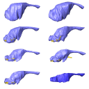
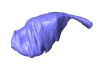
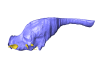
M3#10993D surface model of the cranial endocast of specimen CM 30660 (Sphenodon punctatus). Type: "3D_surfaces"doi: 10.18563/m3.sf.1099 state:published |
Download 3D surface file |
Sphenodon punctatus KCLZJ 001 View specimen
|
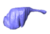
M3#11003D surface models of the cranial endocast and the initial trunks of the cranial nerves of specimen KCLZJ 001 (Sphenodon punctatus). Type: "3D_surfaces"doi: 10.18563/m3.sf.1100 state:published |
Download 3D surface file |
Sphenodon punctatus LDUCZ x0036 View specimen
|
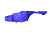
M3#11013D surface models of the cranial endocast and the initial trunks of the cranial nerves of specimen LDUCZ x0036 (Sphenodon punctatus). Type: "3D_surfaces"doi: 10.18563/m3.sf.1101 state:published |
Download 3D surface file |
Sphenodon punctatus LDUCZ x1126 View specimen
|
M3#11023D surface model of the cranial endocast of specimen LDUCZ x1126 (Sphenodon punctatus). Type: "3D_surfaces"doi: 10.18563/m3.sf.1102 state:published |
Download 3D surface file |
Clevosaurus brasiliensis MCN PV 2852 View specimen
|
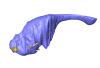
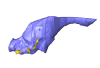
M3#11033D surface model of the cranial endocast of specimen MCN PV 2852 (Clevosaurus brasiliensis). Type: "3D_surfaces"doi: 10.18563/m3.sf.1103 state:published |
Download 3D surface file |
Sphenodon punctatus SAMA 70524 View specimen
|
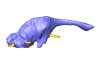
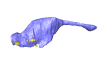
M3#11043D surface models of the cranial endocast, brain, endosseous labyrinth and initial trunks of the cranial nerves of specimen SAMA 70524 (Sphenodon punctatus). Type: "3D_surfaces"doi: 10.18563/m3.sf.1104 state:published |
Download 3D surface file |
Sphenodon punctatus SU1 View specimen
|
M3#11053D surface models of the cranial endocast and the initial trunks of the cranial nerves of specimen SU1 (Sphenodon punctatus). Type: "3D_surfaces"doi: 10.18563/m3.sf.1105 state:published |
Download 3D surface file |
Sphenodon punctatus YPM HERR 009194 View specimen
|
M3#11063D surface models of the cranial endocast and the initial trunks of the cranial nerves of specimen YPM HERR 009194 (Sphenodon punctatus). Type: "3D_surfaces"doi: 10.18563/m3.sf.1106 state:published |
Download 3D surface file |
Balanoff, A.M., and Bever, G.S., 2017. The role of endocasts in the study of brain evolution. In Evolution of Nervous Systems, Second Edition, J. H. Kaas, ed. (Academic Press), pp. 223–241. https://doi.org/10.1016/B978-0-12-804042-3.00023-3
Balanoff, A.M., Bever, G.S., Colbert, M.W., Clarke, J.A., Field, D.J., Gignac, P.M., Ksepka, D.T., Ridgely, R.C., Smith, N.A., Torres, C.R., et al., 2016. Best practices for digitally constructing endocranial casts: examples from birds and their dinosaurian relatives. Journal of Anatomy 229, 173-190. https://doi.org/10.1111/joa.12378
Edinger, T., 1941. The brain of Pterodactylus. American Journal of Science 239 (9), 665-682. https://doi.org/10.2475/ajs.239.9.665
Evans, S.E., and Jones, M.E.H., 2010. The origin, early history and diversification of lepidosauromorph reptiles. In New aspects of Mesozoic biodiversity (Springer Berlin, Heidelberg), pp. 27-44. https://doi.org/10.1007/978-3-642-10311-7_2
Jones, M.E.H., and Cree, A., 2012. Tuatara. Current Biology. 22 (23), R986-R987. https://doi.org/10.1016/j.cub.2012.10.049
Maisano, J. (2001, a). "Sphenodon punctatus" (On-line), Digital Morphology. In http://digimorph.org/specimens/Sphenodon_punctatus/juvenile/
Maisano, J. (2001, b). "Sphenodon punctatus" (On-line), Digital Morphology. In http://digimorph.org/specimens/Sphenodon_punctatus/adult/
Roese-Miron, L., Jones, M.E.H., Ferreira,J.D. and Hsiou., A.S., 2023. Virtual endocasts of Clevosaurus brasiliensis and the tuatara: rhynchocephalian neuroanatomy and the oldest endocranial record for Lepidosauria. https://doi.org/10.1002/ar.25212
Lívia Roese‐Miron, Marc Emyr Huw Jones, José Darival Ferreira and Annie Schmaltz Hsiou (2024). Virtual endocasts of Clevosaurus brasiliensis and the tuatara: Rhynchocephalian neuroanatomy and the oldest endocranial record for Lepidosauria. The Anatomical Record. https://doi.org/10.1002/ar.25212