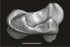
3D models of Cainotheriids Ossicular chain
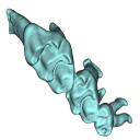
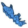
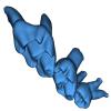
3D models of Kalakocetus, the earliest Cetacea
The specimens of Speothos pacivorus
3D GM dataset of bird skeletal variation
Skeletal embryonic development in the catshark
Bony connexions of the petrosal bone of extant hippos
bony labyrinth (14) , inner ear (11) , Eocene (11) , geometric morphometrics (10) , CT-scan (10) , Oligocene (9) , Micro-CT (9)
Lionel Hautier (25) , Maëva Judith Orliac (24) , Laurent Marivaux (18) , Renaud Lebrun (15) , Rodolphe Tabuce (15) , Pierre-Olivier Antoine (13) , Bastien Mennecart (13)
MorphoMuseuM Volume 09, issue 03:September 2023

|
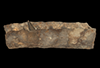
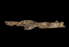
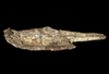
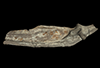
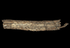
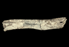
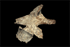
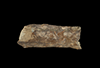
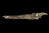
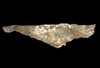
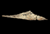
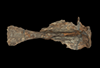
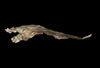
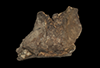
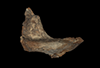
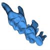
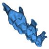
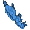
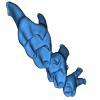
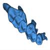
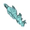
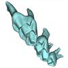
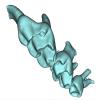
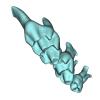
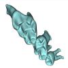
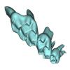
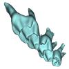
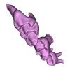
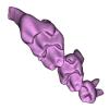
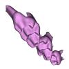
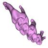
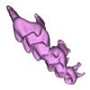
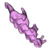
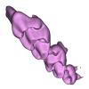
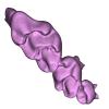
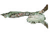
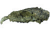
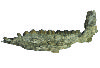
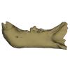
Virtual reconstruction of a Late Jurassic metriorhynchid skull from Switzerland and its use for scientific illustration and paleoartSophie De Sousa Oliveira, Léa Girard
Published online: 19/07/2023 |
|
M3#1037Left dentary (3 meshes) Type: "3D_surfaces"doi: 10.18563/m3.sf.1037 state:published |
Download 3D surface file |
|
M3#1038Right dentary (3 meshes) Type: "3D_surfaces"doi: 10.18563/m3.sf.1038 state:published |
Download 3D surface file |
|
M3#1039Left ramus (2 meshes) Type: "3D_surfaces"doi: 10.18563/m3.sf.1039 state:published |
Download 3D surface file |
|
M3#1040Right ramus (3 meshes) Type: "3D_surfaces"doi: 10.18563/m3.sf.1040 state:published |
Download 3D surface file |
|
M3#1041Left splenial (2 meshes) Type: "3D_surfaces"doi: 10.18563/m3.sf.1041 state:published |
Download 3D surface file |
|
M3#1042Right splenial (2 meshes) Type: "3D_surfaces"doi: 10.18563/m3.sf.1042 state:published |
Download 3D surface file |
|
M3#1043Frontal and left prefrontal Type: "3D_surfaces"doi: 10.18563/m3.sf.1043 state:published |
Download 3D surface file |
|
M3#1044Left maxilla (4 meshes) Type: "3D_surfaces"doi: 10.18563/m3.sf.1044 state:published |
Download 3D surface file |
|
M3#1045Right maxilla Type: "3D_surfaces"doi: 10.18563/m3.sf.1045 state:published |
Download 3D surface file |
|
M3#1046Left nasal Type: "3D_surfaces"doi: 10.18563/m3.sf.1046 state:published |
Download 3D surface file |
|
M3#1047Right nasal Type: "3D_surfaces"doi: 10.18563/m3.sf.1047 state:published |
Download 3D surface file |
|
M3#1048Parietal Type: "3D_surfaces"doi: 10.18563/m3.sf.1048 state:published |
Download 3D surface file |
|
M3#1049Right postorbital Type: "3D_surfaces"doi: 10.18563/m3.sf.1049 state:published |
Download 3D surface file |
|
M3#1050Right prefrontal Type: "3D_surfaces"doi: 10.18563/m3.sf.1050 state:published |
Download 3D surface file |
|
M3#1051Right premaxilla Type: "3D_surfaces"doi: 10.18563/m3.sf.1051 state:published |
Download 3D surface file |
|
M3#1052Left squamosal Type: "3D_surfaces"doi: 10.18563/m3.sf.1052 state:published |
Download 3D surface file |
|
M3#1053Right squamosal Type: "3D_surfaces"doi: 10.18563/m3.sf.1053 state:published |
Download 3D surface file |
|
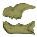
M3#1054Reconstruction of the mandible Type: "3D_surfaces"doi: 10.18563/m3.sf.1054 state:published |
Download 3D surface file |

|
M3#1055Reconstruction of the cranium Type: "3D_surfaces"doi: 10.18563/m3.sf.1055 state:published |
Download 3D surface file |
|
3D model related to the publication: An eosimiid primate of South Asian affinities in the Paleogene of Western Amazonia and the origin of New World monkeysLaurent Marivaux
Published online: 04/07/2023 |
|
M3#1114Right first upper molar (rM1), pristine. Type: "3D_surfaces"doi: 10.18563/m3.sf.1114 state:published |
Download 3D surface file |
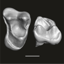
This contribution contains 3D models of upper molar rows of house mice (Mus musculus domesticus) belonging to Western European commensal and Sub-Antarctic feral populations. These two groups are characterized by different patterns of wear and alignment of the three molars along the row, related to contrasted masticatory demand in relation with their diet. These models are analyzed in the following publication: Renaud et al 2023, “Molar wear in house mice, insight into diet preferences at an ecological time scale?”, https://doi.org/10.1093/biolinnean/blad091
Mus musculus G09_06 View specimen
|
M3#1166right upper molar row Type: "3D_surfaces"doi: 10.18563/m3.sf.1166 state:published |
Download 3D surface file |
Mus musculus G09_10 View specimen
|
M3#1168right upper molar row Type: "3D_surfaces"doi: 10.18563/m3.sf.1168 state:published |
Download 3D surface file |
Mus musculus G09_15 View specimen
|
M3#1169right upper molar row Type: "3D_surfaces"doi: 10.18563/m3.sf.1169 state:published |
Download 3D surface file |
Mus musculus G09_16 View specimen
|
M3#1170right upper molar row Type: "3D_surfaces"doi: 10.18563/m3.sf.1170 state:published |
Download 3D surface file |
Mus musculus G09_17 View specimen
|
M3#1171right upper molar row Type: "3D_surfaces"doi: 10.18563/m3.sf.1171 state:published |
Download 3D surface file |
Mus musculus G09_21 View specimen
|
M3#1172right upper molar row Type: "3D_surfaces"doi: 10.18563/m3.sf.1172 state:published |
Download 3D surface file |
Mus musculus G09_26 View specimen
|
M3#1173right upper molar row Type: "3D_surfaces"doi: 10.18563/m3.sf.1173 state:published |
Download 3D surface file |
Mus musculus G09_27 View specimen
|
M3#1174right upper molar row Type: "3D_surfaces"doi: 10.18563/m3.sf.1174 state:published |
Download 3D surface file |
Mus musculus G09_29 View specimen
|
M3#1175right upper molar row Type: "3D_surfaces"doi: 10.18563/m3.sf.1175 state:published |
Download 3D surface file |
Mus musculus G09_65 View specimen
|
M3#1176right upper molar row Type: "3D_surfaces"doi: 10.18563/m3.sf.1176 state:published |
Download 3D surface file |
Mus musculus G09_66 View specimen
|
M3#1177right upper molar row Type: "3D_surfaces"doi: 10.18563/m3.sf.1177 state:published |
Download 3D surface file |
Mus musculus G93_03 View specimen
|
M3#1178right upper molar row Type: "3D_surfaces"doi: 10.18563/m3.sf.1178 state:published |
Download 3D surface file |
Mus musculus G93_04 View specimen
|
M3#1179right upper molar row Type: "3D_surfaces"doi: 10.18563/m3.sf.1179 state:published |
Download 3D surface file |
Mus musculus G93_10 View specimen
|
M3#1180right upper molar row Type: "3D_surfaces"doi: 10.18563/m3.sf.1180 state:published |
Download 3D surface file |
Mus musculus G93_11 View specimen
|
M3#1181right upper molar row Type: "3D_surfaces"doi: 10.18563/m3.sf.1181 state:published |
Download 3D surface file |
Mus musculus G93_13 View specimen
|
M3#1182right upper molar row Type: "3D_surfaces"doi: 10.18563/m3.sf.1182 state:published |
Download 3D surface file |
Mus musculus G93_14 View specimen
|
M3#1183right upper molar row Type: "3D_surfaces"doi: 10.18563/m3.sf.1183 state:published |
Download 3D surface file |
Mus musculus G93_15 View specimen
|
M3#1184right upper molar row Type: "3D_surfaces"doi: 10.18563/m3.sf.1184 state:published |
Download 3D surface file |
Mus musculus G93_24 View specimen
|
M3#1185left molar row Type: "3D_surfaces"doi: 10.18563/m3.sf.1185 state:published |
Download 3D surface file |
Mus musculus Tourch_7819 View specimen
|
M3#1186right upper molar row Type: "3D_surfaces"doi: 10.18563/m3.sf.1186 state:published |
Download 3D surface file |
Mus musculus G93_25 View specimen
|
M3#1187right upper molar row Type: "3D_surfaces"doi: 10.18563/m3.sf.1187 state:published |
Download 3D surface file |
Mus musculus Tourch_7821 View specimen
|
M3#1188right upper molar row Type: "3D_surfaces"doi: 10.18563/m3.sf.1188 state:published |
Download 3D surface file |
Mus musculus Tourch_7839 View specimen
|
M3#1189right upper molar row Type: "3D_surfaces"doi: 10.18563/m3.sf.1189 state:published |
Download 3D surface file |
Mus musculus Tourch_7873 View specimen
|
M3#1190right upper molar row Type: "3D_surfaces"doi: 10.18563/m3.sf.1190 state:published |
Download 3D surface file |
Mus musculus Tourch_7877 View specimen
|
M3#1196right upper molar row Type: "3D_surfaces"doi: 10.18563/m3.sf.1196 state:published |
Download 3D surface file |
Mus musculus Tourch_7922 View specimen
|
M3#1191right upper molar row Type: "3D_surfaces"doi: 10.18563/m3.sf.1191 state:published |
Download 3D surface file |
Mus musculus Tourch_7923 View specimen
|
M3#1192right upper molar row Type: "3D_surfaces"doi: 10.18563/m3.sf.1192 state:published |
Download 3D surface file |
Mus musculus Tourch_7925 View specimen
|
M3#1193right upper molar row Type: "3D_surfaces"doi: 10.18563/m3.sf.1193 state:published |
Download 3D surface file |
Mus musculus Tourch_7927 View specimen
|
M3#1194right upper molar row Type: "3D_surfaces"doi: 10.18563/m3.sf.1194 state:published |
Download 3D surface file |
Mus musculus Tourch_7932 View specimen
|
M3#1195right upper molar row Type: "3D_surfaces"doi: 10.18563/m3.sf.1195 state:published |
Download 3D surface file |
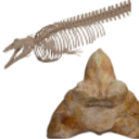
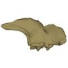
The present 3D Dataset contains the 3D models analyzed in Bianucci et al. 2023, A heavyweight early whale pushes the boundaries of vertebrate morphology, Nature. These include bones of the holotype of new species Perucetus colossus (MUSM 3248), as well as the articulated skeleton of Cynthiacetus peruvianus (holotype, MNHN.F.PRU10). The latter was used to estimate the total skeleton volume of P. colossus.
Perucetus colossus MUSM 3248 View specimen
|
M3#1131Thirteen vertebrae, rib, and innominate of Perucetus colossus (holotype, MUSM NNNN). Type: "3D_surfaces"doi: 10.18563/m3.sf.1131 state:published |
Download 3D surface file |
Cynthiacetus peruvianus MNHN.F.PRU10 View specimen
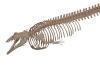
|
M3#1130Articulated skeleton of the holotype of Cynthiacetus peruvianus MNHN.F.PRU10 Type: "3D_surfaces"doi: 10.18563/m3.sf.1130 state:published |
Download 3D surface file |
This contribution contains the 3D models described and figured in: New remains of Neotropical bunodont litopterns and the systematics of Megadolodinae (Mammalia: Litopterna). Geodiversitas.
Megadolodus molariformes VPPLT 974 View specimen
|
M3#1020Partial mandible with the symphysis and left body, bearing the alveoli of ?i2, right and left ?i3, alveolus of right c and p1, roots of left p1, and the left p2–m3 of Megadolodus molariformes (Litopterna, Mammalia) Type: "3D_surfaces"doi: 10.18563/m3.sf.1020 state:published |
Download 3D surface file |
Neodolodus colombianus VPPLT 1696 View specimen
|
M3#1021Almost complete skull with left and right ?I2 and P1–M3 Type: "3D_surfaces"doi: 10.18563/m3.sf.1021 state:published |
Download 3D surface file |
|
M3#1022Partial mandible with complete right and left dentition except for left ?i2 Type: "3D_surfaces"doi: 10.18563/m3.sf.1022 state:published |
Download 3D surface file |
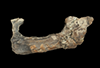
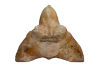
This contribution contains the 3D models described and figured in the following publication: Bonis et al. 2023. A new large pantherine and a sabre-toothed cat (Mammalia, Carnivora, Felidae) from the late Miocene hominoid-bearing Khorat sand pits, Nakhon Ratchasima Province, northeastern Thailand. The Science of Nature 110(5):42. https://doi.org/10.1007/s00114-023-01867-4
Pachypanthera piriyai CUF-KR-1 View specimen
|
M3#1209Holotype of Pachypanthera piriyai, a left hemi-mandible with alveoli for i1-i3 and canine, roots of p3, p4 and partially broken off m1 crown. Type: "3D_surfaces"doi: 10.18563/m3.sf.1209 state:published |
Download 3D surface file |
Pachypanthera piriyai CUF-KR-2 View specimen
|
M3#1210Paratype of Pachypanthera piriyai, a right hemi-maxilla with P3-P4, alveoli of C and M1, root of P2 Type: "3D_surfaces"doi: 10.18563/m3.sf.1210 state:published |
Download 3D surface file |
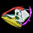
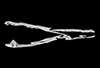
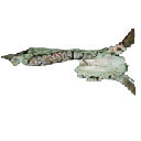
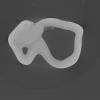
We provide a 3D reconstruction of the skull of Latimeria chalumnae that can be easily accessed and visualized for a better understanding of its cranial anatomy. Different skeletal elements are saved as separate PLY files that can be combined to visualize the entire skull or isolated to virtually dissect the skull. We included some guidelines for a fast and easy visualization of the 3D skull.
Latimeria chalumnae MHNG 1080.070 View specimen

|
M3#1254the skeletal elements of the skull of Latimeria chalumnae included in 26 different PLY files Type: "3D_surfaces"doi: 10.18563/m3.sf.1254 state:published |
Download 3D surface file |
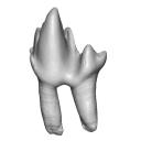
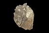
This contribution contains the 3D model described and figured in the following publication: Martin, T., Averianov, A. O., Schultz, J. A., & Schwermann, A. H. (2023). A stem therian mammal from the Lower Cretaceous of Germany. Journal of Vertebrate Paleontology, e2224848.
Spelaeomolitor speratus WMNM P99101 View specimen

|
M3#12573D_model_Spelaeomolitor_lower_molar Type: "3D_surfaces"doi: 10.18563/m3.sf.1257 state:published |
Download 3D surface file |
|
M3#1258CT imagestack (jpgs) and info data sheet (pca file) in one zip folder Type: "3D_CT"doi: 10.18563/m3.sf.1258 state:published |
Download CT data |
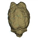
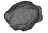
The present 3D Dataset contains the 3D model of the endocranial cast of Palaeolama sp. from the mid-Pleistocene (~1.2 Mya) of South America, analyzed in Balcarcel et al. 2023.
Palaeolama sp. PIMUZ A/V 4091 View specimen
|
M3#11283D model of a natural endocast Type: "3D_surfaces"doi: 10.18563/m3.sf.1128 state:published |
Download 3D surface file |