3D models of Arthropleura sp. from the Montceau-les-Mines Lagerstätte
3D models of Peradectes crocheti from Palette (early Eocene, Provence)
3D models of the masticatory muscles of Anomaluromorpha
3D GM dataset of bird skeletal variation
Skeletal embryonic development in the catshark
Bony connexions of the petrosal bone of extant hippos
bony labyrinth (11) , inner ear (10) , Eocene (8) , South America (8) , Paleobiogeography (7) , skull (7) , phylogeny (6)
Lionel Hautier (21) , Maëva Judith Orliac (19) , Laurent Marivaux (13) , Bastien Mennecart (12) , Pierre-Olivier Antoine (11) , Renaud Lebrun (10) , Leonardo Kerber (10)
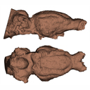
Brain damage: the endocranial cast of Mixtotherium cuspidatum (Mammalia, Artiodactyla) from the Victor Brun Museum (Montauban, France)Maëva J. Orliac , Hugo Bouaziz and Romain WeppePublished online: 25/11/2021Keywords: artiodactyl; Late Eocene; Quercy https://doi.org/10.18563/journal.m3.158 Cite this article: Maëva J. Orliac, Hugo Bouaziz and Romain Weppe, 2021. Brain damage: the endocranial cast of Mixtotherium cuspidatum (Mammalia, Artiodactyla) from the Victor Brun Museum (Montauban, France). MorphoMuseuM 7:e158. doi: 10.18563/journal.m3.158 Export citationAbstract Our knowledge of the external brain morphology of the late Eocene artiodactyl ungulate Mixtotherium, relies on a plaster model realized on a specimen from the Victor Brun Museum in Montauban (France) and described by Dechaseaux (1973). Here, based on micro CT-scan data, we virtually reconstruct the 3D cast of the empty cavity of the partial cranium MA PHQ 716 from the Victor Brun Museum and compare it to the plaster model illustrated and described by Dechaseaux (1973). Indeed, the specimen from which the original plaster endocast originates was not identified by Dechaseaux by a specimen number. We confirm here that the studied specimen was indeed the one described and illustrated by Dechaseaux (1973). We also reconstruct a second, more detailed, model providing additional morphological and quantitative observations made available by micro CT scan investigation such as precisions on the neopallium folding and endocranial volumes. M3 article infos Published in Volume 07, issue 04 (2021) |
|
M3#857endocast of the brain cavity Type: "3D_surfaces"doi: 10.18563/m3.sf.857 Data citation: Maëva J. Orliac, Hugo Bouaziz and Romain Weppe, 2021. M3#857. doi: 10.18563/m3.sf.857 state:published |
Download 3D surface file |