3D models of Cainotheriids Ossicular chain
3D models of Kalakocetus, the earliest Cetacea
The specimens of Speothos pacivorus
3D GM dataset of bird skeletal variation
Skeletal embryonic development in the catshark
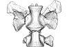
Bony connexions of the petrosal bone of extant hippos
bony labyrinth (14) , inner ear (11) , Eocene (11) , geometric morphometrics (10) , CT-scan (10) , Oligocene (9) , Micro-CT (9)
Lionel Hautier (25) , Maëva Judith Orliac (24) , Laurent Marivaux (18) , Renaud Lebrun (15) , Rodolphe Tabuce (15) , Pierre-Olivier Antoine (13) , Bastien Mennecart (13)
MorphoMuseuM Volume 06, issue 05:December 2020
|
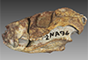
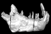
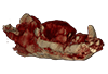
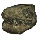
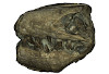
3D models related to the publication: A new traversodontid cynodont with a peculiar postcanine dentition from the Middle/Late Triassic of Namibia and dental evolution in basal gomphodonts.Christophe Hendrickx
Published online: 22/09/2020 |
|
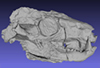
M3#557Surface model derived from µCT data of the holotype of Etjoia dentitransitus Type: "3D_surfaces"doi: 10.18563/m3.sf.557 state:published |
Download 3D surface file |
|
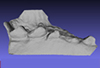
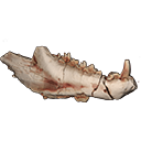
M3#558Photogrammetric 3D surface model of the postcanines of the Holotype of Etjoia dentitransitus Type: "3D_surfaces"doi: 10.18563/m3.sf.558 state:published |
Download 3D surface file |
|
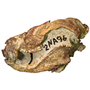
M3#559Photogrammetric 3D surface model of the Holotype of Etjoia dentitransitus Type: "3D_surfaces"doi: 10.18563/m3.sf.559 state:published |
Download 3D surface file |
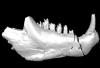
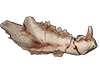
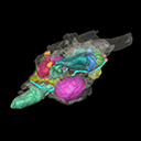
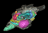
This contribution contains the 3D model(s) described and figured in the following publication: The present 3D Dataset contains the 3D models and CT-Scan slices of the lower jaws and teeth analyzed in “A new prozostrodontian cynodont (Eucynodontia, Probainognathia) from the Upper Triassic of southern Brazil”. https://doi.org/10.1080/02724634.2020.1782415
Agudotherium gassenae CAPPA/UFSM 0262 View specimen
|
M3#546Left lower jaw and cheek teeth Type: "3D_surfaces"doi: 10.18563/m3.sf.546 state:published |
Download 3D surface file |
|
M3#5471578 slices Type: "3D_CT"doi: 10.18563/m3.sf.547 state:published |
Download CT data |
Agudotherium gassenae CAPPA/UFSM 0208 View specimen
|
M3#548right lower jaw Type: "3D_surfaces"doi: 10.18563/m3.sf.548 state:published |
Download 3D surface file |
|
M3#549CT data of CAPPA_UFSM_0208 Type: "3D_CT"doi: 10.18563/m3.sf.549 state:published |
Download CT data |
The present Dataset contains the 3D model of the male genital organs of greater horseshoe bat, Rhinolophus ferrumequinum. This is the first detailed 3D structure of the soft-tissue genital organs of bats. The 3D model was generated using microCT and techniques of virtual reconstruction.
Rhinolophus ferrumequinum JP18-006 View specimen
|
M3#521The genital organs of male greater horseshoe bat. Type: "3D_surfaces"doi: 10.18563/m3.sf.521 state:published |
Download 3D surface file |
The present 3D Dataset contains the 3D model used in in the following publication: Interacting with the inaccessible: utilization of multimedia-based visual contents of Japan’s National Monument, the Taniwhasaurus mikasaensis (Mosasauridae) holotype for educational workshops at Mikasa City Museum.
Taniwhasaurus mikasaensis MCM.M0009 View specimen
|
M3#499Taniwhasaurus mikasaensis, Caldwell et al. 2008 Type: "3D_surfaces"doi: 10.18563/m3.sf.499 state:published |
Download 3D surface file |
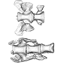
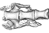
The present 3D Dataset contains the 3D models of the sacral vertebrae analyzed in “Sacral co-ossification in dinosaurs: The oldest record of fused sacral vertebrae in Dinosauria and the diversity of sacral co-ossification patterns in the group”.
Buriolestes schultzi CAPPA/UFSM 0035 View specimen
|
M3#705Sacral vertebrae of Buriolestes schultzi Type: "3D_surfaces"doi: 10.18563/m3.sf.705 state:published |
Download 3D surface file |
indet indet CAPPA/UFSM 0228 View specimen
|
M3#706Sacral vertebrae of a saurischian dinosaur indet. Type: "3D_surfaces"doi: 10.18563/m3.sf.706 state:published |
Download 3D surface file |