Digital reconstruction of the skull of Sarmientosaurus musacchioi
3D models of Arthropleura sp. from the Montceau-les-Mines Lagerstätte
3D models of isolated teeth of a South Asian Paroxyclaenidae
3D GM dataset of bird skeletal variation
Skeletal embryonic development in the catshark
Bony connexions of the petrosal bone of extant hippos
bony labyrinth (11) , inner ear (10) , Eocene (8) , South America (8) , Paleobiogeography (7) , skull (7) , phylogeny (6)
Maëva Judith Orliac (21) , Lionel Hautier (21) , Laurent Marivaux (14) , Bastien Mennecart (12) , Pierre-Olivier Antoine (11) , Renaud Lebrun (10) , Rodolphe Tabuce (10)
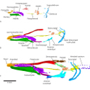
MicroCT survey of larval skeletal mineralization in the Cuban gar Atractosteus tristoechus (Actinopterygii; Lepisosteiformes)Raphaël Scherrer , Andrés Hurtado, Erik Garcia Machado and Mélanie Debiais-ThibaudPublished online: 17/05/2017Keywords: Actinopterygii; development; Lepisosteiformes; mineralization; skeleton https://doi.org/10.18563/m3.3.3.e3 Cite this article: Raphaël Scherrer, Andrés Hurtado, Erik Garcia Machado and Mélanie Debiais-Thibaud, 2017. MicroCT survey of larval skeletal mineralization in the Cuban gar Atractosteus tristoechus (Actinopterygii; Lepisosteiformes). MorphoMuseuM 3 (3)-e3. doi: 10.18563/m3.3.3.e3 Export citationAbstract Using X-ray microtomography, we describe the ossification events during the larval development of a non-teleost actinopterygian species: the Cuban gar Atractosteus tristoechus from the order Lepisosteiformes. We provide a detailed developmental series for each anatomical structure, covering a large sequence of mineralization events going from an early stage (13 days post-hatching, 21mm total length) to an almost fully ossified larval stage (118dph or 87mm in standard length). With this work, we expect to bring new developmental data to be used in further comparative studies with other lineages of bony vertebrates. We also hope that the on-line publication of these twelve successive 3D reconstructions, fully labelled and flagged, will be an educational tool for all students in comparative anatomy. M3 article infos Published in Volume 03, Issue 03 (2017) |
|
M3#96Atractosteus tristoechus larva, 19 dph, 27mm SL. Type: "3D_surfaces"doi: 10.18563/m3.sf.96 Data citation: Raphaël Scherrer, Andrés Hurtado, Erik Garcia Machado and Mélanie Debiais-Thibaud, 2017. M3#96. doi: 10.18563/m3.sf.96 state:published |
Download 3D surface file |