3D models of Cainotheriids Ossicular chain
3D models of Kalakocetus, the earliest Cetacea
Explodable 3D Dog Skull for Veterinary Education
3D GM dataset of bird skeletal variation
Skeletal embryonic development in the catshark
Bony connexions of the petrosal bone of extant hippos
bony labyrinth (14) , inner ear (11) , Eocene (11) , geometric morphometrics (10) , CT-scan (10) , Oligocene (9) , Micro-CT (9)
Lionel Hautier (25) , Maëva Judith Orliac (24) , Laurent Marivaux (18) , Renaud Lebrun (15) , Rodolphe Tabuce (15) , Pierre-Olivier Antoine (13) , Bastien Mennecart (13)
Page 1 of 1, showing 2 record(s) out of 2 total
|
3D model related to the publication: Morphology and distribution of scales, dermal ossifications, and other non-feather integumentary structures in non-avialan theropod dinosaursChristophe Hendrickx
Published online: 10/01/2022 |
|
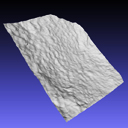
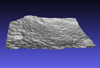
M3#902The material consists of a 3D reconstruction of the counterpart of a 30 cm2 patch of skin impression associated with the anterior dorsal ribs/pectoral region of the specimen of Allosaurus jimmadseni UMNH VP C481. The skin shows a semi-uniform basement of 1-2 mm diameter pebbles with a smaller number of slightly larger (up to 3 mm) ovoid scales. The irregular shape, distribution, and overall small size of these larger scales suggest that they are not classifiable as feature scales but rather as variations in the basement scales. Type: "3D_surfaces"doi: 10.18563/m3.sf.902 state:published |
Download 3D surface file |
The present 3D Dataset contains the 3D model analyzed in the following publication: Solé et al. (2018), Niche partitioning of the European carnivorous mammals during the paleogene. Palaios. https://doi.org/10.2110/palo.2018.022
Hyaenodon leptorhynchus FSL848325 View specimen
|
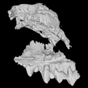
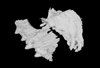
M3#336The specimen FSL848325 is separated in two fragments: the anterior part bears the incisors, the deciduous and permanent canines, while the posterior part bears the right P3, P4, M1 and M2. The P2 is isolated. When combined, the cranium length is approximatively 10.5 cm long. The anterior part is 6.9 cm long and 2.15 cm wide (taken at the level of the P1). The posterior part is 4.8 cm long. The anterior part of the cranium is very narrow. Type: "3D_surfaces"doi: 10.18563/m3.sf.336 state:published |
Download 3D surface file |
Page 1 of 1, showing 2 record(s) out of 2 total


