3D models of Cainotheriids Ossicular chain
3D models of Kalakocetus, the earliest Cetacea
The specimens of Speothos pacivorus
3D GM dataset of bird skeletal variation
Skeletal embryonic development in the catshark
Bony connexions of the petrosal bone of extant hippos
bony labyrinth (14) , inner ear (11) , Eocene (11) , geometric morphometrics (10) , CT-scan (10) , Oligocene (9) , Micro-CT (9)
Lionel Hautier (25) , Maëva Judith Orliac (24) , Laurent Marivaux (18) , Renaud Lebrun (15) , Rodolphe Tabuce (15) , Pierre-Olivier Antoine (13) , Bastien Mennecart (13)
Page 1 of 1, showing 4 record(s) out of 4 total
|
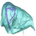
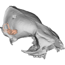
3D models related to the publication: Brain drain: exceptional pattern of calvarial venation in pangolins and its phylogenetic significance for FeraeGuillaume Billet
Published online: 26/03/2026 |
|
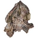
M3#1847cranium & intradiploic canals (sinuses & diploic veins) Type: "3D_surfaces"doi: 10.18563/m3.sf.1847 state:published |
Download 3D surface file |
Manis javanica NHM-UK 9.1.5.858 View specimen
|
M3#1848cranium & intradiploic canals (sinuses & diploic veins) Type: "3D_surfaces"doi: 10.18563/m3.sf.1848 state:published |
Download 3D surface file |
Felis silvestris UM-ZOOL-149N View specimen
|
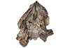
M3#1849cranium & intradiploic canals (sinuses & diploic veins) Type: "3D_surfaces"doi: 10.18563/m3.sf.1849 state:published |
Download 3D surface file |
Erinaceus europaeus SMNS40759 View specimen
|
M3#1850cranium & intradiploic canals (sinuses & diploic veins) Type: "3D_surfaces"doi: 10.18563/m3.sf.1850 state:published |
Download 3D surface file |
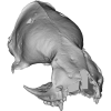
Pterodon dasyuroides MNHN.F.Qu8301 View specimen
|
M3#1851cranium & intradiploic canals (sinuses & diploic veins) Type: "3D_surfaces"doi: 10.18563/m3.sf.1851 state:published |
Download 3D surface file |

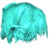
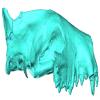
Veterinary education often relies on cadaveric specimens, but there is increasing demand for alternatives due to limited resources and ethical considerations. To address this, we developed a 3D printed ‘explodable’ model of a dog cranium with detachable, magnetized cranial components for teaching anatomy to students. This model was generated from a computed tomographic scan of a juvenile dog cranium for which cranial sutures were still partially open and segmented such that major cranial bones were isolated. All bones are printed at actual size and retain openings for cranial nerves and major vessels. This interactive model enhances anatomical education by supplying a hands-on tool that can be used either in the classroom setting or for independent learning and can be incorporated at the high school, college, or veterinary school level. It is currently being integrated into the first-year anatomy foundation course at Cornell University’s College of Veterinary Medicine. The model can be printed using any hobbyist or specialist 3D printer and we outline assembly instructions on how to attach magnets at prefabricated attachment points. Using both digital and 3D printed resources, we hope to help to address current shortages of anatomical resources and also inspire future generations of practicing veterinarians by making anatomy more accessible and engaging.
Canis lupus familiaris CUHL 9 View specimen

|
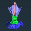
M3#1858PLYs of the segmented cranial bones with pre-fabricated magnetic casings and shelves for assembly following 3D printing Type: "3D_surfaces"doi: 10.18563/m3.sf.1858 state:published |
Download 3D surface file |
|
M3#1859PLYs of the segmented cranial bones of the "BOTTOM" cranial component. Downloadable for additional learning opportunities for students Type: "3D_surfaces"doi: 10.18563/m3.sf.1859 state:published |
Download 3D surface file |
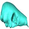
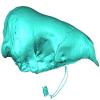
The present 3D Dataset contains 3D models of the cranium surface and of the bony labyrinth endocast of the stem bat Vielasia sigei. They are used by (Hand et al., 2023) to explore the phylogenetic position of this species, to infer its laryngeal echolocating capabilities, and to eventually discuss chiropteran evolution before the crown clade diversification.
Vielasia sigei UM VIE-250 View specimen
|
M3#1269External surface of the cranium Type: "3D_surfaces"doi: 10.18563/m3.sf.1269 state:published |
Download 3D surface file |
|
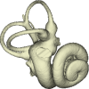
M3#1270Virtual endocast of the right bony labyrinth Type: "3D_surfaces"doi: 10.18563/m3.sf.1270 state:published |
Download 3D surface file |
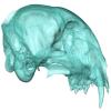
This contribution contains the 3D surface model of the holotype cranium of the Late Jurassic thalassochelydian turtle Solnhofia brachyrhyncha described and figured in the publication of Anquetin and Püntener (2020).
Solnhofia brachyrhyncha MJSN BAN001-2.1 View specimen
|
M3#536Textured 3D surface model of the holotype cranium of the Late Jurassic turtle Solnhofia brachyrhyncha Type: "3D_surfaces"doi: 10.18563/m3.sf.536 state:published |
Download 3D surface file |
Page 1 of 1, showing 4 record(s) out of 4 total


