3D models of Cainotheriids Ossicular chain
3D models of Kalakocetus, the earliest Cetacea
The specimens of Speothos pacivorus
3D GM dataset of bird skeletal variation
Skeletal embryonic development in the catshark
Bony connexions of the petrosal bone of extant hippos
bony labyrinth (14) , inner ear (11) , Eocene (11) , geometric morphometrics (10) , CT-scan (10) , Oligocene (9) , Micro-CT (9)
Lionel Hautier (25) , Maëva Judith Orliac (24) , Laurent Marivaux (18) , Renaud Lebrun (15) , Rodolphe Tabuce (15) , Pierre-Olivier Antoine (13) , Bastien Mennecart (13)
Page 1 of 1, showing 2 record(s) out of 2 total
|
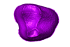
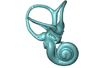
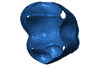
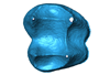
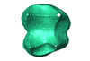
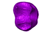
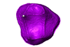
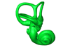
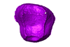
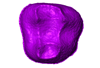
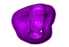
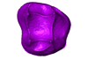
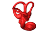
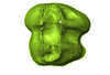
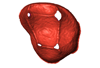
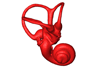
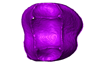
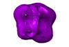
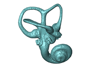
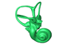
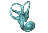
3D models related to the publication: Upper third molar internal structural organization and semicircular canal morphology in Plio-Pleistocene South African cercopithecoids.Amélie Beaudet
Published online: 10/10/2019 |
|
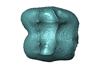
M3#4453D model of the enamel-dentine junction of the right upper third molar. Type: "3D_surfaces"doi: 10.18563/m3.sf.445 state:published |
Download 3D surface file |
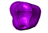
Cercocebus torquatus 73.018-M-0359 View specimen
|
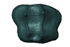
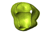
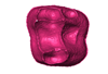
M3#4463D model of the enamel-dentine junction of the right upper third molar. Type: "3D_surfaces"doi: 10.18563/m3.sf.446 state:published |
Download 3D surface file |

|
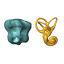
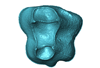
M3#4963D model of the left bony labyrinth. Type: "3D_surfaces"doi: 10.18563/m3.sf.496 state:published |
Download 3D surface file |
Mandrillus leucophaeus 73.029-M-0106 View specimen
|
M3#4473D model of the enamel-dentine junction of the right upper third molar. Type: "3D_surfaces"doi: 10.18563/m3.sf.447 state:published |
Download 3D surface file |

|
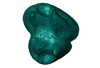
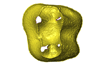
M3#4703D model of the right bony labyrinth. Type: "3D_surfaces"doi: 10.18563/m3.sf.470 state:published |
Download 3D surface file |
Lophocebus albigena 73.029-M-0109 View specimen
|
M3#4483D model of the enamel-dentine junction of the right upper third molar. Type: "3D_surfaces"doi: 10.18563/m3.sf.448 state:published |
Download 3D surface file |
|
M3#4713D model of the right bony labyrinth. Type: "3D_surfaces"doi: 10.18563/m3.sf.471 state:published |
Download 3D surface file |
Piliocolobus foai 91.060-M-0071 View specimen
|
M3#4493D model of the enamel-dentine junction of the right upper third molar. Type: "3D_surfaces"doi: 10.18563/m3.sf.449 state:published |
Download 3D surface file |

|
M3#4723D model of the right bony labyrinth. Type: "3D_surfaces"doi: 10.18563/m3.sf.472 state:published |
Download 3D surface file |
Colobus guereza 1215 View specimen
|
M3#4503D model of the enamel-dentine junction of the right upper third molar. Type: "3D_surfaces"doi: 10.18563/m3.sf.450 state:published |
Download 3D surface file |

|
M3#4733D model of the right bony labyrinth. Type: "3D_surfaces"doi: 10.18563/m3.sf.473 state:published |
Download 3D surface file |
Colobus guereza 2800 View specimen
|
M3#4513D model of the enamel-dentine junction of the right upper third molar. Type: "3D_surfaces"doi: 10.18563/m3.sf.451 state:published |
Download 3D surface file |

|
M3#4743D model of the right bony labyrinth. Type: "3D_surfaces"doi: 10.18563/m3.sf.474 state:published |
Download 3D surface file |
Papio cynocephalus kindae 3503 View specimen
|
M3#4523D model of the enamel-dentine junction of the right upper third molar. Type: "3D_surfaces"doi: 10.18563/m3.sf.452 state:published |
Download 3D surface file |

|
M3#4753D model of the right bony labyrinth. Type: "3D_surfaces"doi: 10.18563/m3.sf.475 state:published |
Download 3D surface file |
Erythrocebus patas 8452 View specimen
|
M3#4533D model of the enamel-dentine junction of the right upper third molar. Type: "3D_surfaces"doi: 10.18563/m3.sf.453 state:published |
Download 3D surface file |

|
M3#4763D model of the right bony labyrinth. Type: "3D_surfaces"doi: 10.18563/m3.sf.476 state:published |
Download 3D surface file |
Papio cynocephalus kindae 17979 View specimen
|
M3#4543D model of the enamel-dentine junction of the right upper third molar. Type: "3D_surfaces"doi: 10.18563/m3.sf.454 state:published |
Download 3D surface file |

|
M3#4773D model of the right bony labyrinth. Type: "3D_surfaces"doi: 10.18563/m3.sf.477 state:published |
Download 3D surface file |
Colobus angolensis 25456 View specimen
|
M3#4553D model of the enamel-dentine junction of the right upper third molar. Type: "3D_surfaces"doi: 10.18563/m3.sf.455 state:published |
Download 3D surface file |

|
M3#4783D model of the right bony labyrinth. Type: "3D_surfaces"doi: 10.18563/m3.sf.478 state:published |
Download 3D surface file |
Chlorocebus pygerythrus 37477 View specimen
|
M3#4563D model of the enamel-dentine junction of the right upper third molar. Type: "3D_surfaces"doi: 10.18563/m3.sf.456 state:published |
Download 3D surface file |
|
M3#4813D model of the right bony labyrinth. Type: "3D_surfaces"doi: 10.18563/m3.sf.481 state:published |
Download 3D surface file |
Chlorocebus pygerythrus 37478 View specimen
|
M3#4573D model of the enamel-dentine junction of the right upper third molar. Type: "3D_surfaces"doi: 10.18563/m3.sf.457 state:published |
Download 3D surface file |

|
M3#4823D model of the right bony labyrinth. Type: "3D_surfaces"doi: 10.18563/m3.sf.482 state:published |
Download 3D surface file |
Lophocebus albigena 37572 View specimen
|
M3#4583D model of the enamel-dentine junction of the right upper third molar. Type: "3D_surfaces"doi: 10.18563/m3.sf.458 state:published |
Download 3D surface file |

|
M3#4833D model of the right bony labyrinth. Type: "3D_surfaces"doi: 10.18563/m3.sf.483 state:published |
Download 3D surface file |
Lophocebus albigena 37579 View specimen
|
M3#4593D model of the enamel-dentine junction of the right upper third molar. Type: "3D_surfaces"doi: 10.18563/m3.sf.459 state:published |
Download 3D surface file |
Erythrocebus patas OST.2002-26 View specimen
|
M3#4603D model of the enamel-dentine junction of the right upper third molar. Type: "3D_surfaces"doi: 10.18563/m3.sf.460 state:published |
Download 3D surface file |
|
M3#4843D model of the right bony labyrinth. Type: "3D_surfaces"doi: 10.18563/m3.sf.484 state:published |
Download 3D surface file |
Mandrillus sphinx OST.AC.488 View specimen
|
M3#4613D model of the enamel-dentine junction of the right upper third molar. Type: "3D_surfaces"doi: 10.18563/m3.sf.461 state:published |
Download 3D surface file |

|
M3#4853D model of the left bony labyrinth. Type: "3D_surfaces"doi: 10.18563/m3.sf.485 state:published |
Download 3D surface file |
Macaca mulatta OST.AC.492 View specimen
|
M3#4623D model of the enamel-dentine junction of the right upper third molar. Type: "3D_surfaces"doi: 10.18563/m3.sf.462 state:published |
Download 3D surface file |

|
M3#4863D model of the right bony labyrinth. Type: "3D_surfaces"doi: 10.18563/m3.sf.486 state:published |
Download 3D surface file |
Chlorocebus aethiops OST.AC.523 View specimen
|
M3#4633D model of the enamel-dentine junction of the right upper third molar. Type: "3D_surfaces"doi: 10.18563/m3.sf.463 state:published |
Download 3D surface file |
|
M3#4913D model of the right bony labyrinth. Type: "3D_surfaces"doi: 10.18563/m3.sf.491 state:published |
Download 3D surface file |
Cercopithecus cephus OST.AC.533 View specimen
|
M3#4643D model of the enamel-dentine junction of the right upper third molar. Type: "3D_surfaces"doi: 10.18563/m3.sf.464 state:published |
Download 3D surface file |
|
M3#4933D model of the right bony labyrinth. Type: "3D_surfaces"doi: 10.18563/m3.sf.493 state:published |
Download 3D surface file |
Chlorocebus aethiops OST.AC.540 View specimen
|
M3#4653D model of the enamel-dentine junction of the right upper third molar. Type: "3D_surfaces"doi: 10.18563/m3.sf.465 state:published |
Download 3D surface file |

|
M3#4943D model of the right bony labyrinth. Type: "3D_surfaces"doi: 10.18563/m3.sf.494 state:published |
Download 3D surface file |
Mandrillus sphinx OST.AC.543 View specimen
|
M3#4663D model of the enamel-dentine junction of the right upper third molar. Type: "3D_surfaces"doi: 10.18563/m3.sf.466 state:published |
Download 3D surface file |
|
M3#4953D model of the right bony labyrinth. Type: "3D_surfaces"doi: 10.18563/m3.sf.495 state:published |
Download 3D surface file |
Cercocebus torquatus 73.018-M-389 View specimen

|
M3#4683D model of the right bony labyrinth. Type: "3D_surfaces"doi: 10.18563/m3.sf.468 state:published |
Download 3D surface file |
Mandrillus leucophaeus 73.029-M-0105 View specimen
|
M3#4693D model of the right bony labyrinth. Type: "3D_surfaces"doi: 10.18563/m3.sf.469 state:published |
Download 3D surface file |
Mandrillus leucophaeus 28425 View specimen

|
M3#4793D model of the right bony labyrinth. Type: "3D_surfaces"doi: 10.18563/m3.sf.479 state:published |
Download 3D surface file |
Cercocebus atys 28998 View specimen
|
M3#4803D model of the right bony labyrinth. Type: "3D_surfaces"doi: 10.18563/m3.sf.480 state:published |
Download 3D surface file |
Macaca sylvanus OST.AC.493 View specimen

|
M3#4873D model of the right bony labyrinth. Type: "3D_surfaces"doi: 10.18563/m3.sf.487 state:published |
Download 3D surface file |
Chlorocebus aethiops OST.AC.508 View specimen

|
M3#4883D model of the left bony labyrinth. Type: "3D_surfaces"doi: 10.18563/m3.sf.488 state:published |
Download 3D surface file |
Cercopithecus cephus OST.AC.515 View specimen

|
M3#4893D model of the right bony labyrinth. Type: "3D_surfaces"doi: 10.18563/m3.sf.489 state:published |
Download 3D surface file |
Colobus guereza OST.AC.519 View specimen

|
M3#4903D model of the right bony labyrinth. Type: "3D_surfaces"doi: 10.18563/m3.sf.490 state:published |
Download 3D surface file |
Macaca sp. OST.AC.532 View specimen

|
M3#4923D model of the left bony labyrinth. Type: "3D_surfaces"doi: 10.18563/m3.sf.492 state:published |
Download 3D surface file |
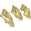
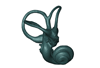
Archaeozoological studies are increasingly using new methods and approaches to explore questions about domestication. Here, we provide 3D models of three archaeological Canis lupus skulls from Belgium originating from the sites of Goyet (31,680±250BP; 31,890+240/-220BP), Trou des Nutons (21,810±90BP) and Trou Balleux (postglacial). Since their identification as either wolves or early dogs is still debated, we present these models as additional tools for further investigating their evolutionary history and the history of dog domestication.
Canis lupus Goyet 2860 View specimen
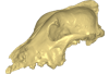
|
M3#213D surface model of the cranium of the Late Pleistocene Canis lupus "Goyet 2860" from the Royal Belgian Institute of Natural Sciences. Type: "3D_surfaces"doi: 10.18563/m3.sf21 state:published |
Download 3D surface file |
Canis lupus Trou Balleux no-nr View specimen
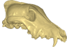
|
M3#223D surface model of the cranium of the Late Pleistocene Canis lupus "Trou Balleux no-nr" from the University of Liège, Belgium Type: "3D_surfaces"doi: 10.18563/m3.sf22 state:published |
Download 3D surface file |
Canis lupus Trou des Nutons 2559-1 View specimen
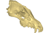
|
M3#233D surface model of the cranium of the Late Pleistocene Canis lupus "Trou des Nutons 2559-1" from the Royal Belgian Institute of Natural Sciences. Type: "3D_surfaces"doi: 10.18563/m3.sf23 state:published |
Download 3D surface file |
Page 1 of 1, showing 2 record(s) out of 2 total


