Explodable 3D Dog Skull for Veterinary Education
3D models of Cainotheriids Ossicular chain
3D models of teeth and neurovascular system in the rostrum of Spinosauridae (theropod dinosaurs)
3D GM dataset of bird skeletal variation
Skeletal embryonic development in the catshark
Bony connexions of the petrosal bone of extant hippos
bony labyrinth (14) , inner ear (11) , Eocene (11) , geometric morphometrics (10) , CT-scan (10) , Oligocene (9) , Micro-CT (9)
Lionel Hautier (25) , Maëva Judith Orliac (24) , Laurent Marivaux (19) , Bastien Mennecart (16) , Renaud Lebrun (15) , Rodolphe Tabuce (15) , Pierre-Olivier Antoine (13)
|
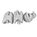
3D reconstructions of dental epithelium during Oryctolagus cuniculus embryonic development related to the publication ”Morphological features of tooth development and replacement in the rabbit Oryctolagus cuniculus”Ludivine Bertonnier-Brouty
Published online: 30/09/2019 |
|
M3#390Right cheek teeth, Left and right incisors at 14 dpf Type: "3D_surfaces"doi: 10.18563/m3.sf.390 state:published |
Download 3D surface file |
Oryctogalus cuniculus E16 View specimen
|
M3#391Left cheek teeth, Left and right incisors at 16 dpf Type: "3D_surfaces"doi: 10.18563/m3.sf.391 state:published |
Download 3D surface file |
Oryctogalus cuniculus E18 View specimen
|
M3#392Left cheek teeth and incisors at 18 dpf Type: "3D_surfaces"doi: 10.18563/m3.sf.392 state:published |
Download 3D surface file |
Oryctogalus cuniculus E20 View specimen
|
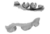
M3#393Left cheek teeth and incisors at 20 dpf Type: "3D_surfaces"doi: 10.18563/m3.sf.393 state:published |
Download 3D surface file |
Oryctogalus cuniculus E22 View specimen
|
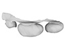
M3#394Left lower cheek teeth and incisors, right upper cheek teeth and incisors at 22 dpf Type: "3D_surfaces"doi: 10.18563/m3.sf.394 state:published |
Download 3D surface file |
Oryctogalus cuniculus E24 View specimen
|
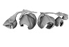
M3#395Left cheek teeth and incisors at 24 dpf Type: "3D_surfaces"doi: 10.18563/m3.sf.395 state:published |
Download 3D surface file |
Oryctogalus cuniculus E28 View specimen
|
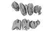
M3#396Right cheek teeth and incisors at 28 dpf Type: "3D_surfaces"doi: 10.18563/m3.sf.396 state:published |
Download 3D surface file |
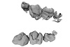
Oryctogalus cuniculus E26 View specimen
|
M3#397Right cheek teeth and incisors at 26 dpf Type: "3D_surfaces"doi: 10.18563/m3.sf.397 state:published |
Download 3D surface file |
 , Chigako Uwabe, Katsumi Kose and Tetsuya Takakuwa
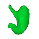
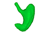
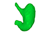
 The present 3D Dataset contains the 3D models analyzed in: Kaigai N et al. Morphogenesis and three-dimensional movement of the stomach during the human embryonic period, Anat Rec (Hoboken). 2014 May;297(5):791-797. doi: 10.1002/ar.22833.
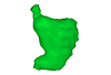
Homo sapiens KC-CS16STM27159 View specimen
|
M3#56computationally reconstructed stomach of the human embryo (M3#56_KC-CS16STM27159) at Carnegie Stage 16 (Crown Rump Length= 9.9mm). Type: "3D_surfaces"doi: 10.18563/m3.sf56 state:published |
Download 3D surface file |
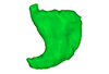
Homo sapiens KC-CS17STM20383 View specimen
|
M3#57computationally reconstructed stomach of the human embryo (M3#57_KC-CS17STM20383) at Carnegie Stage 17 (Crown Rump Length= 12.3mm). Type: "3D_surfaces"doi: 10.18563/m3.sf57 state:published |
Download 3D surface file |
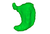
Homo sapiens KC-CS18STM21807 View specimen
|
M3#58computationally reconstructed stomach of the human embryo (M3#58_KC-CS18STM21807) at Carnegie Stage 18 (Crown Rump Length= 14.7mm). Type: "3D_surfaces"doi: 10.18563/m3.sf58 state:published |
Download 3D surface file |
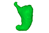
Homo sapiens KC-CS19STM17998 View specimen
|
M3#59computationally reconstructed stomach of the human embryo (M3#59_KC-CS19STM17998) at Carnegie Stage 19 (Crown Rump Length was unmeasured ). Type: "3D_surfaces"doi: 10.18563/m3.sf59 state:published |
Download 3D surface file |
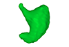
Homo sapiens KC-CS20STM20785 View specimen
|
M3#60computationally reconstructed stomach of the human embryo (M3#60_KC-CS20STM20785) at Carnegie Stage 20 (Crown Rump Length= 18.7 mm). Type: "3D_surfaces"doi: 10.18563/m3.sf60 state:published |
Download 3D surface file |
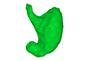
Homo sapiens KC-CS21STM24728 View specimen
|
M3#61computationally reconstructed stomach of the human embryo (M3#61_KC-CS21STM24728) at Carnegie Stage 21 (Crown Rump Length= 20.9 mm). Type: "3D_surfaces"doi: 10.18563/m3.sf61 state:published |
Download 3D surface file |
Homo sapiens KC-CS22STM26438 View specimen
|
M3#62computationally reconstructed stomach of the human embryo (M3#62_KC-CS22STM26438) at Carnegie Stage 22 (Crown Rump Length= 21.5 mm). Type: "3D_surfaces"doi: 10.18563/m3.sf62 state:published |
Download 3D surface file |
Homo sapiens KC-CS23STM20018 View specimen
|
M3#63computationally reconstructed stomach of the human embryo (M3#63_KC-CS23STM20018) at Carnegie Stage 23 (Crown Rump Length= 23.1 mm). Type: "3D_surfaces"doi: 10.18563/m3.sf63 state:published |
Download 3D surface file |
 , Caroline Romestaing and Yoland Savriama
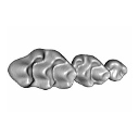
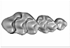
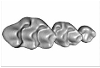
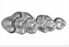
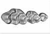
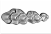
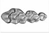
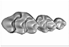
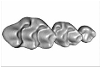
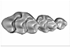
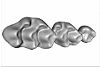
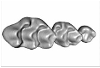
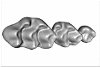
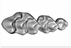
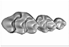
 This contribution contains 3D models of upper molar rows of house mice (Mus musculus domesticus). The erupted part of the right row is presented for specimens belonging to four groups: wild-trapped mice, wild-derived lab offspring, a typical laboratory strain (Swiss) and hybrids between wild-derived and Swiss mice. These models are analyzed in the following publication: Savriama et al 2021: Wild versus lab house mice: Effects of age, diet, and genetics on molar geometry and topography. https://doi.org/10.1111/joa.13529
Mus musculus BW_03 View specimen
|
M3#736BW_03 Type: "3D_surfaces"doi: 10.18563/m3.sf.736 state:published |
Download 3D surface file |
Mus musculus BW_04 View specimen
|
M3#752BW_04 Type: "3D_surfaces"doi: 10.18563/m3.sf.752 state:published |
Download 3D surface file |
Mus musculus BW_06 View specimen
|
M3#753BW_06 Type: "3D_surfaces"doi: 10.18563/m3.sf.753 state:published |
Download 3D surface file |
Mus musculus BW_07 View specimen
|
M3#754BW_07 Type: "3D_surfaces"doi: 10.18563/m3.sf.754 state:published |
Download 3D surface file |
Mus musculus BW_08 View specimen
|
M3#755BW_08 Type: "3D_surfaces"doi: 10.18563/m3.sf.755 state:published |
Download 3D surface file |
Mus musculus BW_11 View specimen
|
M3#756BW_11 Type: "3D_surfaces"doi: 10.18563/m3.sf.756 state:published |
Download 3D surface file |
Mus musculus BW_12 View specimen
|
M3#757BW_12 Type: "3D_surfaces"doi: 10.18563/m3.sf.757 state:published |
Download 3D surface file |
Mus musculus Blab_035 View specimen
|
M3#758Blab_035 Type: "3D_surfaces"doi: 10.18563/m3.sf.758 state:published |
Download 3D surface file |
Mus musculus Blab_046 View specimen
|
M3#759Blab_046 Type: "3D_surfaces"doi: 10.18563/m3.sf.759 state:published |
Download 3D surface file |
Mus musculus Blab_054 View specimen
|
M3#760Blab_054 Type: "3D_surfaces"doi: 10.18563/m3.sf.760 state:published |
Download 3D surface file |
Mus musculus Blab_056 View specimen
|
M3#761Blab_056 Type: "3D_surfaces"doi: 10.18563/m3.sf.761 state:published |
Download 3D surface file |
Mus musculus Blab_082 View specimen
|
M3#762Blab_082 Type: "3D_surfaces"doi: 10.18563/m3.sf.762 state:published |
Download 3D surface file |
Mus musculus Blab_086 View specimen
|
M3#763Blab_086 Type: "3D_surfaces"doi: 10.18563/m3.sf.763 state:published |
Download 3D surface file |
Mus musculus Blab_092 View specimen
|
M3#764Blab_092 Type: "3D_surfaces"doi: 10.18563/m3.sf.764 state:published |
Download 3D surface file |
Mus musculus Blab_319 View specimen
|
M3#751Blab_319 Type: "3D_surfaces"doi: 10.18563/m3.sf.751 state:published |
Download 3D surface file |
Mus musculus Blab_325 View specimen
|
M3#750Blab_325 Type: "3D_surfaces"doi: 10.18563/m3.sf.750 state:published |
Download 3D surface file |
Mus musculus Blab_329 View specimen
|
M3#737Blab_329 Type: "3D_surfaces"doi: 10.18563/m3.sf.737 state:published |
Download 3D surface file |
Mus musculus Blab_330 View specimen
|
M3#738Blab_330 Type: "3D_surfaces"doi: 10.18563/m3.sf.738 state:published |
Download 3D surface file |
Mus musculus Blab_F2a View specimen
|
M3#739Blab_F2a Type: "3D_surfaces"doi: 10.18563/m3.sf.739 state:published |
Download 3D surface file |
Mus musculus Blab_F2b View specimen
|
M3#740Blab_F2b Type: "3D_surfaces"doi: 10.18563/m3.sf.740 state:published |
Download 3D surface file |
Mus musculus Blab_BB3w View specimen
|
M3#741Blab_BB3w Type: "3D_surfaces"doi: 10.18563/m3.sf.741 state:published |
Download 3D surface file |
Mus musculus hyb_BS01 View specimen
|
M3#742hyb_BS01 Type: "3D_surfaces"doi: 10.18563/m3.sf.742 state:published |
Download 3D surface file |
Mus musculus hyb_BS02 View specimen
|
M3#743hyb_BS02 Type: "3D_surfaces"doi: 10.18563/m3.sf.743 state:published |
Download 3D surface file |
Mus musculus hyb_SB01 View specimen
|
M3#744hyb_SB01 Type: "3D_surfaces"doi: 10.18563/m3.sf.744 state:published |
Download 3D surface file |
Mus musculus hyb_SB02 View specimen
|
M3#745hyb_SB02 Type: "3D_surfaces"doi: 10.18563/m3.sf.745 state:published |
Download 3D surface file |
Mus musculus SW_001 View specimen
|
M3#746SW_001 Type: "3D_surfaces"doi: 10.18563/m3.sf.746 state:published |
Download 3D surface file |
Mus musculus SW_002 View specimen
|
M3#747SW_002 Type: "3D_surfaces"doi: 10.18563/m3.sf.747 state:published |
Download 3D surface file |
Mus musculus SW_005 View specimen
|
M3#748SW_005 Type: "3D_surfaces"doi: 10.18563/m3.sf.748 state:published |
Download 3D surface file |
Mus musculus SW_0ter View specimen
|
M3#749SW_0ter Type: "3D_surfaces"doi: 10.18563/m3.sf.749 state:published |
Download 3D surface file |
Mus musculus SW_343 View specimen
|
M3#765SW_343 Type: "3D_surfaces"doi: 10.18563/m3.sf.765 state:published |
Download 3D surface file |
 , Leonardo Kerber and Sérgio Dias-da-Silva
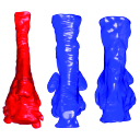
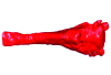
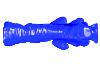
 The present 3D Dataset contains the 3D models of brain endocast of traversodontid cynodonts studied in: Pavanatto et al. 2019. Virtual reconstruction of cranial endocasts of traversodontid cynodonts (Eucynodontia: Gomphodontia) from the upper Triassic of Southern Brazil. Journal of Morphology. https://doi.org/10.1002/jmor.21029
Siriusgnathus niemeyerorum CAPPA/UFSM 0032 View specimen
|
M3#4253D model of the brain endocast Type: "3D_surfaces"doi: 10.18563/m3.sf.425 state:published |
Download 3D surface file |
Exaeretodon riograndensis CAPPA/UFSM 0030 View specimen
|
M3#4263D model of the brain endocast Type: "3D_surfaces"doi: 10.18563/m3.sf.426 state:published |
Download 3D surface file |
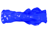
Exaeretodon riograndensis CAPPA/UFSM 0227 View specimen
|
M3#4273D model of the brain endocast Type: "3D_surfaces"doi: 10.18563/m3.sf.427 state:published |
Download 3D surface file |
 and Torsten M. Scheyer
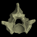
 The present 3D Dataset contains the 3D models analyzed in the following publication: Georgalis, G. L., and T. M. Scheyer. A new species of Palaeopython (Serpentes) and other extinct squamates from the Eocene of Dielsdorf (Zurich, Switzerland). Swiss Journal of Geosciences (in press). https://doi.org/10.1007/s00015-019-00341-6
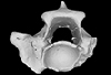
Palaeopython helveticus PIMUZ A/III 631 View specimen
|
M3#399ZIP file containing .ply of vertebra PIMUZ A/III 631 from Palaeopython helveticus n. sp. Type: "3D_surfaces"doi: 10.18563/m3.sf.399 state:published |
Download 3D surface file |
|
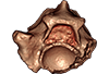
M3#403dataset of snake vertebra PIMUZ A/III 631 Type: "3D_CT"doi: 10.18563/m3.sf.403 state:published |
Download CT data |
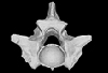
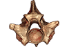
Palaeopython helveticus PIMUZ A/III 634 View specimen
|
M3#400ZIP file containing .ply of vertebra PIMUZ A/III 634 from Palaeopython helveticus n. sp. (holotype) Type: "3D_surfaces"doi: 10.18563/m3.sf.400 state:published |
Download 3D surface file |
|
M3#404dataset of snake vertbra PIMUZ A/III 634 (holotype) Type: "3D_CT"doi: 10.18563/m3.sf.404 state:published |
Download CT data |
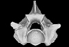
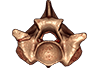
Palaeopython helveticus PIMUZ A/III 636 View specimen
|
M3#401ZIP file containing .ply of vertebra PIMUZ A/III 636 from Palaeopython helveticus n. sp. Type: "3D_surfaces"doi: 10.18563/m3.sf.401 state:published |
Download 3D surface file |
|
M3#406dataset of snake vertebra PIMUZ A/III 636 Type: "3D_CT"doi: 10.18563/m3.sf.406 state:published |
Download CT data |
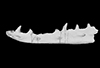
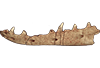
Palaeovaranus sp. PIMUZ A/III 234 View specimen
|
M3#402ZIP file containing .ply of dentary PIMUZ A/III 234 of Palaeovaranus sp. Type: "3D_surfaces"doi: 10.18563/m3.sf.402 state:published |
Download 3D surface file |
|
M3#405dataset of dentary of Palaeovaranus sp. (PIMUZ A/III 234) Type: "3D_CT"doi: 10.18563/m3.sf.405 state:published |
Download CT data |
 , Jürg Jost and Sarah Hilfiker
 This contribution contains the 3D models described and figured in the following publication: Aguirre-Fernández G, Jost J, and Hilfiker S. 2022. First records of extinct kentriodontid and squalodelphinid dolphins from the Upper Marine Molasse (Burdigalian age) of Switzerland and a reappraisal of the Swiss cetacean fauna.
Kentriodon sp. NMBE 5023944 View specimen
|
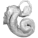
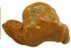
M3#8583D models of left periotic and bony labyrinth of NMBE 5023944 (Kentriodon sp.) Type: "3D_surfaces"doi: 10.18563/m3.sf.858 state:published |
Download 3D surface file |
Kentriodon sp. NMBE 5023945 View specimen
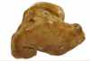
|
M3#8593D models of right periotic and bony labyrinth of NMBE 5023945 (Kentriodontidae indet.) Type: "3D_surfaces"doi: 10.18563/m3.sf.859 state:published |
Download 3D surface file |
Kentriodon sp. NMBE 5023946 View specimen
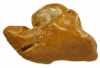
|
M3#8603D models of left periotic and bony labyrinth of NMBE 5023946 (Kentriodon sp.) Type: "3D_surfaces"doi: 10.18563/m3.sf.860 state:published |
Download 3D surface file |
Kentriodon sp. NMBE 5036436 View specimen
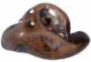
|
M3#8613D models of right periotic and bony labyrinth of NMBE 5036436 (Kentriodontidae indet.) Type: "3D_surfaces"doi: 10.18563/m3.sf.861 state:published |
Download 3D surface file |
indet. indet. NMBE 5023942 View specimen
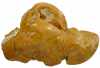
|
M3#8623D models of right periotic and bony labyrinth of NMBE 5023942 (Squalodelphinidae indet.) Type: "3D_surfaces"doi: 10.18563/m3.sf.862 state:published |
Download 3D surface file |
indet. indet. NMBE 5023943 View specimen
|
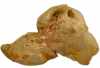
M3#8633D models of left periotic and bony labyrinth of NMBE 5023943 (Squalodelphinidae indet.) Type: "3D_surfaces"doi: 10.18563/m3.sf.863 state:published |
Download 3D surface file |
indet. indet. NMBE 5036437 View specimen
|
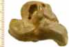
M3#8643D models of left periotic and bony labyrinth of NMBE 5036437 (Physeteridae indet.) Type: "3D_surfaces"doi: 10.18563/m3.sf.864 state:published |
Download 3D surface file |
 , Helder Gomes Rodrigues , Guillaume Billet and Robert J. Asher
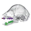
 This contribution contains the 3D models described and figured in the following publication: Hautier L., Gomes Rodrigues H., Billet G., Asher R.J., 2016. The hidden teeth of sloths: evolutionary vestiges and the development of a simplified dentition. Scientific Reports. doi: 10.1038/srep27763
Bradypus variegatus ZMB 33812 View specimen
|
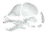
M3#110Three-dimensional reconstruction of the teeth, mandibles, maxillary and premaxillary bones Type: "3D_surfaces"doi: 10.18563/m3.sf.110 state:published |
Download 3D surface file |
Bradypus variegatus ZMB 41122 View specimen
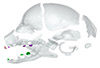
|
M3#109Three-dimensional reconstruction of the teeth, mandibles, maxillary and premaxillary bones Type: "3D_surfaces"doi: 10.18563/m3.sf.109 state:published |
Download 3D surface file |
Bradypus variegatus MNHN-ZM-MO-1995-326A View specimen
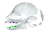
|
M3#111Three-dimensional reconstruction of the teeth, mandibles, maxillary and premaxillary bones Type: "3D_surfaces"doi: 10.18563/m3.sf.111 state:published |
Download 3D surface file |
Bradypus variegatus MNHN-ZM-MO-1995-326B View specimen
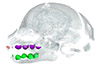
|
M3#112Three-dimensional reconstruction of the teeth, mandibles, maxillary and premaxillary bones Type: "3D_surfaces"doi: 10.18563/m3.sf.112 state:published |
Download 3D surface file |
Bradypus sp. MNHN-ZM-MO-1902-325 View specimen
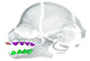
|
M3#113Three-dimensional reconstruction of the teeth, mandibles, maxillary, and premaxillary bones Type: "3D_surfaces"doi: 10.18563/m3.sf.113 state:published |
Download 3D surface file |
Bradypus sp. MNHN-ZM-MO-1995-327 View specimen
|
M3#114Three-dimensional reconstruction of the teeth, mandibles, maxillary and premaxillary bones Type: "3D_surfaces"doi: 10.18563/m3.sf.114 state:published |
Download 3D surface file |
Choloepus didactylus MNHN-ZM-MO-1882-625 View specimen
|
M3#115Three-dimensional reconstruction of the teeth, mandibles, maxillary and premaxillary bones Type: "3D_surfaces"doi: 10.18563/m3.sf.115 state:published |
Download 3D surface file |
 , Rohan Mansuit , Anthony Herrel , Antoine Cuckovic , Dominique Didier, Paul Tafforeau and Alan Pradel
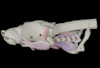
 This contribution contains 3D models of the cranial skeleton and muscles in an elephantfish (Callorhinchus milii) and a catshark (Scyliorhinus canicula), based on synchrotron tomographic scans. These datasets were analyzed and described in Dearden et al. (2021) “The morphology and evolution of chondrichthyan cranial muscles: a digital dissection of the elephantfish Callorhinchus milii and the catshark Scyliorhinus canicula.” Journal of Anatomy.
Callorhinchus milii 001 View specimen
|
M3#7083D models of the cranial skeleton and muscles of Callorhinchus milii, created using Mimics. Type: "3D_surfaces"doi: 10.18563/m3.sf.708 state:published |
Download 3D surface file |
Scyliorhinus canicula 002 View specimen
|
M3#7093D models of the cranial skeleton and muscles of Scyliorhinus canicula, created using Mimics. Type: "3D_surfaces"doi: 10.18563/m3.sf.709 state:published |
Download 3D surface file |
 , Rodolfo Salas-Gismondi and Pierre-Olivier Antoine
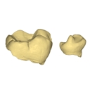
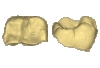
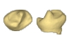
 This contribution contains the 3D models of the isolated teeth attributed to stem representatives of the Cebuella and Cebus lineages (Cebuella sp. and Cebus sp.), described and figured in the following publication: Marivaux et al. (2016), Dental remains of cebid platyrrhines from the earliest late Miocene of Western Amazonia, Peru: macroevolutionary implications on the extant capuchin and marmoset lineages. American Journal of Physical Anthropology. http://dx.doi.org/10.1002/ajpa.23052
Cebus sp. MUSM-3243 View specimen
|
M3#2823D model of left lower m1 (lingual part) Type: "3D_surfaces"doi: 10.18563/m3.sf.282 state:published |
Download 3D surface file |
Cebuella sp. MUSM-3239 View specimen
|
M3#2833D model of left lower p4 Type: "3D_surfaces"doi: 10.18563/m3.sf.283 state:published |
Download 3D surface file |
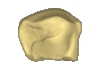
Cebuella sp. MUSM-3240 View specimen
|
M3#2943D model of right upper P3 or P4 (buccal part) Type: "3D_surfaces"doi: 10.18563/m3.sf.294 state:published |
Download 3D surface file |
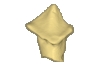
Cebuella sp. MUSM-3241 View specimen
|
M3#2953D model of right upper P2 Type: "3D_surfaces"doi: 10.18563/m3.sf.295 state:published |
Download 3D surface file |
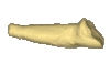
Cebuella sp. MUSM-3242 View specimen
|
M3#2963D model of upper I2 Type: "3D_surfaces"doi: 10.18563/m3.sf.296 state:published |
Download 3D surface file |
 , Damien Becker , Vlad Codrea , Loïc Costeur , Cristina Fărcaş, Alexandru Solomon , Marton Venczel and Olivier Maridet
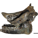
 This contribution contains the 3D models described and figured in the following publication: Tissier et al. (in prep.).
Sellamynodon zimborensis UBB MPS 15795 View specimen
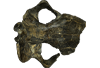
|
M3#297Incomplete skull with left M3. Type: "3D_surfaces"doi: 10.18563/m3.sf.297 state:published |
Download 3D surface file |
Sellamynodon zimborensis UBB MPS 15795 View specimen
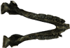
|
M3#298Mandible with complete molar and premolar rows, lacking symphysis. Type: "3D_surfaces"doi: 10.18563/m3.sf.298 state:published |
Download 3D surface file |
Amynodontopsis aff. bodei UBB MPS V545 View specimen
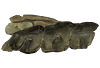
|
M3#299Maxillary fragment with M1-3. Type: "3D_surfaces"doi: 10.18563/m3.sf.299 state:published |
Download 3D surface file |
Amynodontopsis aff. bodei UBB MPS V546 View specimen
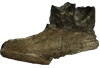
|
M3#300Unworn m1/2 on mandible fragment. Type: "3D_surfaces"doi: 10.18563/m3.sf.300 state:published |
Download 3D surface file |
 , Ricardo Araújo and Fabrice Lihoreau
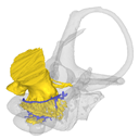
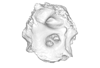
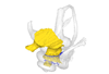
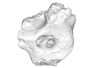
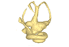
 This contribution contains the 3D models described and figured in the publication entitled "The petrosal and bony labyrinth of Diplobune minor, an enigmatic Artiodactyla from the Oligocene of Western Europe" by Orliac, Araújo, and Lihoreau published in Journal of Morphology (Orliac et al. 2017) https://doi.org/10.1002/jmor.20702.
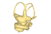
Diplobune minor UM ITD 1079 View specimen
|
M3#138right bony labyrinth of Diplobune minor from Itardies, France Type: "3D_surfaces"doi: 10.18563/m3.sf.138 state:published |
Download 3D surface file |
|
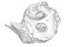
M3#139right isolated petrosal of Diplobune minor from Itardies, France Type: "3D_surfaces"doi: 10.18563/m3.sf.139 state:published |
Download 3D surface file |
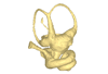
Diplobune minor UM ITD 1080 View specimen
|
M3#140left bony labyrinth of Diplobune minor from Itardies, France Type: "3D_surfaces"doi: 10.18563/m3.sf.140 state:published |
Download 3D surface file |
|
M3#141left isolated petrosal of Diplobune minor from Itardies, France Type: "3D_surfaces"doi: 10.18563/m3.sf.141 state:published |
Download 3D surface file |
Diplobune minor UM ITD 1081 View specimen
|
M3#142right bony labyrinth and associated nerves and veins of Diplobune minor from Itardies, France Type: "3D_surfaces"doi: 10.18563/m3.sf.142 state:published |
Download 3D surface file |
|
M3#143right isolated petrosal of Diplobune minor from Itardies, France Type: "3D_surfaces"doi: 10.18563/m3.sf.143 state:published |
Download 3D surface file |
Diplobune minor UM ITD 1083 View specimen
|
M3#144left bony labyrinth of Diplobune minor from Itardies, France Type: "3D_surfaces"doi: 10.18563/m3.sf.144 state:published |
Download 3D surface file |
|
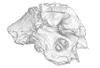
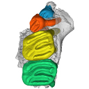
M3#145left petrosal of Diplobune minor from Itardies, France Type: "3D_surfaces"doi: 10.18563/m3.sf.145 state:published |
Download 3D surface file |
 , Olivier Maridet and Jérôme Priéto
 This contribution contains the 3D model of the holotype of Simplomys hugi, the new dormouse species from the locality of Glovelier described and figured in the following publication: New data on the Miocene dormouse Simplomys García-Paredes, 2009 from the peri-alpin basins of Switzerland and Germany: palaeodiversity of a rare genus in Central Europe. https://doi.org/10.1007/s12549-018-0339-y
Simplomys hugi MJSN-GLM017-0001 View specimen
|
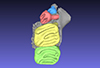
M3#385the left maxilla with four teeth ( DP4, P4, M1 and M2) Type: "3D_surfaces"doi: 10.18563/m3.sf.385 state:published |
Download 3D surface file |
 , Morgane Dubied , Kévin Le Verger and Bastien Mennecart
 The present 3D Dataset contains the 3D model analyzed in the following publication: Solé et al. (2018), Niche partitioning of the European carnivorous mammals during the paleogene. Palaios. https://doi.org/10.2110/palo.2018.022
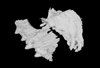
Hyaenodon leptorhynchus FSL848325 View specimen
|
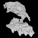
M3#336The specimen FSL848325 is separated in two fragments: the anterior part bears the incisors, the deciduous and permanent canines, while the posterior part bears the right P3, P4, M1 and M2. The P2 is isolated. When combined, the cranium length is approximatively 10.5 cm long. The anterior part is 6.9 cm long and 2.15 cm wide (taken at the level of the P1). The posterior part is 4.8 cm long. The anterior part of the cranium is very narrow. Type: "3D_surfaces"doi: 10.18563/m3.sf.336 state:published |
Download 3D surface file |
 and Bastien Mennecart
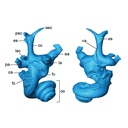
 The present 3D Dataset contains the 3D models analyzed in Costeur L., Mennecart B., Müller B., Schulz G., 2016. Prenatal growth stages show the development of the ruminant bony labyrinth and petrosal bone. Journal of Anatomy. https://doi.org/10.1111/joa.12549
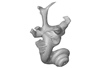
Bos taurus NMB3038 View specimen
|
M3#124Right bony labyrinth of a Bos taurus foetus (gestational age 115 days) Type: "3D_surfaces"doi: 10.18563/m3.sf.124 state:published |
Download 3D surface file |
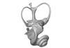
Bos taurus NMB3367 View specimen
|
M3#125Right bony labyrinth of a Bos taurus foetus (gestational age 165 days) Type: "3D_surfaces"doi: 10.18563/m3.sf.125 state:published |
Download 3D surface file |
Bos taurus NMB3365 View specimen
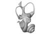
|
M3#126Right bony labyrinth of a Bos taurus foetus (gestational age 210 days) Type: "3D_surfaces"doi: 10.18563/m3.sf.126 state:published |
Download 3D surface file |
Bos taurus NMB2855 View specimen
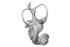
|
M3#127Right bony labyrinth of a Bos taurus foetus (gestational age 260 days) Type: "3D_surfaces"doi: 10.18563/m3.sf.127 state:published |
Download 3D surface file |
Bos taurus NMB1037 View specimen
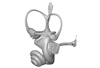
|
M3#128Left bony labyrinth of an adult Bos taurus Type: "3D_surfaces"doi: 10.18563/m3.sf.128 state:published |
Download 3D surface file |
 , Pierre-Olivier Antoine and Damien Becker
 The present 3D Dataset contains two 3D models described in Tissier et al. (https://doi.org/10.1098/rsos.200633): the only known complete mandible of the early-branching rhinocerotoid Epiaceratherium magnum Uhlig, 1999, and a hypothetical reconstruction of the complete archetypic skull of Epiaceratherium Heissig, 1969, created by merging three cranial parts from three distinct Epiaceratherium species.
Epiaceratherium magnum NMB.O.B.928 View specimen
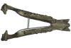
|
M3#5343D surface model of the mandible NMB.O.B.928 of Epiaceratherium magnum, with texture file. Type: "3D_surfaces"doi: 10.18563/m3.sf.534 state:published |
Download 3D surface file |
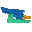
Epiaceratherium magnum NMB.O.B.928 + MJSN POI007–245 + NMB.I.O.43 View specimen
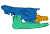
|
M3#535Archetypal reconstruction of the skull of Epiaceratherium, generated by 3D virtual association of the cranium of E. delemontense (MJSN POI007–245, in blue), mandible of E. magnum (NMB.O.B.928, green) and snout of E. bolcense (NMB.I.O.43, in orange). Type: "3D_surfaces"doi: 10.18563/m3.sf.535 state:published |
Download 3D surface file |
 , Carlo Corradini and Catherine Girard
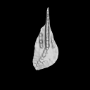
The present 3D Dataset contains the 3D models of the holotype and the paratypes of the new species Siphonodella leiosa described and analyzed in the following publication: L. Souquet, C. Corradini, C. Girard: Siphonodella leiosa (Conodonta), a new unornamented species from the Tournaisian (lower Carboniferous) of Puech de la Suque (Montagne Noire, France). Geobios, https://doi.org/10.1016/j.geobios.2020.06.004.
Siphonodella leiosa UM PSQ 1 View specimen
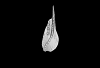
|
M3#525Siphonodella leiosa, paratype, dextral P1 element Type: "3D_surfaces"doi: 10.18563/m3.sf.525 state:published |
Download 3D surface file |
Siphonodella leiosa UM PSQ 2 View specimen
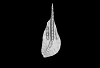
|
M3#526Siphonodella leiosa, holotype, dextral P1 element Type: "3D_surfaces"doi: 10.18563/m3.sf.526 state:published |
Download 3D surface file |
Siphonodella leiosa UM PSQ 3 View specimen
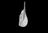
|
M3#527Siphonodella leiosa, paratype, dextral P1 element Type: "3D_surfaces"doi: 10.18563/m3.sf.527 state:published |
Download 3D surface file |
Siphonodella leiosa UM PSQ 4 View specimen
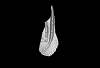
|
M3#528Siphonodella leiosa, paratype, dextral P1 element Type: "3D_surfaces"doi: 10.18563/m3.sf.528 state:published |
Download 3D surface file |
Siphonodella leiosa UM PSQ 5 View specimen
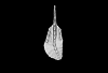
|
M3#529Siphonodella leiosa, paratype, sinistral P1 element Type: "3D_surfaces"doi: 10.18563/m3.sf.529 state:published |
Download 3D surface file |
Siphonodella leiosa UM PSQ 6 View specimen
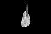
|
M3#530Siphonodella leiosa, paratype, dextral P1 element Type: "3D_surfaces"doi: 10.18563/m3.sf.530 state:published |
Download 3D surface file |
Siphonodella leiosa UM PSQ 7 View specimen
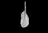
|
M3#531Siphonodella leiosa, paratype, dextral P1 element Type: "3D_surfaces"doi: 10.18563/m3.sf.531 state:published |
Download 3D surface file |
Siphonodella leiosa UM PSQ 8 View specimen
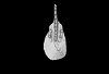
|
M3#532Siphonodella leiosa, paratype, sinistral P1 element Type: "3D_surfaces"doi: 10.18563/m3.sf.532 state:published |
Download 3D surface file |
Siphonodella leiosa UM PSQ 9 View specimen
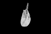
|
M3#533Siphonodella leiosa, paratype, dextral P1 element Type: "3D_surfaces"doi: 10.18563/m3.sf.533 state:published |
Download 3D surface file |
 , Jens N. Lallensack , Junhong Mou, Lin Liu , Shan Jiang, Qi Qi , Anthony Romilio , Yong Ye, Xin Shen and Ting Xu
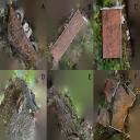
The present 3D dataset contains the 3D models analyzed in Xing et al. (in press), Newly discovered Cretaceous dinosaur track sites from the Jiaguan Formation, Sichuan Province, China: ichnotaxonomy and preservation. The dataset includes photogrammetric 3D models of the specimens shown in Figures 6–9 and 16–17 of the original article. These correspond to track-bearing slabs and selected footprints from the Hongyangoukou and Jinyuxi tracksites and the Shimiaogou 2025 tracksite in the Gulin area, Sichuan Province, China. The Hongyangoukou material comprises three reddish-brown siltstone slabs preserving at least 61 theropod tracks, including nine trackways (HYGK-T1 to HYGK-T9) and isolated footprints (HYGK-TI1 onward). The Jinyuxi material comprises one isolated theropod natural cast (JYX-TI5) assigned to cf. Eubrontes, associated with microbially induced sedimentary structures (MISS). The Shimiaogou 2025 material comprises two slabs preserving at least 25 dinosaur footprints, including ornithopod trackways SMG25-O1 and SMG25-O2, theropod trackway SMG25-T1, and isolated footprints; all occur as natural positive reliefs. The 3D surface models are provided in PLY format.
Eubrontes isp. HYGK Slab 1 View specimen
|
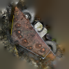
M3#1982All footprints in Fig. 6 are tridactyl theropod footprints assigned to Morphotype I and tentatively identified as Eubrontes-like. Fig. 6 (Hongyangoukou Slab 1) preserves at least 31 footprints, comprising seven trackways (HYGK-T1–T7) and 15 isolated footprints (HYGK-TI1–TI15). They are preserved as natural molds in reddish-brown siltstone and are heavily weathered, but they generally retain distinct tridactyl outlines and U- or V-shaped metatarsophalangeal regions. Claw impressions are present in some specimens (e.g., T3, T7-L1, and TI9), whereas digital pad impressions are absent. Footprint length ranges from 10.8 to 20.9 cm (mean ≈16.0 cm); the length-to-width ratio ranges from 1.1 to 1.7 (mean ≈1.4); mesaxony is weak (mean ≈0.47); and the divarication angle between digits II and IV ranges from 41° to 73° (mean ≈60°). The stride lengths of T1, T3, T4, and T6 are 144.5, 104.8, 122.3, and 64.5 cm, respectively, corresponding to 9.7, 5.6, 9.4, and 3.3 times their respective mean footprint lengths. T1 and T5 show outward rotations of about 4° and 9°, respectively. T3 is among the best-preserved examples, with digit III the longest and stoutest, digit IV the shortest, and clear claw impressions present on digits II and III, whereas TI10 is a complete tridactyl track. These footprints are therefore small theropod tracks of Morphotype I, tentatively assigned to Eubrontes-like. Type: "3D_surfaces"doi: 10.18563/m3.sf.1982 state:in_press |
Download 3D surface file |
Eubrontes isp. HYGK Slab 2 View specimen
|
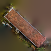
M3#1983All footprints in Fig. 7 (Hongyangoukou Slab 2) are tridactyl theropod footprints assigned to Morphotype II and tentatively identified as Eubrontes-like. Slab 2 preserves at least 28 footprints, including two trackways (HYGK-T8 and HYGK-T9) and 18 isolated footprints (HYGK-TI16–TI33). Preservation is poorer than on Slab 1: impressions are shallower and more rounded overall; in T9 and in some isolated footprints (e.g., TI17, TI18, TI20, TI23, TI24, and TI26), discrete digit impressions are not recognizable and instead appear as oval depressions, whereas the remaining footprints show three discernible digit impressions. No digital pad impressions were observed; the metatarsophalangeal region is U-shaped, and relatively clear claw impressions are preserved in some tracks, such as T8-R1, TI18, and TI31. Footprint length ranges from 11.2 to 21.6 cm (mean 15.1 cm); the length-to-width ratio ranges from 0.9 to 1.7 (mean 1.4); mesaxony is weak (mean 0.45); and the divarication angle between digits II and IV ranges from 46° to 62° (mean 54°). The stride lengths of T8 and T9 are 60 cm and 116 cm, corresponding to approximately 4 and 7.7 times the average footprint length, respectively; the pace angulation of T9 is 166°. TI31 is relatively well preserved and markedly symmetrical, with digit III being the stoutest and longest, and with a distinct, sharp claw impression. Type: "3D_surfaces"doi: 10.18563/m3.sf.1983 state:in_press |
Download 3D surface file |
Eubrontes isp. HYGK Slab 3 View specimen
|
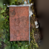
M3#1984All footprints in Fig. 8 (Hongyangoukou Slab 3) are tridactyl theropod footprints assigned to Morphotype I and tentatively identified as Eubrontes-like. Slab 3 preserves at least seven isolated footprints (HYGK-TI34–TI40) and no trackways; these footprints have relatively distinct tridactyl outlines and U- or V-shaped metatarsophalangeal regions, with some specimens (e.g., TI37 and TI38) preserving claw impressions but lacking digital pad impressions. The divarication angle between digits II and IV on Slab 3 ranges from 53° to 57° (mean 56°). As members of Morphotype I, they fall within the morphotype ranges for footprint length (10.8–20.9 cm; mean 16.0 cm), length-to-width ratio (1.1–1.7; mean 1.4), and mesaxony (weak; mean 0.47). Type: "3D_surfaces"doi: 10.18563/m3.sf.1984 state:in_press |
Download 3D surface file |
Eubrontes isp. JYX-TI5 View specimen
|
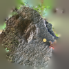
M3#1985JYX-TI5 is tentatively assigned to cf. Eubrontes. It is preserved as a natural cast on dark red quartz sandstone, with a preservation grade of 0.5–1.5. It shows largely complete digit impressions and a metatarsophalangeal region, as well as some claw impressions, but no digital pad impressions. The footprint has three distinct digit impressions, a relatively distinct U-shaped metatarsophalangeal region, a separation between the proximal region of digit III and the metatarsophalangeal region, and relatively clear claw impressions; its surface bears irregular linear ridges that likely represent microbially induced sedimentary structures (MISS). JYX-TI5 is 22.1 cm long, with a length-to-width ratio of 1.5, weak mesaxony (0.45), and a divarication angle between digits II and IV of 50°; the interdigital angle between digits II and III is slightly larger than that between digits III and IV. Type: "3D_surfaces"doi: 10.18563/m3.sf.1985 state:in_press |
Download 3D surface file |
Grallator isp. SMG25 Slab 1 View specimen
|
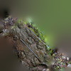
M3#1986Fig. 16 includes ornithopod footprints assigned to Caririchnium isp. and theropod footprints tentatively assigned to cf. Grallator. The ornithopod tracks are natural casts, clearest on Slab 1, and show a distinct quadripartite structure. Preservation grade is 1–2; footprint length is 23.5–31.2 cm (mean 27.3 cm), and length-to-width ratio is 1.2–1.5 (mean 1.4). They are highly symmetrical, with three clear rounded digit impressions and a sub-circular metatarsophalangeal region separated by a depression; claw impressions are absent, mesaxony is weak (0.24–0.38; mean 0.30), and the mean divarication angle between digits II and IV is 49°. The potential trackway SMG25-O2 has a pace length about 2.7 times the footprint length, a pace angulation of 111°, and inward rotation of about 15°. The theropod tracks are mostly isolated, with only one potential trackway, SMG25-T1, on Slab 1. Preservation grade is 1–2; footprints are small (8.5–20.4 cm) and generally slender, mostly complete tridactyl impressions with digit III longest and digit II shortest, V-shaped metatarsophalangeal regions, and faint claw impressions or digital pads in some tracks. SMG25-T1 comprises two footprints (mean length 19.9 cm, length-to-width ratio 1.9, mesaxony 0.54, mean divarication angle between digits II and IV 41°). Isolated footprint TI9 is the best preserved (length 9.8 cm, length-to-width ratio 2.1, mesaxony 0.72, divarication angle between digits II and IV 38°), with the metatarsophalangeal pad of digit IV aligned along the axis of digit III. Type: "3D_surfaces"doi: 10.18563/m3.sf.1986 state:in_press |
Download 3D surface file |
Grallator isp. SMG25 Slab 2 View specimen
|
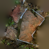
M3#1987cf. Grallator is the dominant ichnotaxon in Fig. 17, which preserves 12 isolated cf. Grallator footprints, all preserved as natural positive reliefs with preservation grade 1–2. These small theropod tracks are 8.5–20.4 cm long and generally slender, and are mostly complete tridactyl impressions with digit III longest and digit II shortest, V-shaped metatarsophalangeal regions, and faint claw impressions or digital pads in some specimens. Type: "3D_surfaces"doi: 10.18563/m3.sf.1987 state:in_press |
Download 3D surface file |
 , Charles Leriche, Mohd Waqas , Rajendra Rana , Ashok Sahni and Thierry Smith
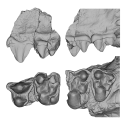
 This 3D dataset contains the 3D models of the fossil specimens of Lilliputia gujjarorum, a new tiny dichobunoid artiodactyl from the early middle Eocene of northwestern India. The material includes the holotype, GU/RJ/003 (Rajouri Collection, H.N.B. Garhwal University), consisting of an associated left mandible fragment preserving the distal part of p4 and m1–3 (GU/RJ/003a) and a left maxillary fragment preserving P4 and M1–3 (GU/RJ/003b), as well as the referred specimen GU/RJ/005, a right mandible fragment preserving m1–2. The exceptionally small body size of Lilliputia, among the smallest known in Artiodactyla, together with its mosaic of primitive and derived dental characters, provides new insights into the early evolution, palaeobiogeography, and ecological diversification of artiodactyls during the Eocene.
Lilliputia gujjarorum GU/RJ/003a View specimen
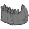
|
M3#1949left mandible fragment preserving the distal part of p4 and m1–3 Type: "3D_surfaces"doi: 10.18563/m3.sf.1949 state:in_press |
Download 3D surface file |
Lilliputia gujjarorum GU/RJ/003b View specimen
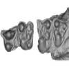
|
M3#1950left maxillary fragment preserving P4 and M1–3 Type: "3D_surfaces"doi: 10.18563/m3.sf.1950 state:in_press |
Download 3D surface file |
Lilliputia gujjarorum GU/RJ/005 View specimen
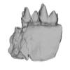
|
M3#1951right mandible fragment preserving m1–2 Type: "3D_surfaces"doi: 10.18563/m3.sf.1951 state:in_press |
Download 3D surface file |
 , Emilie A. Hardouin , Georges P. Mitsainas and Sabrina Renaud
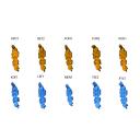
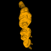
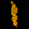
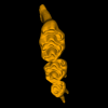
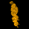
 This contribution contains 3D models of upper molar rows of Cypriot mice (Mus cypriacus) and house mice (Mus musculus domesticus) from the island of Cyprus. Although morphologically similar and occasionally syntopic, the two species differ in habitat use and dietary strategies, ranging from anthropogenic resources in M. m. domesticus to a more natural and diversified trophic niche in M. cypriacus. Mandibular and molar shape variation was quantified through 3D geometric morphometrics, enabling the detection of interspecific differences, allometric trends, and age-related dental wear trajectories. The morphometric descriptors were evaluated for their capacity to capture phylogenetic divergence, ecological specialization, and ontogenetic dynamics. Results highlight that subtle morphological signals can expose fine-scale ecological processes and species-specific adaptive strategies.
Mus cypriacus APO1 View specimen
|
M3#1897Upper molar row APO1 Mus cypriacus Type: "3D_surfaces"doi: 10.18563/m3.sf.1897 state:in_press |
Download 3D surface file |
Mus cypriacus BET2 View specimen
|
M3#1898Upper molar row BET2 Mus cypriacus Type: "3D_surfaces"doi: 10.18563/m3.sf.1898 state:in_press |
Download 3D surface file |
Mus cypriacus FON1 View specimen
|
M3#1899Upper molar row FON1 Mus cypriacus Type: "3D_surfaces"doi: 10.18563/m3.sf.1899 state:in_press |
Download 3D surface file |
Mus cypriacus FON2 View specimen
|
M3#1900Upper molar row FON2 Mus cypriacus Type: "3D_surfaces"doi: 10.18563/m3.sf.1900 state:in_press |
Download 3D surface file |
Mus cypriacus KOU1 View specimen
|
M3#1901Upper molar row KOU1 Mus cypriacus Type: "3D_surfaces"doi: 10.18563/m3.sf.1901 state:in_press |
Download 3D surface file |
Mus musculus KOF1 View specimen
|
M3#1902Upper molar row KOF1 Mus musculus domesticus Type: "3D_surfaces"doi: 10.18563/m3.sf.1902 state:in_press |
Download 3D surface file |
Mus musculus LEF1 View specimen
|
M3#1903Upper molar row LEF1 Mus musculus domesticus Type: "3D_surfaces"doi: 10.18563/m3.sf.1903 state:in_press |
Download 3D surface file |
Mus musculus MEN1 View specimen
|
M3#1904Upper molar row MEN1 Mus musculus domesticus Type: "3D_surfaces"doi: 10.18563/m3.sf.1904 state:in_press |
Download 3D surface file |
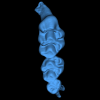
Mus musculus TSE2 View specimen
|
M3#1905Upper molar row TSE2 Mus musculus domesticus Type: "3D_surfaces"doi: 10.18563/m3.sf.1905 state:in_press |
Download 3D surface file |
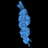
Mus musculus XYL5 View specimen
|
M3#1906Upper molar row XYL5 Mus musculus domesticus Type: "3D_surfaces"doi: 10.18563/m3.sf.1906 state:in_press |
Download 3D surface file |