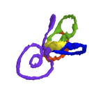
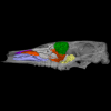
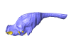
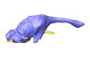
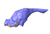
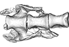
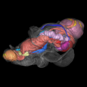
3D models of Kalakocetus, the earliest Cetacea
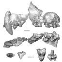
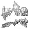
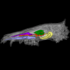
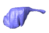
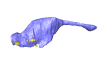
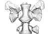
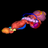
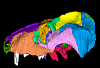
3D models of Ocnotherium skull
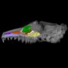
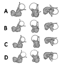
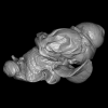
Explodable 3D Dog Skull for Veterinary Education
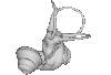
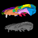
3D GM dataset of bird skeletal variation
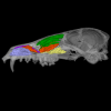
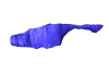
Skeletal embryonic development in the catshark
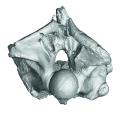
Bony connexions of the petrosal bone of extant hippos
bony labyrinth (14) , inner ear (11) , Eocene (11) , geometric morphometrics (10) , CT-scan (10) , Oligocene (9) , Micro-CT (9)
Maëva Judith Orliac (24) , Lionel Hautier (24) , Laurent Marivaux (18) , Renaud Lebrun (15) , Rodolphe Tabuce (14) , Pierre-Olivier Antoine (13) , Bastien Mennecart (13)
|
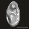
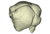
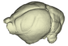
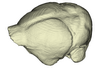
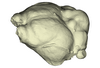
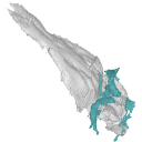
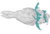
3D models related to the publication: Morphogenesis of the inner ear at different stages of normal human developmentSaki Toyoda, Naoto Shiraki, Shigehito Yamada
Published online: 22/10/2015 |

|
M3#36Computationally reconstructed membranous labyrinth of a human embryo (KC-CS17IER29248) at Carnegie Stage 17 (Crown Rump Length= 7mm). Type: "3D_surfaces"doi: 10.18563/m3.sf36 state:published |
Download 3D surface file |
Homo sapiens KC-CS18IER17746 View specimen

|
M3#37Computationally reconstructed membranous labyrinth of a human embryo (KC-CS18IER17746) at Carnegie Stage 18 (Crown Rump Length= 12mm). Type: "3D_surfaces"doi: 10.18563/m3.sf37 state:published |
Download 3D surface file |
Homo sapiens KC-CS19IER16127 View specimen

|
M3#38Computationally reconstructed membranous labyrinth of a human embryo (KC-CS19IER16127) at Carnegie Stage 19 (Crown Rump Length= 13mm). Type: "3D_surfaces"doi: 10.18563/m3.sf38 state:published |
Download 3D surface file |
Homo sapiens KC-CS20IER20268 View specimen

|
M3#39Computationally reconstructed membranous labyrinth of a human embryo (KC-CS20IER20268) at Carnegie Stage 20 (Crown Rump Length= 13.7mm). Type: "3D_surfaces"doi: 10.18563/m3.sf39 state:published |
Download 3D surface file |
Homo sapiens KC-CS21IER28066 View specimen

|
M3#40Computationally reconstructed membranous labyrinth of a human embryo (KC-CS21IER28066) at Carnegie Stage 21 (Crown Rump Length= 16.7mm). Type: "3D_surfaces"doi: 10.18563/m3.sf40 state:published |
Download 3D surface file |
Homo sapiens KC-CS22IER35233 View specimen

|
M3#41Computationally reconstructed membranous labyrinth of a human embryo (KC-CS22IER35233) at Carnegie Stage 22 (Crown Rump Length= 22mm). Type: "3D_surfaces"doi: 10.18563/m3.sf41 state:published |
Download 3D surface file |
Homo sapiens KC-CS23IER15919 View specimen

|
M3#42Computationally reconstructed membranous labyrinth of a human embryo (KC-CS23IER15919) at Carnegie Stage 23 (Crown Rump Length= 32.3mm). Type: "3D_surfaces"doi: 10.18563/m3.sf42 state:published |
Download 3D surface file |
Homo sapiens KC-FIER52730 View specimen

|
M3#43Computationally reconstructed human membranous labyrinth in post embryonic phase (KC-FIER52730). Crown Rump Length: 43.5mm. Type: "3D_surfaces"doi: 10.18563/m3.sf43 state:published |
Download 3D surface file |
This contribution contains the three-dimensional models of the most complete and/or informative fossil materials attributed to Peradectes crocheti Gernelle, 2024, the earliest peradectid metatherian species of Europe, from its type locality (Palette, Provence, ~55 Ma). These specimens were analyzed and discussed in: Gernelle et al. (2024), Taxonomy and evolutionary history of peradectids (Metatheria): new data from the early Eocene of France. https://doi.org/10.1007/s10914-024-09724-5
Peradectes crocheti MHN.AIX.PV.2018.26.14 View specimen
|
M3#14993D surface model of MHN.AIX.PV.2018.26.14, fragmentary left maxilla with C-P1, anterior root of P2, and M1-M3 Type: "3D_surfaces"doi: 10.18563/m3.sf.1499 state:published |
Download 3D surface file |
Peradectes crocheti MHN.AIX.PV.2017.6.6 View specimen
|
M3#15003D surface model of MHN.AIX.PV.2017.6.6, left P2 Type: "3D_surfaces"doi: 10.18563/m3.sf.1500 state:published |
Download 3D surface file |
Peradectes crocheti MHN.AIX.PV.2017.6.7 View specimen
|
M3#15013D surface model of MHN.AIX.PV.2017.6.7, left M3 Type: "3D_surfaces"doi: 10.18563/m3.sf.1501 state:published |
Download 3D surface file |
Peradectes crocheti MHN.AIX.PV.2017.6.8 View specimen
|
M3#15023D surface model of MHN.AIX.PV.2017.6.8, right hemi-mandible fragment with canine alveolus, posterior root of p1, partial p2, p3, partial m1, and m2-m3 Type: "3D_surfaces"doi: 10.18563/m3.sf.1502 state:published |
Download 3D surface file |
Peradectes crocheti MHN.AIX.PV.2017.6.9 View specimen
|
M3#15033D surface model of MHN.AIX.PV.2017.6.9, leftm1-m4 row with fragments of dentary Type: "3D_surfaces"doi: 10.18563/m3.sf.1503 state:published |
Download 3D surface file |
Peradectes crocheti MHN.AIX.PV.2017.6.14 View specimen
|
M3#15043D surface model of MHN.AIX.PV.2017.6.14, right astragalus Type: "3D_surfaces"doi: 10.18563/m3.sf.1504 state:published |
Download 3D surface file |
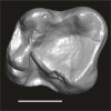
This contribution contains the three-dimensional models of the turbinal complex of 10 myrmecophagous and 10 non-myrmecophagous placental species. These specimens were analyzed and discussed in: Wright et. al (2024), Sniffing out morphological convergence in the turbinal complex of myrmecophagous placentals. https://doi.org/10.1002/ar.25603
Priodontes maximus NHMUK 732-a View specimen

|
M3#1536Turbinals of Priodontes maximus Type: "3D_surfaces"doi: 10.18563/m3.sf.1536 state:published |
Download 3D surface file |
Dasypus pilosus NHMUK 94-10-1-13 View specimen
|
M3#1537Turbinals of Dasypus pilosus Type: "3D_surfaces"doi: 10.18563/m3.sf.1537 state:published |
Download 3D surface file |
Dasypus novemcinctus AMNH 263287 View specimen
|
M3#1538Turbinals of Dasypus novemcinctus Type: "3D_surfaces"doi: 10.18563/m3.sf.1538 state:published |
Download 3D surface file |
Bradypus tridactylus UM 789N View specimen
|
M3#1539Turbinals of Bradypus tridactylus Type: "3D_surfaces"doi: 10.18563/m3.sf.1539 state:published |
Download 3D surface file |
Choloepus didactylus UM 767V View specimen
|
M3#1540Turbinals of Choloepus didactylus Type: "3D_surfaces"doi: 10.18563/m3.sf.1540 state:published |
Download 3D surface file |
Cyclopes didactylus NHMUK 88-8-8-14 View specimen

|
M3#1541Turbinals of Cyclopes didactylus Type: "3D_surfaces"doi: 10.18563/m3.sf.1541 state:published |
Download 3D surface file |
Myrmecophaga tridactyla UM 065V View specimen
|
M3#1542Turbinals of Myrmecophaga tridactyla Type: "3D_surfaces"doi: 10.18563/m3.sf.1542 state:published |
Download 3D surface file |
Tamandua tetradactyla NHMUK 3-7-7-135 View specimen
|
M3#1543Turbinals of Tamandua tetradactyla Type: "3D_surfaces"doi: 10.18563/m3.sf.1543 state:published |
Download 3D surface file |
Tamandua mexicana NHMUK 79-1-6-1 View specimen
|
M3#1544Turbinals of Tamandua mexicana Type: "3D_surfaces"doi: 10.18563/m3.sf.1544 state:published |
Download 3D surface file |
Orycteropus afer NHMUK 2-9-9-58 View specimen
|
M3#1545Turbinals of Orycteropus afer Type: "3D_surfaces"doi: 10.18563/m3.sf.1545 state:published |
Download 3D surface file |
Tenrec eucaudatus UM N439 View specimen
|
M3#1546Turbinals of Tenrec eucaudatus Type: "3D_surfaces"doi: 10.18563/m3.sf.1546 state:published |
Download 3D surface file |
Elephantulus rozeti UM N227 View specimen
|
M3#1547Turbinals of Elephantulus rozeti Type: "3D_surfaces"doi: 10.18563/m3.sf.1547 state:published |
Download 3D surface file |
Phataginus tetradactyla NHMUK 1-11-21-35 View specimen

|
M3#1548Turbinals of Phataginus tetradactyla Type: "3D_surfaces"doi: 10.18563/m3.sf.1548 state:published |
Download 3D surface file |
Smutsia gigantea KMMA 25479 View specimen

|
M3#1549Turbinals of Smutsia gigantea Type: "3D_surfaces"doi: 10.18563/m3.sf.1549 state:published |
Download 3D surface file |
Manis culionensis MNHN ZM-MO 1884-1822 View specimen

|
M3#1550Turbinals of Manis culionensis Type: "3D_surfaces"doi: 10.18563/m3.sf.1550 state:published |
Download 3D surface file |
Vulpes vulpes UM N140 View specimen
|
M3#1551Turbinals of Vulpes vulpes Type: "3D_surfaces"doi: 10.18563/m3.sf.1551 state:published |
Download 3D surface file |
Alopex lagopus UM N110 View specimen

|
M3#1552Turbinals of Alopex lagopus Type: "3D_surfaces"doi: 10.18563/m3.sf.1552 state:published |
Download 3D surface file |
Felis silvestris UM N149 View specimen
|
M3#1553Turbinals of Felis sylvestris Type: "3D_surfaces"doi: 10.18563/m3.sf.1553 state:published |
Download 3D surface file |
Hyaena hyaena UM N109 View specimen
|
M3#1554Turbinals of Hyaena hyaena Type: "3D_surfaces"doi: 10.18563/m3.sf.1554 state:published |
Download 3D surface file |
Proteles cristata NHMUK 4-3-1-58 View specimen

|
M3#1555Turbinals of Proteles cristata Type: "3D_surfaces"doi: 10.18563/m3.sf.1555 state:published |
Download 3D surface file |
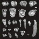
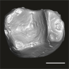
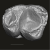
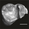
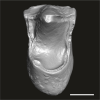
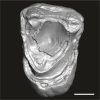
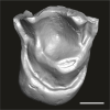
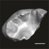
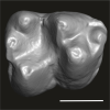
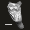
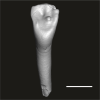
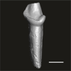
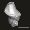
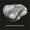
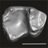
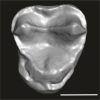
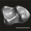
This contribution contains the three-dimensional digital models of the dental fossil material of anthropoid and strepsirrhine primates, discovered in Lower Oligocene detrital deposits outcropping in the Porto Rico and El Argoub areas, east of the Dakhla peninsula region (Atlantic Sahara; in the south of Morocco, near the northern border of Mauritania). These fossils were described, figured and discussed in the following publication: Marivaux et al. (2024), A new primate community from the earliest Oligocene of the Atlantic margin of Northwest Africa: Systematic, paleobiogeographic and paleoenvironmental implications. Journal of Human Evolution. https://doi.org/10.1016/j.jhevol.2024.103548
Catopithecus aff. browni DAK-Arg-087 View specimen

|
M3#1211Isolated right lower m3 (worn) Type: "3D_surfaces"doi: 10.18563/m3.sf.1211 state:published |
Download 3D surface file |
Catopithecus aff. browni DAK-Arg-088 View specimen
|
M3#1212Isolated right lower m2 (abraded/corroded) Type: "3D_surfaces"doi: 10.18563/m3.sf.1212 state:published |
Download 3D surface file |
Catopithecus aff. browni DAK-Arg-089 View specimen
|
M3#1213Isolated left lower m1 (worn) Type: "3D_surfaces"doi: 10.18563/m3.sf.1213 state:published |
Download 3D surface file |
Catopithecus aff. browni DAK-Pto-052 View specimen
|
M3#1214Isolated right lower m1 (pristine but lacking the mesiobuccal region) Type: "3D_surfaces"doi: 10.18563/m3.sf.1214 state:published |
Download 3D surface file |
Catopithecus aff. browni DAK-Arg-090 View specimen
|
M3#1215Isolated left upper P4 Type: "3D_surfaces"doi: 10.18563/m3.sf.1215 state:published |
Download 3D surface file |
Catopithecus aff. browni DAK-Arg-091 View specimen
|
M3#1216Isolated left upper M2 (worn and corroded) Type: "3D_surfaces"doi: 10.18563/m3.sf.1216 state:published |
Download 3D surface file |
Catopithecus aff. browni DAK-Pto-053 View specimen
|
M3#1217Isolated right upper M1 (lacking the buccal region) Type: "3D_surfaces"doi: 10.18563/m3.sf.1217 state:published |
Download 3D surface file |
Abuqatrania cf. basiodontos DAK-Arg-092 View specimen
|
M3#1218Isolated left lower c1 Type: "3D_surfaces"doi: 10.18563/m3.sf.1218 state:published |
Download 3D surface file |
?Propliopithecus sp. DAK-Pto-056 View specimen
|
M3#1219Isolated right lower m3 (fragment of talonid of a germ) Type: "3D_surfaces"doi: 10.18563/m3.sf.1219 state:published |
Download 3D surface file |
Abuqatrania cf. basiodontos DAK-Arg-093 View specimen
|
M3#1469Isolated right lower m1 Type: "3D_surfaces"doi: 10.18563/m3.sf.1469 state:published |
Download 3D surface file |
Abuqatrania cf. basiodontos DAK-Arg-094 View specimen
|
M3#1221Isolated left upper M1 or M2 (corroded, lacking the enamel cap [exposed dentine]) Type: "3D_surfaces"doi: 10.18563/m3.sf.1221 state:published |
Download 3D surface file |
Abuqatrania cf. basiodontos DAK-Arg-095 View specimen
|
M3#1222Isolated right lower i1 or i2 Type: "3D_surfaces"doi: 10.18563/m3.sf.1222 state:published |
Download 3D surface file |
Abuqatrania cf. basiodontos DAK-Arg-096 View specimen
|
M3#1223Isolated right lower p2 (worn apex) Type: "3D_surfaces"doi: 10.18563/m3.sf.1223 state:published |
Download 3D surface file |
Abuqatrania cf. basiodontos DAK-Arg-097 View specimen
|
M3#1224Isolated right lower p2 (worn apex and broken root) Type: "3D_surfaces"doi: 10.18563/m3.sf.1224 state:published |
Download 3D surface file |
Afrotarsius sp. DAK-Arg-098 View specimen
|
M3#1225Isolated left lower p3 Type: "3D_surfaces"doi: 10.18563/m3.sf.1225 state:published |
Download 3D surface file |
Afrotarsius sp. DAK-Pto-054 View specimen
|
M3#1226Isolated right lower m1 (abraded/corroded) Type: "3D_surfaces"doi: 10.18563/m3.sf.1226 state:published |
Download 3D surface file |
Orolemur mermozi DAK-Pto-055 View specimen
|
M3#1227Isolated right upper M1 or M2 (pristine, Holotype) Type: "3D_surfaces"doi: 10.18563/m3.sf.1227 state:published |
Download 3D surface file |
Wadilemur cf. elegans DAK-Arg-099 View specimen
|
M3#1228Isolated right lower m2 Type: "3D_surfaces"doi: 10.18563/m3.sf.1228 state:published |
Download 3D surface file |
cf. 'Anchomomys' milleri DAK-Arg-100 View specimen

|
M3#1229Isolated right lower c1 Type: "3D_surfaces"doi: 10.18563/m3.sf.1229 state:published |
Download 3D surface file |
Abuqatrania cf. basiodontos DAK-Arg-101 View specimen
|
M3#1396Isolated left upper M3 (abraded) Type: "3D_surfaces"doi: 10.18563/m3.sf.1396 state:published |
Download 3D surface file |
Orogalago saintexuperyi DAK-Arg-102 View specimen
|
M3#1397Isolated left lower m2 Type: "3D_surfaces"doi: 10.18563/m3.sf.1397 state:published |
Download 3D surface file |
Wadilemur cf. elegans DAK-Arg-103 View specimen

|
M3#1473Isolated right upper M1 or M2 (lacking the mesial and buccal regions) Type: "3D_surfaces"doi: 10.18563/m3.sf.1473 state:published |
Download 3D surface file |
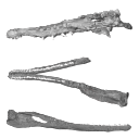
The present 3D dataset contains the 3D models of the holotype of Proterochampsa nodosa that were built and analysed in “Redescription, taxonomic revaluation, and phylogenetic affinities of Proterochampsa nodosa (Archosauriformes: Proterochampsidae), early Late Triassic of Candelaria Sequence (Santa Maria Supersequence)”.
Proterochampsa nodosa MCP 1694-PV View specimen
|
M3#9743D models of Proterochampsa nodosa. Model 1: skull. Model 2: mandible. Model 3: left mandibular ramus. Type: "3D_surfaces"doi: 10.18563/m3.sf.974 state:published |
Download 3D surface file |
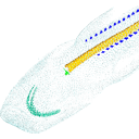
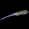
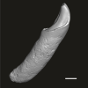
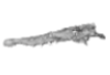
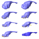
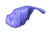
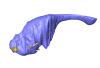
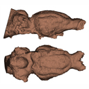
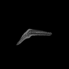
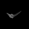
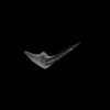
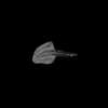
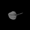
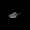
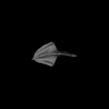
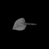
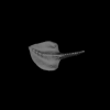
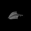
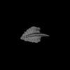
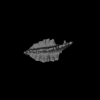
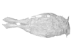
Current knowledge on the skeletogenesis of Chondrichthyes is scarce compared with their extant sister group, the bony fishes. Most of the previously described developmental tables in Chondrichthyes have focused on embryonic external morphology only. Due to its small body size and relative simplicity to raise eggs in laboratory conditions, the small-spotted catshark Scyliorhinus canicula has emerged as a reference species to describe developmental mechanisms in the Chondrichthyes lineage. Here we investigate the dynamic of mineralization in a set of six embryonic specimens using X-ray microtomography and describe the developing units of both the dermal skeleton (teeth and dermal scales) and endoskeleton (vertebral axis). This preliminary data on skeletogenesis in the catshark sets the first bases to a more complete investigation of the skeletal developmental in Chondrichthyes. It should provide comparison points with data known in osteichthyans and could thus be used in the broader context of gnathostome skeletal evolution.
Scyliorhinus canicula SC6_2_2015_03_20 View specimen

|
M3#50Mineralized skeleton of a 6,2 cm long embryo of Scyliorhinus canicula Type: "3D_surfaces"doi: 10.18563/m3.sf.50 state:published |
Download 3D surface file |
Scyliorhinus canicula SC6_7_2015_03_20 View specimen

|
M3#51Mineralized skeleton of a 6,7 cm long embryo of Scyliorhinus canicula Type: "3D_surfaces"doi: 10.18563/m3.sf.51 state:published |
Download 3D surface file |
Scyliorhinus canicula SC7_1_2015_04_03 View specimen

|
M3#52Mineralized skeleton of a 7,1 cm long embryo of Scyliorhinus canicula Type: "3D_surfaces"doi: 10.18563/m3.sf.52 state:published |
Download 3D surface file |
Scyliorhinus canicula SC7_5_2015_03_13 View specimen

|
M3#53Mineralized skeleton of a 7,5 cm long embryo of Scyliorhinus canicula Type: "3D_surfaces"doi: 10.18563/m3.sf.53 state:published |
Download 3D surface file |
Scyliorhinus canicula SC8_2015_03_20 View specimen

|
M3#54Mineralized skeleton of a 8 cm long embryo of Scyliorhinus canicula Type: "3D_surfaces"doi: 10.18563/m3.sf.54 state:published |
Download 3D surface file |
Scyliorhinus canicula SC10_2015_02_27 View specimen

|
M3#55Mineralized skeleton of a 10 cm long embryo of Scyliorhinus canicula Type: "3D_surfaces"doi: 10.18563/m3.sf.55 state:published |
Download 3D surface file |
The present 3D Dataset contains the 3D models analyzed in the following manuscript: L. Roese-Miron, M.E.H. Jones, J.D. Ferreira and A.S. Hsiou., 2023. Virtual endocasts of Clevosaurus brasiliensis and the tuatara: Rhynchocephalian neuroanatomy and the oldest endocranial record for Lepidosauria.
Sphenodon punctatus CM 30660 View specimen
|
M3#10993D surface model of the cranial endocast of specimen CM 30660 (Sphenodon punctatus). Type: "3D_surfaces"doi: 10.18563/m3.sf.1099 state:published |
Download 3D surface file |
Sphenodon punctatus KCLZJ 001 View specimen
|
M3#11003D surface models of the cranial endocast and the initial trunks of the cranial nerves of specimen KCLZJ 001 (Sphenodon punctatus). Type: "3D_surfaces"doi: 10.18563/m3.sf.1100 state:published |
Download 3D surface file |
Sphenodon punctatus LDUCZ x0036 View specimen
|
M3#11013D surface models of the cranial endocast and the initial trunks of the cranial nerves of specimen LDUCZ x0036 (Sphenodon punctatus). Type: "3D_surfaces"doi: 10.18563/m3.sf.1101 state:published |
Download 3D surface file |
Sphenodon punctatus LDUCZ x1126 View specimen
|
M3#11023D surface model of the cranial endocast of specimen LDUCZ x1126 (Sphenodon punctatus). Type: "3D_surfaces"doi: 10.18563/m3.sf.1102 state:published |
Download 3D surface file |
Clevosaurus brasiliensis MCN PV 2852 View specimen
|
M3#11033D surface model of the cranial endocast of specimen MCN PV 2852 (Clevosaurus brasiliensis). Type: "3D_surfaces"doi: 10.18563/m3.sf.1103 state:published |
Download 3D surface file |
Sphenodon punctatus SAMA 70524 View specimen
|
M3#11043D surface models of the cranial endocast, brain, endosseous labyrinth and initial trunks of the cranial nerves of specimen SAMA 70524 (Sphenodon punctatus). Type: "3D_surfaces"doi: 10.18563/m3.sf.1104 state:published |
Download 3D surface file |
Sphenodon punctatus SU1 View specimen
|
M3#11053D surface models of the cranial endocast and the initial trunks of the cranial nerves of specimen SU1 (Sphenodon punctatus). Type: "3D_surfaces"doi: 10.18563/m3.sf.1105 state:published |
Download 3D surface file |
Sphenodon punctatus YPM HERR 009194 View specimen
|
M3#11063D surface models of the cranial endocast and the initial trunks of the cranial nerves of specimen YPM HERR 009194 (Sphenodon punctatus). Type: "3D_surfaces"doi: 10.18563/m3.sf.1106 state:published |
Download 3D surface file |
The present 3D Dataset contains the 3D model analyzed in Wazir, W. A., Sehgal, R. K., Čerňanský, A., Patnaik, R., Kumar, N., Singh, A. P. and Singh, N. P. 2022. A find from the Ladakh Himalaya reveals a survival of madtsoiid snakes (Serpentes, Madtsoiidae) in India through the late Oligocene. Journal of Vertebrate Paleontology, 41(6), e2058401. https://doi.org/10.1080/02724634.2021.2058401
indet. indet. WIMF/A 4816 View specimen

|
M3#1754Vertebra Type: "3D_surfaces"doi: 10.18563/m3.sf.1754 state:published |
Download 3D surface file |
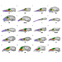
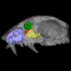
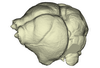
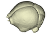
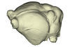
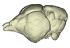
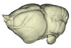
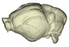
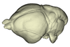
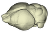
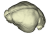
The present 3D Dataset contains the 3D models of extant Chiropteran endocranial casts, documenting 16 of the 19 extant bat families. They are used by Maugoust & Orliac (2023) to assess the correspondences between the brain and brain-surrounding tissues (i.e., neural tissues, blood vessels, meninges) and their imprint on the braincase, allowing for eventually proposing a Chiroptera-scale nomenclature of the endocast.
Balantiopteryx plicata UMMZ 102659 View specimen
|
M3#1132Endocranial cast of the corresponding cranium of Balantiopteryx plicata Type: "3D_surfaces"doi: 10.18563/m3.sf.1132 state:published |
Download 3D surface file |
Idiurus macrotis AMNH M-187705 View specimen
|
M3#1133Endocranial cast of the corresponding cranium of Nycteris macrotis Type: "3D_surfaces"doi: 10.18563/m3.sf.1133 state:published |
Download 3D surface file |
Thyroptera tricolor UMMZ 53240 View specimen
|
M3#1134Endocranial cast of the corresponding cranium of Thyroptera tricolor Type: "3D_surfaces"doi: 10.18563/m3.sf.1134 state:published |
Download 3D surface file |
Noctilio albiventris UMMZ 105827 View specimen
|
M3#1135Endocranial cast of the corresponding cranium of Noctilio albiventris Type: "3D_surfaces"doi: 10.18563/m3.sf.1135 state:published |
Download 3D surface file |
Mormoops blainvillii AMNH M-271513 View specimen
|
M3#1136Endocranial cast of the corresponding cranium of Mormoops blainvillii Type: "3D_surfaces"doi: 10.18563/m3.sf.1136 state:published |
Download 3D surface file |
Macrotus waterhousii UMMZ 95718 View specimen
|
M3#1137Endocranial cast of the corresponding cranium of Macrotus waterhousii Type: "3D_surfaces"doi: 10.18563/m3.sf.1137 state:published |
Download 3D surface file |
Nyctiellus lepidus UMMZ 105767 View specimen
|
M3#1138Endocranial cast of the corresponding cranium of Nyctiellus lepidus Type: "3D_surfaces"doi: 10.18563/m3.sf.1138 state:published |
Download 3D surface file |
Cheiromeles torquatus AMNH M-247585 View specimen
|
M3#1139Endocranial cast of the corresponding cranium of Cheiromeles torquatus Type: "3D_surfaces"doi: 10.18563/m3.sf.1139 state:published |
Download 3D surface file |
Miniopterus schreibersii UMMZ 156998 View specimen
|
M3#1140Endocranial cast of the corresponding cranium of Miniopterus schreibersii Type: "3D_surfaces"doi: 10.18563/m3.sf.1140 state:published |
Download 3D surface file |
Kerivoula pellucida UMMZ 161396 View specimen
|
M3#1141Endocranial cast of the corresponding cranium of Kerivoula pellucida Type: "3D_surfaces"doi: 10.18563/m3.sf.1141 state:published |
Download 3D surface file |
Scotophilus kuhlii UMMZ 157013 View specimen
|
M3#1142Endocranial cast of the corresponding cranium of Scotophilus kuhlii Type: "3D_surfaces"doi: 10.18563/m3.sf.1142 state:published |
Download 3D surface file |
Rhinolophus luctus MNHN CG-2006-87 View specimen
|
M3#1143Endocranial cast of the corresponding cranium of Rhinolophus luctus Type: "3D_surfaces"doi: 10.18563/m3.sf.1143 state:published |
Download 3D surface file |
Triaenops persicus AM RG-38552 View specimen
|
M3#1144Endocranial cast of the corresponding cranium of Triaenops persicus Type: "3D_surfaces"doi: 10.18563/m3.sf.1144 state:published |
Download 3D surface file |
Hipposideros armiger UM ZOOL-762-V View specimen
|
M3#1145Endocranial cast of the corresponding cranium of Hipposideros armiger Type: "3D_surfaces"doi: 10.18563/m3.sf.1145 state:published |
Download 3D surface file |
Lavia frons AM RG-12268 View specimen
|
M3#1146Endocranial cast of the corresponding cranium of Lavia frons Type: "3D_surfaces"doi: 10.18563/m3.sf.1146 state:published |
Download 3D surface file |
Rhinopoma hardwickii AM RG-M31166 View specimen
|
M3#1147Endocranial cast of the corresponding cranium of Rhinopoma hardwickii Type: "3D_surfaces"doi: 10.18563/m3.sf.1147 state:published |
Download 3D surface file |
Sphaerias blanfordi AMNH M-274330 View specimen
|
M3#1148Endocranial cast of the corresponding cranium of Sphaerias blanfordi Type: "3D_surfaces"doi: 10.18563/m3.sf.1148 state:published |
Download 3D surface file |
Rousettus aegyptiacus UMMZ 161026 View specimen
|
M3#1149Endocranial cast of the corresponding cranium of Rousettus aegyptiacus Type: "3D_surfaces"doi: 10.18563/m3.sf.1149 state:published |
Download 3D surface file |
Pteropus pumilus UMMZ 162253 View specimen
|
M3#1150Endocranial cast of the corresponding cranium of Pteropus pumilus Type: "3D_surfaces"doi: 10.18563/m3.sf.1150 state:published |
Download 3D surface file |
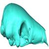
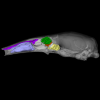
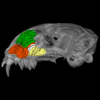
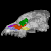
Our knowledge of the external brain morphology of the late Eocene artiodactyl ungulate Mixtotherium, relies on a plaster model realized on a specimen from the Victor Brun Museum in Montauban (France) and described by Dechaseaux (1973). Here, based on micro CT-scan data, we virtually reconstruct the 3D cast of the empty cavity of the partial cranium MA PHQ 716 from the Victor Brun Museum and compare it to the plaster model illustrated and described by Dechaseaux (1973). Indeed, the specimen from which the original plaster endocast originates was not identified by Dechaseaux by a specimen number. We confirm here that the studied specimen was indeed the one described and illustrated by Dechaseaux (1973). We also reconstruct a second, more detailed, model providing additional morphological and quantitative observations made available by micro CT scan investigation such as precisions on the neopallium folding and endocranial volumes.
Mixtotherium cuspidatum MA PHQ 716 View specimen
|
M3#857endocast of the brain cavity Type: "3D_surfaces"doi: 10.18563/m3.sf.857 state:published |
Download 3D surface file |
This contribution contains the 3D models described and figured in the following publication: Paulina-Carabajal, A. and Nieto, M. N. In press. Brief comment on the brain and inner ear of Giganotosaurus carolinii (Dinosauria: Theropoda) based on CT scans. Ameghiniana. https://doi.org/10.5710/AMGH.25.10.2019.3237
Giganotosaurus carolinii MUCPv-CH-1 View specimen
|
M3#504The current file contents 3D models of the braincase, brain, left and right inner ears Type: "3D_surfaces"doi: 10.18563/m3.sf.504 state:published |
Download 3D surface file |
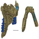
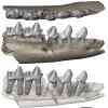
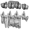
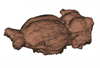
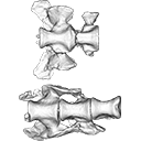
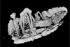
This contribution contains the 3D models described and figured in the following publication: Gaetano, L. C., Abdala, F., Mancuso, C, and Vega N.2025. New traversodontid cynodont from the Late Triassic Chañares Formation. Publicación Electrónica de la Asociación Paleontológica Argentina.
Pontognathus ignotus PULR-V 287 View specimen
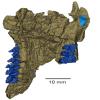
|
M3#1647partial snout preserving the lateralmost incisor, the base of the canine, and several postcanines Type: "3D_surfaces"doi: 10.18563/m3.sf.1647 state:published |
Download 3D surface file |
Massetognathus pascuali PULR-V 289 View specimen
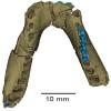
|
M3#1646partial lower jaw Type: "3D_surfaces"doi: 10.18563/m3.sf.1646 state:published |
Download 3D surface file |
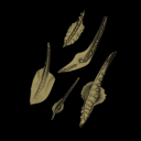
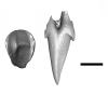
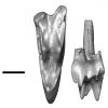
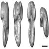
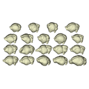
The present 3D Dataset contains the 3D models analyzed in Assemat et al. 2023: Shape diversity in conodont elements, a quantitative study using 3D topography. Marine Micropaleontology 184. https://doi.org/10.1016/j.marmicro.2023.102292
P1 elements represent dental components of the conodont apparatus that perform the final stage of food processing before ingestion. Consequently, quantifying the shape of P1 elements across the topographic indices of different conodont species becomes crucial for deciphering the diversity in feeding behavior within this group.
Bispathodus aculeatus UM CTB 082 View specimen
|
M3#1404P element Type: "3D_surfaces"doi: 10.18563/m3.sf.1404 state:published |
Download 3D surface file |
Bispathodus aculeatus UM CTB 083 View specimen

|
M3#1405P element Type: "3D_surfaces"doi: 10.18563/m3.sf.1405 state:published |
Download 3D surface file |
Bispathodus aculeatus UM CTB 086 View specimen
|
M3#1406P element Type: "3D_surfaces"doi: 10.18563/m3.sf.1406 state:published |
Download 3D surface file |
Bispathodus ultimus UM CTB 088 View specimen

|
M3#1407P element Type: "3D_surfaces"doi: 10.18563/m3.sf.1407 state:published |
Download 3D surface file |
Bispathodus aculeatus UM CTB 089 View specimen
|
M3#1408P element Type: "3D_surfaces"doi: 10.18563/m3.sf.1408 state:published |
Download 3D surface file |
Bispathodus costatus UM CTB 090 View specimen
|
M3#1409P element Type: "3D_surfaces"doi: 10.18563/m3.sf.1409 state:published |
Download 3D surface file |
Bispathodus ultimus UM CTB 092 View specimen
|
M3#1410P element Type: "3D_surfaces"doi: 10.18563/m3.sf.1410 state:published |
Download 3D surface file |
Bispathodus costatus UM CTB 093 View specimen
|
M3#1411P element Type: "3D_surfaces"doi: 10.18563/m3.sf.1411 state:published |
Download 3D surface file |
Bispathodus spinulicostatus UM CTB 094 View specimen
|
M3#1412P element Type: "3D_surfaces"doi: 10.18563/m3.sf.1412 state:published |
Download 3D surface file |
Bispathodus aculeatus UM CTB 096 View specimen

|
M3#1413P element Type: "3D_surfaces"doi: 10.18563/m3.sf.1413 state:published |
Download 3D surface file |
Bispathodus ultimus UM CTB 098 View specimen

|
M3#1414P element Type: "3D_surfaces"doi: 10.18563/m3.sf.1414 state:published |
Download 3D surface file |
Bispathodus costatus UM CTB 060 View specimen
|
M3#1415P element Type: "3D_surfaces"doi: 10.18563/m3.sf.1415 state:published |
Download 3D surface file |
Bispathodus spinulicostatus UM CTB 073 View specimen

|
M3#1416P element Type: "3D_surfaces"doi: 10.18563/m3.sf.1416 state:published |
Download 3D surface file |
Branmehla suprema UM CTB 049 View specimen

|
M3#1417P element Type: "3D_surfaces"doi: 10.18563/m3.sf.1417 state:published |
Download 3D surface file |
Branmehla inornata UM CTB 100 View specimen
|
M3#1418P element Type: "3D_surfaces"doi: 10.18563/m3.sf.1418 state:published |
Download 3D surface file |
Bispathodus stabilis (morphe 1) UM CTB 101 View specimen

|
M3#1419P element Type: "3D_surfaces"doi: 10.18563/m3.sf.1419 state:published |
Download 3D surface file |
Branmehla suprema UM CTB 102 View specimen
|
M3#1420P element Type: "3D_surfaces"doi: 10.18563/m3.sf.1420 state:published |
Download 3D surface file |
Branmehla suprema UM CTB 103 View specimen

|
M3#1421P element Type: "3D_surfaces"doi: 10.18563/m3.sf.1421 state:published |
Download 3D surface file |
Branmehla suprema UM CTB 104 View specimen

|
M3#1422P element Type: "3D_surfaces"doi: 10.18563/m3.sf.1422 state:published |
Download 3D surface file |
Branmehla suprema UM CTB 105 View specimen
|
M3#1423P element Type: "3D_surfaces"doi: 10.18563/m3.sf.1423 state:published |
Download 3D surface file |
Branmehla suprema UM CTB 106 View specimen
|
M3#1424P element Type: "3D_surfaces"doi: 10.18563/m3.sf.1424 state:published |
Download 3D surface file |
Branmehla suprema UM CTB 072 View specimen

|
M3#1425P element Type: "3D_surfaces"doi: 10.18563/m3.sf.1425 state:published |
Download 3D surface file |
Branmehla suprema UM CTB 107 View specimen
|
M3#1426P element Type: "3D_surfaces"doi: 10.18563/m3.sf.1426 state:published |
Download 3D surface file |
Branmehla suprema UM CTB 108 View specimen

|
M3#1427P element Type: "3D_surfaces"doi: 10.18563/m3.sf.1427 state:published |
Download 3D surface file |
Branmehla suprema UM CTB 109 View specimen
|
M3#1428P element Type: "3D_surfaces"doi: 10.18563/m3.sf.1428 state:published |
Download 3D surface file |
Bispathodus stabilis (morphe 1) UM CTB 110 View specimen

|
M3#1429P element Type: "3D_surfaces"doi: 10.18563/m3.sf.1429 state:published |
Download 3D surface file |
Palmatolepis gracilis UM CTB 112 View specimen
|
M3#1430P element Type: "3D_surfaces"doi: 10.18563/m3.sf.1430 state:published |
Download 3D surface file |
Palmatolepis gracilis UM CTB 061 View specimen

|
M3#1431P element Type: "3D_surfaces"doi: 10.18563/m3.sf.1431 state:published |
Download 3D surface file |
Palmatolepis gracilis UM CTB 115 View specimen
|
M3#1432P element Type: "3D_surfaces"doi: 10.18563/m3.sf.1432 state:published |
Download 3D surface file |
Palmatolepis gracilis UM CTB 116 View specimen
|
M3#1433P element Type: "3D_surfaces"doi: 10.18563/m3.sf.1433 state:published |
Download 3D surface file |
Palmatolepis gracilis UM CTB 117 View specimen
|
M3#1434P element Type: "3D_surfaces"doi: 10.18563/m3.sf.1434 state:published |
Download 3D surface file |
Palmatolepis gracilis UM CTB 062 View specimen

|
M3#1435P element Type: "3D_surfaces"doi: 10.18563/m3.sf.1435 state:published |
Download 3D surface file |
Palmatolepis gracilis UM CTB 118 View specimen

|
M3#1436P element Type: "3D_surfaces"doi: 10.18563/m3.sf.1436 state:published |
Download 3D surface file |
Palmatolepis gracilis UM CTB 119 View specimen
|
M3#1437P element Type: "3D_surfaces"doi: 10.18563/m3.sf.1437 state:published |
Download 3D surface file |
Palmatolepis gracilis UM CTB 120 View specimen
|
M3#1438P element Type: "3D_surfaces"doi: 10.18563/m3.sf.1438 state:published |
Download 3D surface file |
Polygnathus communis UM CTB 075 View specimen
|
M3#1439P element Type: "3D_surfaces"doi: 10.18563/m3.sf.1439 state:published |
Download 3D surface file |
Polygnathus communis UM CTB 121 View specimen
|
M3#1440P element Type: "3D_surfaces"doi: 10.18563/m3.sf.1440 state:published |
Download 3D surface file |
Polygnathus communis UM CTB 122 View specimen
|
M3#1441P element Type: "3D_surfaces"doi: 10.18563/m3.sf.1441 state:published |
Download 3D surface file |
Polygnathus communis UM CTB 123 View specimen
|
M3#1442P element Type: "3D_surfaces"doi: 10.18563/m3.sf.1442 state:published |
Download 3D surface file |
Polygnathus communis UM CTB 125 View specimen
|
M3#1443P element Type: "3D_surfaces"doi: 10.18563/m3.sf.1443 state:published |
Download 3D surface file |
Polygnathus communis UM CTB 126 View specimen
|
M3#1444P element Type: "3D_surfaces"doi: 10.18563/m3.sf.1444 state:published |
Download 3D surface file |
Polygnathus communis UM CTB 128 View specimen
|
M3#1445P element Type: "3D_surfaces"doi: 10.18563/m3.sf.1445 state:published |
Download 3D surface file |
Polygnathus communis UM CTB 130 View specimen
|
M3#1446P element Type: "3D_surfaces"doi: 10.18563/m3.sf.1446 state:published |
Download 3D surface file |
Polygnathus communis UM CTB 131 View specimen
|
M3#1447P element Type: "3D_surfaces"doi: 10.18563/m3.sf.1447 state:published |
Download 3D surface file |
Polygnathus communis UM CTB 132 View specimen

|
M3#1448P element Type: "3D_surfaces"doi: 10.18563/m3.sf.1448 state:published |
Download 3D surface file |
Polygnathus communis UM CTB 133 View specimen
|
M3#1449P element Type: "3D_surfaces"doi: 10.18563/m3.sf.1449 state:published |
Download 3D surface file |
Polygnathus symmetricus UM CTB 139 View specimen
|
M3#1450P element Type: "3D_surfaces"doi: 10.18563/m3.sf.1450 state:published |
Download 3D surface file |
Polygnathus symmetricus UM CTB 140 View specimen

|
M3#1451P element Type: "3D_surfaces"doi: 10.18563/m3.sf.1451 state:published |
Download 3D surface file |
Polygnathus symmetricus UM CTB 141 View specimen
|
M3#1452P element Type: "3D_surfaces"doi: 10.18563/m3.sf.1452 state:published |
Download 3D surface file |
Polygnathus symmetricus UM CTB 142 View specimen
|
M3#1453P element Type: "3D_surfaces"doi: 10.18563/m3.sf.1453 state:published |
Download 3D surface file |
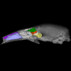
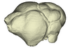
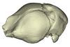
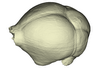
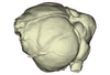
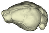
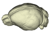
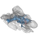
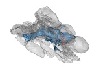
The present 3D Dataset contains the 3D models of the endocranial cast of two specimens of Indohyus indirae described in the article entitled “The endocranial cast of Indohyus (Artiodactyla, Raoellidae): the origin of the cetacean brain” (Orliac and Thewissen, 2021). They represent the cast of the main cavity of the braincase as well as associated intraosseous sinuses.
Indohyus indirae RR 207 View specimen
|
M3#710cast of the main endocranial cavity and associated intraosseous sinuses Type: "3D_surfaces"doi: 10.18563/m3.sf.710 state:published |
Download 3D surface file |
Indohyus indirae RR 601 View specimen
|
M3#711casts of the main endocranial cavity Type: "3D_surfaces"doi: 10.18563/m3.sf.711 state:published |
Download 3D surface file |
The present 3D Dataset contains the 3D models of the sacral vertebrae analyzed in “Sacral co-ossification in dinosaurs: The oldest record of fused sacral vertebrae in Dinosauria and the diversity of sacral co-ossification patterns in the group”.
Buriolestes schultzi CAPPA/UFSM 0035 View specimen
|
M3#705Sacral vertebrae of Buriolestes schultzi Type: "3D_surfaces"doi: 10.18563/m3.sf.705 state:published |
Download 3D surface file |
indet indet CAPPA/UFSM 0228 View specimen
|
M3#706Sacral vertebrae of a saurischian dinosaur indet. Type: "3D_surfaces"doi: 10.18563/m3.sf.706 state:published |
Download 3D surface file |
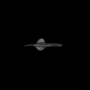
The present 3D Dataset contains the 3D models analyzed in the following publication: Size variation under domestication: Conservatism in the inner ear shape of wolves, dogs and dingoes. Scientific Reports 7, Article number: 13330, https://doi.org/10.1038/s41598-017-13523-9.
Canis lupus familiaris NMBE 16 View specimen

|
M3#2293D virtual endocast of the left inner ear Type: "3D_surfaces"doi: 10.18563/m3.sf.229 state:published |
Download 3D surface file |
Canis lupus familiaris NMBE-LAT-1136 View specimen

|
M3#2423D virtual endocast of the left inner ear Type: "3D_surfaces"doi: 10.18563/m3.sf.242 state:published |
Download 3D surface file |
Canis lupus familiaris NMBE-LAT-1119 View specimen

|
M3#2433D virtual endocast of the left inner ear Type: "3D_surfaces"doi: 10.18563/m3.sf.243 state:published |
Download 3D surface file |
Canis lupus familiaris NMBE-BUR-1057 View specimen

|
M3#2443D virtual endocast of the left inner ear Type: "3D_surfaces"doi: 10.18563/m3.sf.244 state:published |
Download 3D surface file |
Canis lupus familiaris NMBE-LUS-1102 View specimen

|
M3#2453D virtual endocast of the left inner ear Type: "3D_surfaces"doi: 10.18563/m3.sf.245 state:published |
Download 3D surface file |
Canis lupus familiaris NMBE-LUS-1095 View specimen

|
M3#2463D virtual endocast of the left inner ear Type: "3D_surfaces"doi: 10.18563/m3.sf.246 state:published |
Download 3D surface file |
Canis lupus familiaris NMBE-DUR-1124 View specimen

|
M3#2473D virtual endocast of the left inner ear Type: "3D_surfaces"doi: 10.18563/m3.sf.247 state:published |
Download 3D surface file |
Canis lupus chanco ZMUZH 17603 View specimen

|
M3#2483D virtual endocast of the left inner ear Type: "3D_surfaces"doi: 10.18563/m3.sf.248 state:published |
Download 3D surface file |
Canis lupus chanco ZMUZH 20201 View specimen

|
M3#2493D virtual endocast of the left inner ear Type: "3D_surfaces"doi: 10.18563/m3.sf.249 state:published |
Download 3D surface file |
Canis lupus chanco ZMUZH 17602 View specimen

|
M3#2503D virtual endocast of the left inner ear Type: "3D_surfaces"doi: 10.18563/m3.sf.250 state:published |
Download 3D surface file |
Canis lupus ZMUZH 13854 View specimen

|
M3#2403D virtual endocast of the left inner ear Type: "3D_surfaces"doi: 10.18563/m3.sf.240 state:published |
Download 3D surface file |
Canis lupus chanco ZMUZH 20202 View specimen

|
M3#2393D virtual endocast of the left inner ear Type: "3D_surfaces"doi: 10.18563/m3.sf.239 state:published |
Download 3D surface file |
Canis lupus chanco ZMUZH 17612 View specimen

|
M3#2303D virtual endocast of the left inner ear Type: "3D_surfaces"doi: 10.18563/m3.sf.230 state:published |
Download 3D surface file |
Canis lupus chanco ZMUZH 18082 View specimen

|
M3#2313D virtual endocast of the left inner ear Type: "3D_surfaces"doi: 10.18563/m3.sf.231 state:published |
Download 3D surface file |
Canis lupus ZMUZH 17118 View specimen

|
M3#2323D virtual endocast of the left inner ear Type: "3D_surfaces"doi: 10.18563/m3.sf.232 state:published |
Download 3D surface file |
Canis lupus ZMUZH 15858 View specimen

|
M3#2333D virtual endocast of the left inner ear Type: "3D_surfaces"doi: 10.18563/m3.sf.233 state:published |
Download 3D surface file |
Canis lupus familiaris ZMUZH 17712 View specimen

|
M3#2343D virtual endocast of the left inner ear Type: "3D_surfaces"doi: 10.18563/m3.sf.234 state:published |
Download 3D surface file |
Canis lupus familiaris ZMUZH 17713 View specimen

|
M3#2353D virtual endocast of the left inner ear Type: "3D_surfaces"doi: 10.18563/m3.sf.235 state:published |
Download 3D surface file |
Canis lupus familiaris ZMUZH 10166 View specimen

|
M3#2363D virtual endocast of the left inner ear Type: "3D_surfaces"doi: 10.18563/m3.sf.236 state:published |
Download 3D surface file |
Canis lupus familiaris ZMUZH 10175 View specimen

|
M3#2373D virtual endocast of the left inner ear Type: "3D_surfaces"doi: 10.18563/m3.sf.237 state:published |
Download 3D surface file |
Canis lupus familiaris ZMUZH 14842 View specimen

|
M3#2383D virtual endocast of the left inner ear Type: "3D_surfaces"doi: 10.18563/m3.sf.238 state:published |
Download 3D surface file |
Canis lupus familiaris ZMUZH 10342 View specimen

|
M3#2513D virtual endocast of the left inner ear Type: "3D_surfaces"doi: 10.18563/m3.sf.251 state:published |
Download 3D surface file |
Canis lupus familiaris ZMUZH 10343 View specimen

|
M3#2523D virtual endocast of the left inner ear Type: "3D_surfaces"doi: 10.18563/m3.sf.252 state:published |
Download 3D surface file |
Canis lupus familiaris ZMUZH 13766 View specimen

|
M3#2533D virtual endocast of the left inner ear Type: "3D_surfaces"doi: 10.18563/m3.sf.253 state:published |
Download 3D surface file |
Canis lupus familiaris ZMUZH 17717 View specimen

|
M3#2653D virtual endocast of the left inner ear Type: "3D_surfaces"doi: 10.18563/m3.sf.265 state:published |
Download 3D surface file |
Canis lupus familiaris ZMUZH 17711 View specimen

|
M3#2663D virtual endocast of the left inner ear Type: "3D_surfaces"doi: 10.18563/m3.sf.266 state:published |
Download 3D surface file |
Canis lupus familiaris ZMUZH 17714 View specimen

|
M3#2673D virtual endocast of the left inner ear Type: "3D_surfaces"doi: 10.18563/m3.sf.267 state:published |
Download 3D surface file |
Canis lupus familiaris ZMUZH 17715 View specimen

|
M3#2683D virtual endocast of the left inner ear Type: "3D_surfaces"doi: 10.18563/m3.sf.268 state:published |
Download 3D surface file |
Canis lupus familiaris PIMUZ A/V 2835 View specimen

|
M3#2693D virtual endocast of the left inner ear Type: "3D_surfaces"doi: 10.18563/m3.sf.269 state:published |
Download 3D surface file |
Canis lupus familiaris PIMUZ A/V 2834 View specimen

|
M3#2703D virtual endocast of the left inner ear Type: "3D_surfaces"doi: 10.18563/m3.sf.270 state:published |
Download 3D surface file |
Canis lupus familiaris PIMUZ A/V 2837 View specimen

|
M3#2713D virtual endocast of the left inner ear Type: "3D_surfaces"doi: 10.18563/m3.sf.271 state:published |
Download 3D surface file |
Canis lupus familiaris PIMUZ A/V 2831 View specimen

|
M3#2723D virtual endocast of the left inner ear Type: "3D_surfaces"doi: 10.18563/m3.sf.272 state:published |
Download 3D surface file |
Canis lupus familiaris PIMUZ A/V 2845 View specimen

|
M3#2733D virtual endocast of the left inner ear Type: "3D_surfaces"doi: 10.18563/m3.sf.273 state:published |
Download 3D surface file |
Canis lupus familiaris PIMUZ A/V 3001 View specimen

|
M3#2643D virtual endocast of the left inner ear Type: "3D_surfaces"doi: 10.18563/m3.sf.264 state:published |
Download 3D surface file |
Canis lupus familiaris PIMUZ A/V 2832 View specimen

|
M3#2633D virtual endocast of the left inner ear Type: "3D_surfaces"doi: 10.18563/m3.sf.263 state:published |
Download 3D surface file |
Canis lupus familiaris PIMUZ A/V 3000 View specimen

|
M3#2543D virtual endocast of the left inner ear Type: "3D_surfaces"doi: 10.18563/m3.sf.254 state:published |
Download 3D surface file |
Canis lupus familiaris PIMUZ A/V 2847 View specimen

|
M3#2553D virtual endocast of the left inner ear Type: "3D_surfaces"doi: 10.18563/m3.sf.255 state:published |
Download 3D surface file |
Canis lupus familiaris PIMUZ A/V 2846 View specimen

|
M3#2563D virtual endocast of the left inner ear Type: "3D_surfaces"doi: 10.18563/m3.sf.256 state:published |
Download 3D surface file |
Canis lupus familiaris PIMUZ A/V 2836 View specimen

|
M3#2573D virtual endocast of the left inner ear Type: "3D_surfaces"doi: 10.18563/m3.sf.257 state:published |
Download 3D surface file |
Canis lupus familiaris NMB 12080 View specimen

|
M3#2583D virtual endocast of the left inner ear Type: "3D_surfaces"doi: 10.18563/m3.sf.258 state:published |
Download 3D surface file |
Canis lupus familiaris NMB 12081 View specimen

|
M3#2593D virtual endocast of the left inner ear Type: "3D_surfaces"doi: 10.18563/m3.sf.259 state:published |
Download 3D surface file |
Canis lupus familiaris NMB 12079 View specimen

|
M3#2603D virtual endocast of the left inner ear Type: "3D_surfaces"doi: 10.18563/m3.sf.260 state:published |
Download 3D surface file |
Canis lupus familiaris NMB 12078 View specimen

|
M3#2613D virtual endocast of the left inner ear Type: "3D_surfaces"doi: 10.18563/m3.sf.261 state:published |
Download 3D surface file |
Canis lupus familiaris NMBE 1051209 View specimen

|
M3#2623D virtual endocast of the left inner ear Type: "3D_surfaces"doi: 10.18563/m3.sf.262 state:published |
Download 3D surface file |
Canis lupus familiaris NMBE 1051226 View specimen

|
M3#2283D virtual endocast of the left inner ear Type: "3D_surfaces"doi: 10.18563/m3.sf.228 state:published |
Download 3D surface file |
Canis lupus familiaris NMBE 1051381 View specimen

|
M3#2213D virtual endocast of the left inner ear Type: "3D_surfaces"doi: 10.18563/m3.sf.221 state:published |
Download 3D surface file |
Canis lupus familiaris NMBE 1051418 View specimen

|
M3#1843D virtual endocast of the left inner ear Type: "3D_surfaces"doi: 10.18563/m3.sf.184 state:published |
Download 3D surface file |
Canis lupus familiaris ZMUZH A.II. View specimen

|
M3#1973D virtual endocast of the left inner ear Type: "3D_surfaces"doi: 10.18563/m3.sf.197 state:published |
Download 3D surface file |
Canis lupus familiaris ZMUZH A.VII. View specimen

|
M3#1983D virtual endocast of the left inner ear Type: "3D_surfaces"doi: 10.18563/m3.sf.198 state:published |
Download 3D surface file |
Canis lupus familiaris ZMUZH We.6. View specimen

|
M3#1993D virtual endocast of the left inner ear Type: "3D_surfaces"doi: 10.18563/m3.sf.199 state:published |
Download 3D surface file |
Canis lupus familiaris ZMUZH Ez.2. View specimen

|
M3#2003D virtual endocast of the left inner ear Type: "3D_surfaces"doi: 10.18563/m3.sf.200 state:published |
Download 3D surface file |
Canis lupus familiaris ZMUZH Ez.E. View specimen

|
M3#2013D virtual endocast of the left inner ear Type: "3D_surfaces"doi: 10.18563/m3.sf.201 state:published |
Download 3D surface file |
Canis lupus familiaris ZMUZH A.6. View specimen

|
M3#2023D virtual endocast of the left inner ear Type: "3D_surfaces"doi: 10.18563/m3.sf.202 state:published |
Download 3D surface file |
Canis lupus familiaris ZMUZH Wyn.9. View specimen

|
M3#2033D virtual endocast of the left inner ear Type: "3D_surfaces"doi: 10.18563/m3.sf.203 state:published |
Download 3D surface file |
Canis lupus familiaris ZMUZH F.48. View specimen

|
M3#2043D virtual endocast of the left inner ear Type: "3D_surfaces"doi: 10.18563/m3.sf.204 state:published |
Download 3D surface file |
Canis lupus familiaris ZMUZH Terp.1. View specimen

|
M3#2053D virtual endocast of the left inner ear Type: "3D_surfaces"doi: 10.18563/m3.sf.205 state:published |
Download 3D surface file |
Canis lupus familiaris ZMUZH A.VIII. View specimen

|
M3#1963D virtual endocast of the left inner ear Type: "3D_surfaces"doi: 10.18563/m3.sf.196 state:published |
Download 3D surface file |
Canis lupus familiaris ZMUZH A.VI. View specimen

|
M3#1953D virtual endocast of the left inner ear Type: "3D_surfaces"doi: 10.18563/m3.sf.195 state:published |
Download 3D surface file |
Canis lupus familiaris ZMUZH A.IV. View specimen

|
M3#1853D virtual endocast of the left inner ear Type: "3D_surfaces"doi: 10.18563/m3.sf.185 state:published |
Download 3D surface file |
Canis lupus familiaris NMBE A.403. View specimen

|
M3#1873D virtual endocast of the left inner ear Type: "3D_surfaces"doi: 10.18563/m3.sf.187 state:published |
Download 3D surface file |
Canis lupus familiaris NMBE A.5.a. View specimen

|
M3#1883D virtual endocast of the left inner ear Type: "3D_surfaces"doi: 10.18563/m3.sf.188 state:published |
Download 3D surface file |
Canis lupus NMB 8381 View specimen

|
M3#1893D virtual endocast of the left inner ear Type: "3D_surfaces"doi: 10.18563/m3.sf.189 state:published |
Download 3D surface file |
Canis lupus lycaon NMB C.1362 View specimen

|
M3#1903D virtual endocast of the left inner ear Type: "3D_surfaces"doi: 10.18563/m3.sf.190 state:published |
Download 3D surface file |
Canis lupus NMB Z309 View specimen

|
M3#1913D virtual endocast of the left inner ear Type: "3D_surfaces"doi: 10.18563/m3.sf.191 state:published |
Download 3D surface file |
Canis lupus NMB 2761 View specimen

|
M3#1923D virtual endocast of the left inner ear Type: "3D_surfaces"doi: 10.18563/m3.sf.192 state:published |
Download 3D surface file |
Canis lupus occidentalis NMB No Nb View specimen

|
M3#1933D virtual endocast of the left inner ear Type: "3D_surfaces"doi: 10.18563/m3.sf.193 state:published |
Download 3D surface file |
Canis lupus NMB 5258 View specimen

|
M3#1943D virtual endocast of the left inner ear Type: "3D_surfaces"doi: 10.18563/m3.sf.194 state:published |
Download 3D surface file |
Canis lupus NMB SCM320 View specimen

|
M3#2063D virtual endocast of the left inner ear Type: "3D_surfaces"doi: 10.18563/m3.sf.206 state:published |
Download 3D surface file |
Canis lupus arabs NMB 11019 View specimen

|
M3#2073D virtual endocast of the left inner ear Type: "3D_surfaces"doi: 10.18563/m3.sf.207 state:published |
Download 3D surface file |
Canis lupus UMZC K.3141 View specimen

|
M3#2083D virtual endocast of the left inner ear Type: "3D_surfaces"doi: 10.18563/m3.sf.208 state:published |
Download 3D surface file |
Canis lupus UMZC K.3150.1 View specimen

|
M3#2193D virtual endocast of the left inner ear Type: "3D_surfaces"doi: 10.18563/m3.sf.219 state:published |
Download 3D surface file |
Canis lupus UMZC K.3152 View specimen

|
M3#2203D virtual endocast of the left inner ear Type: "3D_surfaces"doi: 10.18563/m3.sf.220 state:published |
Download 3D surface file |
Canis lupus UMZC K.3149 View specimen

|
M3#2223D virtual endocast of the left inner ear Type: "3D_surfaces"doi: 10.18563/m3.sf.222 state:published |
Download 3D surface file |
Canis lupus familiaris UMZC K.3016 View specimen

|
M3#2233D virtual endocast of the left inner ear Type: "3D_surfaces"doi: 10.18563/m3.sf.223 state:published |
Download 3D surface file |
Canis lupus occidentalis ZMUZH 17210 View specimen

|
M3#2243D virtual endocast of the left inner ear Type: "3D_surfaces"doi: 10.18563/m3.sf.224 state:published |
Download 3D surface file |
Canis lupus familiaris SZ 7961 View specimen

|
M3#2253D virtual endocast of the left inner ear Type: "3D_surfaces"doi: 10.18563/m3.sf.225 state:published |
Download 3D surface file |
Canis lupus familiaris SZ 7959 View specimen

|
M3#2263D virtual endocast of the left inner ear Type: "3D_surfaces"doi: 10.18563/m3.sf.226 state:published |
Download 3D surface file |
Canis lupus familiaris SZ 7958 View specimen

|
M3#2173D virtual endocast of the left inner ear Type: "3D_surfaces"doi: 10.18563/m3.sf.217 state:published |
Download 3D surface file |
Canis lupus familiaris SZ 7930 View specimen

|
M3#2163D virtual endocast of the left inner ear Type: "3D_surfaces"doi: 10.18563/m3.sf.216 state:published |
Download 3D surface file |
Canis lupus familiaris SZ 7926 View specimen

|
M3#2183D virtual endocast of the left inner ear Type: "3D_surfaces"doi: 10.18563/m3.sf.218 state:published |
Download 3D surface file |
Canis lupus familiaris SZ 7929 View specimen

|
M3#2093D virtual endocast of the left inner ear Type: "3D_surfaces"doi: 10.18563/m3.sf.209 state:published |
Download 3D surface file |
Canis lupus dingo M6297 View specimen

|
M3#1863D virtual endocast of the left inner ear Type: "3D_surfaces"doi: 10.18563/m3.sf.186 state:published |
Download 3D surface file |
Canis lupus dingo M24153 View specimen

|
M3#2103D virtual endocast of the left inner ear Type: "3D_surfaces"doi: 10.18563/m3.sf.210 state:published |
Download 3D surface file |
Canis lupus dingo M33608 View specimen

|
M3#2113D virtual endocast of the left inner ear Type: "3D_surfaces"doi: 10.18563/m3.sf.211 state:published |
Download 3D surface file |
Canis lupus dingo M38587 View specimen

|
M3#2123D virtual endocast of the left inner ear Type: "3D_surfaces"doi: 10.18563/m3.sf.212 state:published |
Download 3D surface file |
Canis lupus dingo Blumenbach UMZC K.3221 View specimen

|
M3#2133D virtual endocast of the left inner ear Type: "3D_surfaces"doi: 10.18563/m3.sf.213 state:published |
Download 3D surface file |
Canis lupus dingo Blumenbach UMZC K.3223 View specimen

|
M3#2143D virtual endocast of the left inner ear Type: "3D_surfaces"doi: 10.18563/m3.sf.214 state:published |
Download 3D surface file |
Canis lupus dingo UniSyd FVS 45 View specimen

|
M3#2153D virtual endocast of the left inner ear Type: "3D_surfaces"doi: 10.18563/m3.sf.215 state:published |
Download 3D surface file |
Canis lupus dingo UNSW Z354 View specimen

|
M3#2273D virtual endocast of the left inner ear Type: "3D_surfaces"doi: 10.18563/m3.sf.227 state:published |
Download 3D surface file |
Canis lupus familiaris TMM M-150 View specimen
|
M3#2413D virtual endocast of the left inner ear Type: "3D_surfaces"doi: 10.18563/m3.sf.241 state:published |
Download 3D surface file |
Canis lupus M39960 View specimen

|
M3#2743D virtual endocast of the left inner ear Type: "3D_surfaces"doi: 10.18563/m3.sf.274 state:published |
Download 3D surface file |
Canis lupus NMB 8635 View specimen

|
M3#2753D virtual endocast of the left inner ear Type: "3D_surfaces"doi: 10.18563/m3.sf.275 state:published |
Download 3D surface file |
The present 3D dataset contains the 3D models analyzed in the publication: Rosa, R. M., Salvador, R. B., & Cavallari, D. C. (2025). The disappearing act of the magician tree snail: anatomy, distribution, and phylogenetic relationships of Drymaeus magus (Gastropoda: Bulimulidae), a long-lost species hidden in plain sight. Zoological Journal of the Linnean Society.
Drymaeus magus CMRP 1049 View specimen
|
M3#1597Internal organs of Drymaeus magus Type: "3D_surfaces"doi: 10.18563/m3.sf.1597 state:published |
Download 3D surface file |
|
M3#1598External surface of Drymaeus magus Type: "3D_surfaces"doi: 10.18563/m3.sf.1598 state:published |
Download 3D surface file |
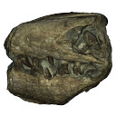
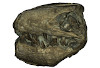
The present 3D Dataset contains the 3D model analyzed in the following publication: Carolina A. Hoffmann, A. G. Martinelli & M. B. Andrade. 2023. Anatomy of the holotype of “Probelesodon” kitchingi revisited, a chiniquodontid cynodont (Synapsida, Probainognathia) from the early Late Triassic of southern Brazil, Journal of Paleontology
Probelesodon kitchingi MCP 1600 PV View specimen
|
M3#11513D models of the skull with segmented bones and without the segmentation. colormap and orientation files also added. Type: "3D_surfaces"doi: 10.18563/m3.sf.1151 state:published |
Download 3D surface file |

This project presents a µCT dataset and an associated 3D surface model of the holotype of Donrussellia magna (UM PAT 17; Primates, Adapiformes). UM PAT17 is the only known specimen for the species and consists of a well-preserved left lower jaw with p4-m3. It documents one of the oldest European primates, eventually dated near the Paleocene Eocene Thermal Maximum.
Donrussellia magna UM PAT 17 View specimen

|
M3#173D surface file model of UM PAT 17 (type specimen of Donrussellia magna), which is a well preserved left lower jaw with p4-m3. The teeth (and roots) were manually segmented. Type: "3D_surfaces"doi: 10.18563/m3.sf17 state:published |
Download 3D surface file |
|
M3#18CT Scan Data of Donrussellia magna UM PAT 17. Voxel size (in µm): 36µm (isotropic voxels). Dimensions in x,y,z : 594 pixels, 294 pixels, 1038 pixels. Image type : 8-bit voxels. Image format : raw data format (no header). Type: "3D_CT"doi: 10.18563/m3.sf18 state:published |
Download CT data |
The present 3D Dataset contains the 3D model used in in the following publication: Interacting with the inaccessible: utilization of multimedia-based visual contents of Japan’s National Monument, the Taniwhasaurus mikasaensis (Mosasauridae) holotype for educational workshops at Mikasa City Museum.
Taniwhasaurus mikasaensis MCM.M0009 View specimen
|
M3#499Taniwhasaurus mikasaensis, Caldwell et al. 2008 Type: "3D_surfaces"doi: 10.18563/m3.sf.499 state:published |
Download 3D surface file |